Sharif University of Technology
Scientia IranicaTransactions B: Mechanical Engineering www.scientiairanica.com
A computational model for estimation of mechanical
parameters in chemotactic endothelial cells
A. Kiyoumarsioskouei, A. Shamloo
, S. Azimi, M. Abeddoust and M.S. Saidi
School of Mechanical Engineering, Sharif University of Technology, Tehran, Iran.Received 9 September 2014; received in revised form 29 December 2014; accepted 25 April 2015
KEYWORDS Cell motility; Chemotaxis; Reaction-diusion model;
Cell cytoeskeleton simulation.
Abstract. A cell migration numerical simulation is presented to mimic the motility of endothelial cells subjected to the concentration gradients of a Forebrain embryoniccortical neuron Conditioned Medium (CM). This factor was previously shown to induce the directional chemotaxis of endothelial cells with an over-expressed G protein coupled receptor 124 (GPR 124). A cell simulator program incorporates basic elements of the cell cytoskeleton, including membrane, nucleus and cytoskeleton. The developed 2D cell model is capable of responding to concentration gradients of biochemical factors by changing the cytoskeleton arrangement. Random walk force, cell drag force and cell inertial eects are also implemented into the cell migration to complete the simulation of the phenomenon. The obtained results of cell migration were calibrated with experimental cell chemotaxis data. This model can be implemented for prediction of cell behavior during cell chemotaxis and also provides a powerful tool to explain the cell migration phenomenon, mechanistically. © 2016 Sharif University of Technology. All rights reserved.
1. Introduction
Cell migration plays a pivotal role in numerous biolog-ical phenomena such as wound healing, embryogenesis, cancer invasion and inammatory response. It is a highly complex process in which dierent elements of the cell, including actin laments, surface signaling receptors and microtubules, are involved. Though highly investigated by dierent researchers in biology and engineering sciences, the mechanisms underlying the migration of cells are still unidentied in many aspects. Finding the answers to some fundamental questions in this area would, undoubtedly, have a great impact on dierent elds of biology.
Dierent types of cell demonstrate diverse be-havior during migration and thus, proposing a single unique mechanism that can be generalized for all types
*. Corresponding author. Tel.: +98 21 66165691; Fax: +98 21 66165599
E-mail address: [email protected] (A. Shamloo)
of cell is impossible in practice. However, the cells are similar in exhibiting cyclic intracellular processes while migrating [1]. After being stimulated by a migration promoting agent, such as the concentration gradient of a biochemical factor, at the rst step, the migrating cell polarizes and extends some protrusions in the direction of the imposed gradient. Actin lament polymerization creates lamellipodia and lopodia that become stabilized by adhering to the extracellular matrix (ECM) or the neighboring cell surfaces [2]. At the second step, the lamellipodia and lopodia at the rear part of the cell attenuate their adhesion to the ECM. Finally, the polarized cell moves forward. The cells repeat this cycle until the external signal diminishes and they receive no stimuli to polarize.
Among the signals which stimulate directional migration of the cells, the gradient of the biochemical species in the external media of the cell is of high importance. The chemotaxis of neutrophils is the underlying factor in the body's immune reaction. They migrate to the location of infection or injury because
of a chemical signal they receive from other cells in the immune system or the microbes at the infection site [3]. As a matter of fact, almost all cells sense extracellular biochemical signals, and many of them react by migrating in response [4].
Various mathematical models have been proposed to simulate the cell migration process and also various computational models have been developed based on these mathematical models. Meinhardt proposed one of the basic and primary computational models for cell chemotaxis simulation [5]. He attributed the cells directional sensitivity to an inherent system inside the cells that translates the extracellular signals of the gradients of chemo attractants into the formation of an intracellular pattern, which, in turn, results in the extension of lamellipodia and lopodia towards the source of biochemical factors. Similar to the model of Gieier and Meinhardt, many researchers, studying the chemotaxis in eukaryotic cells, considered a hypo-thetical \compass needle" mechanism inside the cells that governs the formation of new pseudopods towards the chemo attractant [6-9]. While these compass based models are able to represent some aspects of cell chemotaxis successfully, many observations show that they are usually limited to steep gradients of the chemo attractant since, in shallow gradients, the cell are more controlled by their internal dynamics rather than chemo attractants. In order to overcome this shortcoming, Neilson et al. proposed a pseudopod-based computational model that simply attributes cell chemotaxis to the movement of pseudopods on the cell surface, not a compass inside the cells [10]. In their model, the pseudopods themselves respond to the chemo attractant molecules through a positive or negative feedback loop, and, so, orient cell migra-tion.
Also, Andrew and Insall have shown that many chemotactic cells form a protrusion that splits in a Y-like manner into two branches, only one of which eventually survives [11]. Their study and related works have demonstrated very complex behavior during cell movements and have claimed that it is not possible to consider all the details of cell chemotaxis in a mathematical model [12,13]. Therefore, in this work, the rst target is to simulate cell chemotaxis using a simple mathematical model in such a way that this phenomenon is consistent with experimental data.
In the present study, the numerical simulation of cell motility during the chemotaxis of endothelial cells is performed. The constant biochemical factor concentration gradient around the cell stimulates the cell simulator. This simulator is a simple model for a cell which includes nucleus, cytoskeleton, and membrane. Intracellular mechanics explains how the cell simulator is developed, and, nally, the results are discussed.
2. Interacellular mechanics
In order to investigate the eects of an imposed constant concentration gradient, a model is required to simulate cell polarization and migration. The study of cell chemotaxis might be possible by considering every cell as a single point. However, simulation of the cytoskeletal components would not only give a better physical representation of the phenomenon, but the details of the migrating cell's cytoskeleton would also be appreciable, and, thus, helpful for future studies like cell proliferation and group migration phenomena. Kiyoumarsioskouei et al. have oered a 3-dimensional endothelial cell model to describe a numerical method for simulating the cell cytoskeleton [14]. In the current study, three basic elements of a cell; elastic plasma membrane, cell cytoskeleton, and cell nucleus, are considered in the model. The cell membrane and nucleus are initially divided into N0 elements, each of
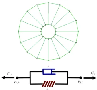
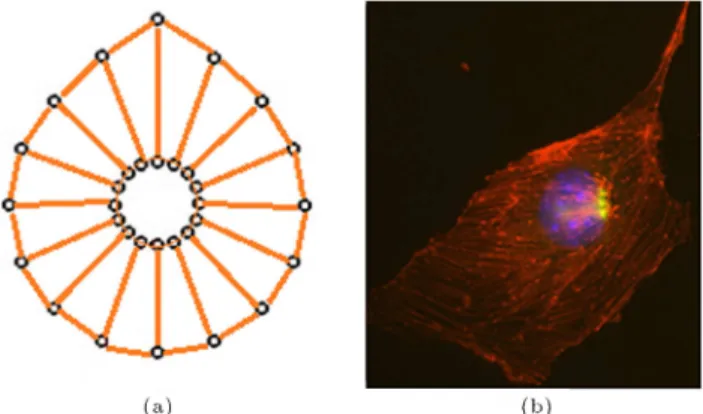
which is modeled by a damper and a spring arranged in parallel (Voigt Model). The damper accounts for the viscous eects, while the spring is responsible for mimicking the elastic behavior of cell elements. This model for investigating cellular and subcellular behavior was previously utilized by Jamali et al. [15] and showed strong capabilities in representation of subcellular behavior, including cell motility. Each node on the cell is connected to the corresponding point on the cell nucleus using a Voigt unit, as depicted in Figure 1. In the same manner, each node on the cell membrane and cell nucleus are connected to their two neighboring nodes.
Acting forces on each element are presented in Eq. (1):
Figure 1. Basic discretized elements of a cell (up) by Voigt units (down). Each element of the discretized cell that connects two nodes consists of a spring and a damper in parallel.
Ftot
i;k = Fi;kinner+ Fi;kcell-cell+ Fi;kcell-ECM+ F(i;k)ext : (1)
Ftot
i;k is the total force acting on a node, Fi;kinner is the
total force resulting from the inner elements of the cell, Fcell-cell
i;k is the force due to a neighboring cell, Fi;kcell-ECM
is the force exerted on the node by the ECM, and Fext i;k
is the result of an external force like a magnetic eld. All mentioned forces will be discussed in detail in the following sections.
2.1. Inner force calculation
The total force acting on a node on the membrane resulting from interaction with other nodes of the inner parts is the sum of three forces: The membrane Voigt units' forces, the cytoskeleton forces and the force due to the inside pressure of the cell. The forces due to the membrane and cytoskeleton are the sum of spring and damper forces. The spring force, Eq. (2), is proportional to changes in its length, and the proportionality factor was adopted from [15]:
Fspring= k
q
(Xj+1 Xj)2+ (Yj+1 Yj)2 l0
; (2) where l0 is the initial length of the Voigt springs, and
X and Y are the coordinates of nodes connected by the Voigt member.
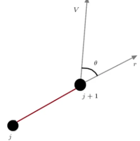
The damper force is proportional to the inner product of velocity and position vectors, as shown schematically in Figure 2.
F = ~V:^r; (3)
where ~V is the velocity vector and ^r is the normalized position vector.
Moreover the membrane is subjected to a force that is a result of the pressure dierence between the inside and outside of the cell. The magnitude of this force is:
Fi;jm;n= (Pin Pout)R=2; (4)
and its direction is normal to the cell surface.
Figure 2. In calculating viscous forces, only the component of velocity in the direction of Voigt nodes contributes to viscous forces.
2.2. Components of cell motility
Cell shape and position change continuously as a result of dierent cell processes, like cell growth, cell migration, mitosis, apoptosis etc.
During cell migration, the cell is polarized and specic protrusions form around its body. The mi-gration of the cell in this study is the result of the concentration gradient of biochemical factors. The important question that arises here is how do the cells polarize, make protrusions and nally migrate in the direction of the imposed concentration gradient.
It is agreed by many researchers that cell mi-gration during chemotaxis is the outcome of four components, Eq. (5) [1]. The rst three components account for cell activities as a living organism. The rst component that contributes to cell migration is the eect of biochemical factors. Another component is cell memory while migrating. In other words, cells are willing to continue movement in their former direction of migrating while subjected to a gradient in a new direction. The third component is the random motion cells demonstrate while migrating. Finally, the last component is the drag force that aects every object in a uid ow. In other words, the rst three components will disappear if we hypothetically assume that the cells are nonliving objects, while the last component is always present, regardless of cells being assumed living or nonliving.
*
Vm= *
Vc+ (1 ) *
Vm+ *
Vr+ *
Vd;
: Blending factor: (5)
When the gradient of a biochemical factor is im-posed on the cells, the density of related receptors for that factor in the membrane is amplied in the leading edge [16]. As an example, Narang et al. utilized the reaction-diusion model, established by Gierer and Meinhardt, to simulate the accumulation of PI(3,4,5)P3 [6]. However, the set of partial dierential
equations they have established is sti. They have proposed four equations, which all are nonlinear and highly coupled. As a result, their model is highly expensive in terms of computational cost. For the current study, a simple linear model is employed in which only the gradient direction is considered. For the sake of simplicity, the gradient magnitude is assumed to be constant through the extracellular medium. This assumption is reasonable for the current study, as the gradient of biochemical species is constant. Therefore, the*Vc component of velocity can be generated by the
following scheme:
1. The gradient direction is sensed by the cell simula-tor;
2. The location of the leading edge due to the external signal is dened;
Figure 3. The procedure for calculating the
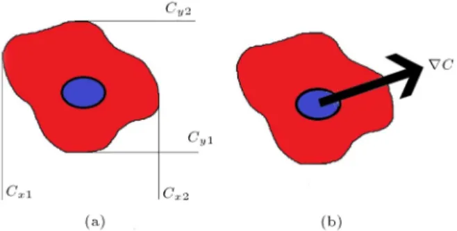
concentration gradient: a) Cell concentration limits; and b) total gradient resulted from the superposition principle.
3. The spring connecting the nucleus to the leading edge is designated;
4. The stiness of this spring is increased, causing a net force acting on the cell;
5. *Vc, the velocity of the cell, resulting from forces
acting upon it, due to external signals, is calculated. In the mentioned cell simulator, the eld of biochemical factor concentration has been solved using the compu-tational uid dynamics method, so the simulator would be able to check the position of the cell and calculate the concentration dierence between the two ends of the cell in two independent directions, as shown in Figure 3.
Therefore, the direction of the gradient can be calculated by the superposition of the gradients in the two perpendicular directions (see Figure 3(b)).
Based on experimental observations, migrating cells tend to maintain their direction of motion, [17]. It means that the cells have a resistance against changing their velocities, so, this force is like inertia inside the cells to maintain its instantaneous velocity. In this simulation, when the velocity of the cell is refreshed at each time step, the new velocity is calculated, as the weighted mean of cell velocity from the previous time step. The*Vm component appearing in Eq. (5) is
the velocity of migration from the previous time step,
*
Vpastm .
The last factor contributing to cell migration is the random walks of the cell. Random motions are considered in the modeling of cell migration in order to include the eects of unknown factors. In this study, at each time step, two random numbers are generated which are used to build a random velocity component,
*
Vr.5
3. Tuning the migration components coecients
After dening various components accounting for cell migration, there are a few constants associated with
each component that should be determined before running the cell migration simulation. Translating the chemo attractant concentration gradient direction into a specied force, and identifying random con-stants, memory constants and drag force constants are important steps that should be determined to complete the cell migration simulator code. As no specic documented data is available in the literature, in this regard, we chose an alternative method in order to nd the relevant constant parameters. For doing this, we used experimental movies of chemotacting cells. The experimental videos are regarding brain derived endothelial cells migrating for a period of 12 hours in response to concentration gradients of the forebrain embryonic cortical neuron Conditioned Medium (CM). This factor was previously shown to induce the directional chemotaxis of endothelial cells with over-expressed G protein coupled protein 124 (GPR 124) [18]. By comparing various cell movement parameters, like cell displacement and its mean veloc-ity, with those of the cell simulator code, we calculated the mentioned constant parameters.
3.1. Random walk constant
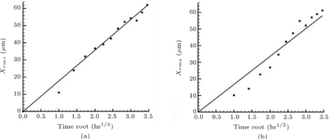
In order to nd the random walk constant, we analyzed the experimental video of chemotacting cells for 12 hours. Since the imposed concentration gradient is in a Y -direction, any movement in the X-direction is a result of random movements of the cell. The root mean square of positions of the cells after each one hour was calculated for a total time of 12 hours, and was plotted against the square root of time. As we know, the root mean square variation of the positions of a random walker is linear against the square root of time, which is evident in both the experimental data and simulation results in Figure 4. Afterwards, a straight line was tted to the plot and its slope was found. Using a trial and error approach, we ran the simulation for dierent values of random walk constant, and chose the value in which the slope of xrms plot
versus square root of time for the simulation matched that of experimental data. As a result, we came up with a random walk constant in which the cell random migration of simulation was consistent with that of experimental videos (see Figure 4).
3.2. Chemotaxis tuning
After setting up the constant of the random walk in the simulation, the relevant parameter of the chemotaxis was identied. As mentioned previously, the chemo-taxis is simulated by identifying the leading edge nodes and then increasing the relevant spring stiness by a constant chemotaxis factor. In order to nd a reason-able value for this factor, we repeated a trial and error approach similar to that done for the random walk.
concentra-Figure 4. Comparison of random migration displacement, xrmsversus square root of time: (a) Experimental data (slope
of the tted line = 17.96 m
Hr0:5); and (b) simulation data (slope of the tted line = 17.52 mHr0:5).
Figure 5. Comparison of average vertical displacement of chemotacting cell versus time in order to tune the chemotaxis eect coecient: (a) Experimental data; and (b) simulation data.
tion gradient is in the Y -direction, it makes sense to nd the chemotaxis constant by comparing the displacement of migrating cells in the movie and simulation in the Y -direction. Since the random walk constant has already been calculated and referred to the code, there is no concern regarding the eects of random walk in the chemotaxis of the cells in the Y -direction. The average vertical displacement of the migrating cells versus time is shown in Figure 5 for experimental data and simulation results. Again, we found the chemotaxis constant using a trial and error approach, so that the simulation curves converge to experimental ones.
It could be concluded from Figures 4 and 5 that the cell migrating simulator has an acceptable agree-ment with experiagree-mental results. As the chemotaxis phenomenon is semi-stochastic, a better agreement can be obtained by further simulations and experiment comparisons.
3.3. Drag force coecient
The nal step in tuning the cell simulator code is nding the viscous drag coecient. In choosing this
constant, we turned our attention to the mean velocity of migrating cells instead of their displacement. The drag force was set to be proportional to the maximum of random walk force by an unknown factor. The unknown coecient was found in such a way that limiting the velocity of migrating cells in the simulation converges to the average velocity of the migrating cells in the experimental video.
At this step, all the necessary parameters and coecients of the cell migration model are identied and xed. By obtaining the velocity of the cells, their motions are simply demonstrated by applying the velocity-Verlet method.
The obtained constants are displayed in Table 1.
Table 1. Numerical values of parameters calibrated in the cell simulator.
Parameters Order of magnitude of the constant value Random force e-14 (N)
Drag force 10 14 N
50mh
It should be considered that these constants are strongly correlated with the type of cell, as well as the type of biochemical factor used. Indeed, in order to nd the calibrated constants in the cell simulator for other cells with dierent types of biochemical factor, specic related experiments are needed.
4. Migration results and discussion
In this study, a cell simulator is developed to model the cell responses to the imposed chemical gradients in the extracellular medium. The positioning of the nodes related to the simulated migrating cell have been shown in Figure 6. It can be seen in this gure that the cell has a tendency to migrate in the gradient direction, but the random walk is able to change the direction of motion. Figure 7 displays the migrating cellmorphology
Figure 6. First few steps of a single cell migration. Membrane and nucleus nodes are shown in this gure. The cell tends to migrate in a vertical direction as a result of imposed gradient, while the random walk eects deviates the cell direction from pure vertical movement.
Figure 7. The morphology of a migrating cell obtained from cell simulation.
Figure 8. Trajectories of migrating cells, all initially at the same location. The cell's tendency to migrate in the direction of the imposed concentration gradient is evident.
obtained from simulator results, in which the reor-ganization of the cytoskeleton is observable. Indeed, this gure makes a qualitative demonstration of the formation of the lamellipodia due to cytoskeleton reor-ganization. In Figure 8, the trajectories of the cells that were initially positioned in the same place are illustrated. As can be seen in this gure, the overall motion of the cells lies in the direction of the gradient, but when exposed to a constant gradient, the cell would not migrate through a unique path. This is the result of the random walk navigations of the cells. The cell trajectories in Figure 8 are not the same because of the stochastic velocity component, *Vr. However,
all trajectories are, generally, in the direction of the gradient when observed for a considerable time. 5. Conclusion
In this project, cell motility during chemotaxis was studied by computational methods. The Voigt model was applied and a cell model, including nucleus, cytoskeleton, and membrane, was developed. The migration of this cell model through the extracellular gradient was attributed to four factors discussed.
The presented method could be used in the study of the mechanisms of cardiovascular diseases and their potential treatment, such as cancer treatment. Thus, studying the mechanism of anti-angiogenic and pro-angiogenic therapies is one of the basic aims of this work. Also, the cell migration simulator code developed in this study, can be extended to study the migration of similar types of cell with other vary-ing biochemical factors, or by varyvary-ing concentration proles by updating various cell migration parameters introduced in this study. The method proposed in this study can make it unnecessary to undertake expensive
experiments to investigate the response of cells in the presence of various biochemical concentration gradi-ents.
As shown in Figures 6 and 8, the method pre-sented in this work has the capability of oering distinct software for cell migration. Thus, one aspect of future work to complete this simulation is to develop a software pack in cell migration. Implementing the eects of cell-cell interactions in the simulator, in order to study group migration of cells, is further possible work in this eld. Enhancing cell cytoskeletal detail is another modication that improves the precision of the simulation that is to be performed.
Conict of interest
There is no conict of interest regarding this study. References
1. Lauenburger, D.A. and Horwitz, A.F. \Cell migra-tion: A physically integrated molecular process", Cell, 84(3), pp. 359-369 (1996).
2. Ridley, A.J., Schwartz, M.A., Burridge, K., Firtel, R.A., Ginsberg, M.H., Borisy, G., Parsons, J.T. and Horwitz, A.R. \Cell migration: Integrating signals from front to back", Science, 302(5651), pp. 1704-1709 (2003).
3. Servant, G., Weiner, O.D., Herzmark, P., Balla, T., Se-dat, J.W. and Bourne, H.R. \Polarization of chemoat-tractant receptor signaling during neutrophil chemo-taxis", Science, 287(5455), pp. 1037-1040 (2000).
4. Franz, C.M., Jones, G.E. and Ridley, A.J. \Cell migration in development and disease", Developmental Cell, 2(2), pp. 153-158 (2002).
5. Meinhardt, H. \Orientation of chemotactic cells and growth cones: models and mechanisms", Journal of Cell Science, 112(17), pp. 2867-2874 (1999).
6. Narang, A., Subramanian, K. and Lauenburger, D.A. \A mathematical model for chemoattractant gradient sensing based on receptor-regulated membrane phos-pholipid signaling dynamics", Annals of Biomedical Engineering, 29(8), pp. 677-691 (2001).
7. Ma, L., Janetopoulos, C., Yang, L., Devreotes, P.N. and Iglesias, P.A. \Two complementary, local excita-tion, global inhibition mechanisms acting in parallel can explain the chemoattractant-induced regulation of PI (3, 4, 5) P3 response in dictyostelium cells", Biophysical Journal, 87(6), pp. 3764-3774 (2004).
8. Levine, H., Kessler, D.A. and Rappel, W.J. \Di-rectional sensing in eukaryotic chemotaxis: A bal-anced inactivation model", Proceedings of the National Academy of Sciences, 103(26), pp. 9761-9766 (2006).
9. Swaney, K.F., Huang, C.H. and Devreotes, P.N. \Eu-karyotic chemotaxis: A network of signaling pathways
controls motility, directional sensing, and polarity", Annual Review of Biophysics, 39, pp. 265-289 (2010).
10. Neilson, M.P., Veltman, D.M., van Haastert, P.J., Webb, S.D., Mackenzie, J.A. and Insall, R.H. \Chemo-taxis: A feedback-based computational model robustly predicts multiple aspects of real cell behaviour", PLoS Biology, 9(5), e1000618 (2011).
11. Andrew, N. and Insall, R.H. \Chemotaxis in shallow gradients is mediated independently of PtdIns 3-kinase by biased choices between random protrusions", Na-ture Cell Biology, 9(2), pp. 193-200 (2007).
12. Killich, T., Plath, P.J., Ha, E.C., Xiang, W., Bult-mann, H., Rensing, L. and Vicker, M.G. \Cell move-ment and shape are non-random and determined by in-tracellular, oscillatory rotating waves in Dictyostelium amoebae", Biosystems, 33(2), pp. 75-87 (1994).
13. Killich, T., Plath, P.J., Wei, X., Bultmann, H., Rens-ing, L. and Vicker, M.G. \The locomotion, shape and pseudopodial dynamics of unstimulated Dictyostelium cells are not random", Journal of Cell Science, 106(4), pp. 1005-1013 (1993).
14. Kiyoumarsioskouei, A., Saidi, M.S. and Firoozabadi, B. \An endothelial cell model containing cytoskeletal components: Suspension and adherent states", Journal of Biomedical Science & Engineering, 5(12), pp. 737-742 (2012).
15. Jamali, Y., Azimi, M. and Mofrad, M.R. \A sub-cellular viscoelastic model for cell population mechan-ics", PloS One, 5(8), e12097 (2010).
16. Whitman, M., Downes, C.P., Keeler, M., Keller, T. and Cantley, L. \Type I phosphatidylinositol ki-nase makes a novel inositol phospholipid, phospha-tidylinositol-3-phosphate", Nature, 332(6165), pp. 644-646 (1988).
17. Zaman, M.H., Kamm, R.D., Matsudaira, P. and Lauenburger, D.A. \Computational model for cell migration in three-dimensional matrices", Biophysical Journal, 89(2), pp. 1389-1397 (2005).
18. Shamloo, A. \Cell-cell interactions mediate cytoskele-ton organization and collective endothelial cell chemo-taxis", Cytoskeleton, 71(9), pp. 501-512 (2014).
Biographies
Amir Kiyoumarsioskouei was born in Osku, Iran. He obtained his BS degree in Mechanical Engineering from Amir Kabir University of Technology, in 2009, and is currently a PhD degree candidate in Sharif University of Technology, Tehran, Iran.
Amir Shamloo received his PhD degree from Stan-ford University, StanStan-ford, CA, USA, in 2010, and became a postdoctoral scholar at the University of Cal-ifornia, Berkeley, CA, USA. He is currently Assistant Professor in the Mechanical Engineering Department of Sharif University of Technology, Tehran, Iran. His
research involves the design and fabrication of microu-idic devices and their application in biology, and cell and tissue mechanics.
Sajjad Azimi obtained his BS degree in Mechan-ical Engineering, in 2012, from Sharif University of Technology, Tehran, Iran, where he is currently an MS degree student. His eld of research study is the modeling of biological systems and microfabrication. Mohammad Abeddust received his BS degree in Me-chanical Engineering from Shiraz University, in 2012, followed by an MS degree in the same subject from Sharif University of Technology, Tehran, Iran, in 2015.
His research interests include numerical investigation of thermo-uidic systems, with particular interest in biological systems. He is also familiar with optical measurement techniques in uid mechanics and heat transfers.
Mohammad Said Saidi received his PhD degree from Massachusetts Institute of Technology, USA, in 1979, and is currently Professor of Mechanical Engi-neering at Sharif University of Technology, Tehran, Iran. His research interests include modeling and numerical analysis of the transport and deposition of aerosol particles, biouids, thermal-hydraulics of porous media, and microchannels