CELLULAR & MOLECULAR BIOLOGY LETTERS http://www.cmbl.org.pl
Received: 02 June 2013 Volume 18 (2013) pp 579-594 Final form accepted: 25 November 2013 DOI: 10.2478/s11658-013-0108-x Published online: November 2013 © 2013 by the University of Wrocław, Poland
* Author for correspondence. e-mail: [email protected], tel.: 48-77-4016045, fax: 48-77-4016051
Abbreviations used: diff – potassium diffusion potential, S – surface potential; DP – degree of polymerization; ODA – octadecylamine; polySia – polysialic acid; OX-V – oxonol V, TB – toluidine blue
Research article
MEMBRANE POTENTIAL-DEPENDENT BINDING OF POLYSIALIC ACID TO LIPID MONOLAYERS AND BILAYERS
KRZYSZTOF NOWOTARSKI, KAROLINA SAPOŃ, MONIKA KOWALSKA, TADEUSZ JANAS and TERESA JANAS*
Department of Biotechnology and Molecular Biology, University of Opole, Kominka 6, 45-032 Opole, Poland
Abstract: Polysialic acids are linear polysaccharides composed of sialic acid monomers. These polyanionic chains are usually membrane-bound, and are expressed on the surfaces of neural, tumor and neuroinvasive bacterial cells. We used toluidine blue spectroscopy, the Langmuir monolayer technique and fluorescence spectroscopy to study the effects of membrane surface potential and transmembrane potential on the binding of polysialic acids to lipid bilayers and monolayers. Polysialic acid free in solution was added to the bathing solution to assess the metachromatic shift in the absorption spectra of toluidine blue, the temperature dependence of the fluorescence anisotropy of DPH in liposomes, the limiting molecular area in lipid monolayers, and the fluorescence spectroscopy of oxonol V in liposomes. Our results show that both a positive surface potential and a positive transmembrane potential inside the vesicles can facilitate the binding of polysialic acid chains to model lipid membranes. These observations suggest that these membrane potentials can also affect the polysialic acid-mediated interaction between cells.
INTRODUCTION
The superposition of the transmembrane potential, the dipole potentials, and the difference in the surface potentials gives rise to the potential inside the membrane (intramembrane potential) [1]. Transmembrane potential is the potential difference between the inner and outer sides of a membrane. It arises from the transfer of charges between two water compartments separated by the membrane. The surface potential is localized at the membrane-solution interface and is generated by a net charge of membrane lipids, proteins or compounds adsorbed on the membrane surface. The dipole potential is generated inside the membrane by the intramolecular dipole moments of lipids, proteins and water molecules, e.g. by the ester linkage of the hydrocarbon chains of phospholipids. Besides its well-known functions in ion transport across the membrane, voltage-gated channel regulation and protein transmembrane translocation, the transmembrane potential also affects the uptake of biologically active peptides [2], the transmembrane translocation of polysialic acid [3, 4], the regulation of cell proliferation and differentiation [5], mitochondrially mediated cell apoptosis [6], the binding of the peptide hormone angiotensin to renal epithelial cells [7], the action of G-protein coupled receptors [8], the dynamics of dolichyl phosphate in membranes [9], and cardiac excitation-contraction coupling [10].
Surface potential plays an important role in cell adhesion, endo-exocytosis, the binding of biologically active ionic species [11], the activity of membrane-bound enzymes [12], and the localization of proteins in the cell [13]. The dipole potential affects the translocation rates of ions across lipid membranes and the structure and function of membrane-incorporated proteins [14].
Polysialic acid (polySia) chains are linear polysaccharides composed of sialic acid monomers. They are usually anchored to a membrane via a phospholipid, an integral membrane glycoprotein or a glycoprotein attached to the membrane via glycosylphosphatidylinositol. They are unique because, besides certain glycosaminoglycans, polySia chains are the only polyanionic molecules permanently attached to the plasma membranes of eukaryotic cells.
the value of negative transmembrane potential [22-23]. Although cellular membranes usually have a negative net charge, the positively charged lipid sphingosine is also present in membranes [24] and can be associated with rafts [25]. In this study, we use the metachromatic shift in the maximum absorption of toluidine blue, the Langmuir monolayer technique, fluorescence anisotropy of DPH, and transmembrane potential-dependent fluorescence of oxonol V in lipid vesicles to study the effects of experimentally generated membrane surface potential and transmembrane potential on the binding of polysialic acid to model lipid membranes.
MATERIALS AND METHODS
Chemicals
DOPC (1,2-dioleoyl-sn-glycero-3-phosphocholine), polySia (poly-2,8-acetylneuraminic acid, sodium salt, from Escherichia coli K1) and TB (toluidine blue) were purchased from Sigma. DPPC (dipalmitoylphosphatidylcholine) and ODA (octadecylamine) were purchased from Fluka. 1,6 diphenyl-1,3,5-hexatriene (DPH) and oxonol V (bis-(3-phenyl-5-oxoisoxazol-4-yl)pentamethine oxonol) were purchased from Invitrogen. Sephacryl S-200 and S-300 were purchased from Amersham Pharmacia.
Purification and characterization of polySia
The polysialic acids were purified and characterized as previously described [26]. Briefly, polySia was treated at 60ºC for 1 h at pH 5 to remove any residual lipids and then purified via chromatography on Sephacryl S-300 and precipitation with 80% ethanol. Sephacryl S-200 and S-300 gel filtration was used to estimate the average degree of polymerization (DP) of the purified polySia using molecular weight markers [27]. A spectrum of chain lengths was found, with the maximum DP in the range of 40 to 60, and an average of ~50.
Preparation of liposomes
Binding of polySia to liposomes, measured using toluidine blue absorption
PolySia in water at a final concentration of 0.05 mg/ml was mixed with DOPC or ODA/DOPC (molar ratio of 0.05) liposomes prepared in water with a final concentration before gel filtration of 10 mg/ml. After incubation for 5 min at room temperature, the suspension was applied to a 1 ml Sephacryl S-1000 column that voids liposomes but retains polyanions [29], and eluted with water. The samples were collected as ca. 30 μl fractions. Co-elution of polyanions and liposomes indicates binding. The presence of polySia in the liposomal fractions was detected by immediate titration of the liposomal fraction (liposome concentration: 1 mg/ml) into a solution of the cationic dye toluidine blue (TB). After prolonged measurement times, liposome aggregation could be detected in the liposomal fraction. Lipid vesicles in the absence of polySia were used to correct for light scattering. TB was previously used to detect sialoglycopeptides [30]. Upon binding of TB to a polySia chain, the absorption maximum shifts from 630 nm to 560 nm (Fig. 1A).
Fluorescence anisotropy of DPH in liposomes
Steady-state fluorescence anisotropy experiments were performed on DPH in 100 nm DPPC liposomes (1:1000 mol ratio of probe:lipid) or ODA/DPPC liposomes as described previously [23]. The excitation and emission wavelengths were 384 nm and 429 nm, respectively. Fluorescence anisotropy (r) was calculated as described in [28].
Measurements of the limiting molecular area in DOPC monolayers
Monolayer experiments were performed as previously described [26]. Briefly, the monolayers were deposited by spreading an appropriate volume of ODA/DOPC mixture in chloroform. Prior to isotherm recording, the monolayers were equilibrated at zero pressure for 5 min to allow the chloroform to evaporate. The experiments were performed both in the presence of polySia (at the indicated concentrations) and in the absence of polySia in the subphase (Fig. 3). The data for the surface pressure – area curves (isotherms) were recorded while the monolayer was being compressed. During this compression, a transition from the gaseous state (surface pressure equals zero) to the liquid expanded state occurs. This small increase in the surface pressure represents the beginning of observable intermolecular forces between adjacent molecules in the monolayer [31, 32]. The area over which this increase occurs defines the limiting molecular area.
Fluorescence of oxonol V in DOPC liposomes
concentration of choline chloride. Experiments were also performed with equal internal and external concentrations of KCl.
RESULTS
The effect of the surface potential on polySia binding to the lipid bilayer and monolayer
To study the effect of the surface potential of liposomal membranes on the binding of polysialic acid (polySia) to liposomes, we mixed polySia with DOPC liposomes modified by positively charged ODA at an ODA/DOPC molar ratio of 0.05. The positive surface potential (S) arises from the presence of a positively charged amino-group of ODA molecules in the lipid bilayer [reviewed in 26]. Then we separated free polySia from bound polySia using gel filtration, collected the fractions under the liposomal peak, and measured the toluidine blue (TB) absorption of the liposomal fractions. TB is a basic dye that is used to determine the surface charge of cells [33]. It has a maximum absorption at 630 nm. The binding of TB to the negative charges of polySia results in a metachromatic shift in its maximum absorption to 560 nm (Fig. 1A). As the concentration of polyanionic polySia increases, the absorption at 630 nm (A630) decreases and the absorption at 560 nm (A560) increases (Fig. 1A). Therefore, the difference (A630 – A560) is inversely related to the presence of polySia in the liposomal fractions. The smaller the absolute value of the difference, the higher the concentration of polySia bound to the liposomes.
Fig. 1. Toluidine blue spectroscopy. A – Absorption spectra for 10 µM toluidine blue (TB). The arrow indicates the increasing concentration of polySia corresponding to each curve: 0, 6, 12, 18, 24, 30, 36 µg/ml. B – The ratio of the absorbance difference (R) as a function of the polySia concentration. R = (A630 – A560 for the sample with ODA/DOPC liposomes)/(A630 – A560 for the sample with DOPC liposomes).
positive charge (DOPC-only liposomes). We determined the ratio of the absorbance difference (R) as:
R A630 – A560 for the sample with ODA/DOPC liposomes
A630 – A560 for the sample with DOPC liposomes
If the binding of polySia to ODA/DOPC liposomal membranes is greater than the binding to pure DOPC membranes, the ratio is less than 1. As shown in Fig. 1B, the ratio R decreases linearly with increasing volume of the liposomal fraction added to the TB solution. The negative slope of the line is equal to (5.2 ± 0.6) x 10-4 g/ml-1 (data from three experiments). The results presented in Fig. 1B show a higher affinity of polySia chains to liposomes with a positive surface charge than to liposomes with a zero net surface charge (DOPC-only liposomes). We estimate from the TB experiments that with a polySia concentration of 0.05 mg/ml, ca. 30% of polySia chains are bound to the ODA/DOPC liposomes. To further test the surface potential-dependent binding of polySia chains to liposomes, we applied a fluorescence spectroscopy technique. We measured the effect of polySia on the temperature dependence of the fluorescence anisotropy of DPH in DPPC liposomes and ODA/DPPC liposomes (molar ratio of 0.05). For a bilayer composed of one lipid, the fluorescence anisotropy is usually inversely related to the membrane fluidity. We calculated the rate of DPH anisotropy change at the phase transition temperature (Fig. 2). This rate is directly related to the cooperativity of the phase transition, i.e. whether the conformation and dynamics of molecules change synchronously or non-synchronously during the phase transitions.
A small decrease in this rate for DPPC bilayers is observed in the presence of polySia. Apparently, polySia chains bound to the DPPC bilayers lessen overall lipid cooperativity during the gel–fluid phase transition. In the case of ODA/DPPC bilayers, only marginal changes in this rate are observed as the concentration of polySia increases up to 0.5 mg/ml. It is possible that polySia induces clustering of oppositely charged ODA in the bilayers and DPH molecules may be withdrawn from these clusters. At the polySia maximal concentration of 0.5 mg/ml, the values of the rates are the same for DPPC and ODA/DPPC bilayers. Within experimental error, no changes in the temperature of transition were observed.
Fig. 2. The main image shows the rate of DPH anisotropy change at the phase transition temperature (absolute values) during the gel-fluid phase transition of DPPC bilayers (circles) or ODA/DPPC bilayers with a 0.5 molar ratio (squares) as a function of polySia concentration. The rate of anisotropy change was found by extrapolating the linear part of the plot of fluorescence anisotropy vs. temperature (shown in the inset).
increased concentrations of polySia. By contrast, adding polySia to the positively charged ODA/DOPC monolayer caused a gradual increase in the limiting molecular area. These data suggest that the presence of the positive surface potential facilitates the binding of polySia chains to the surface, and these bound polySia chains can shield the repulsive interactions within the monolayer and shift the transition of the monolayer from a gaseous state to the liquid expanded state. Our results are in agreement with the data observed earlier for binding of polyacrylate and DNA to a positively charged monolayer, including the effect of polyanions on charged lipid clustering [34].
Effect of the transmembrane potential on polySia binding to lipid bilayers
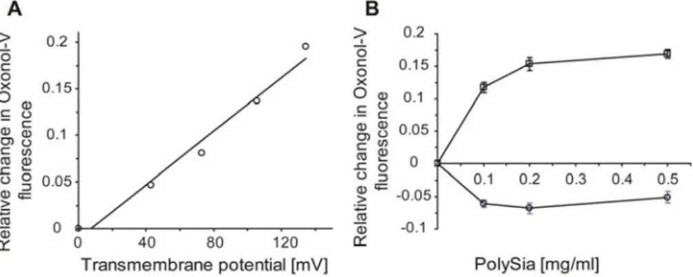
In addition to the surface potential, we studied the effect of the transmembrane potential on polySia binding to the liposomal surface using the fluorescent probe oxonol V (OX-V), which is sensitive to the transmembrane potential [35]. The maximum of the emission fluorescence spectra of OX-V is shifted from 625 nm to 650 nm in the presence of phospholipid vesicles [36]. When a valinomycin/ potassium ion-induced transmembrane gradient (valinomycin-induced potassium diffusion potential, diff) is created in lipid vesicles (positive inside the vesicles), an increase in fluorescence at 650 nm is observed in the case of a low OX-V/phospholipid molar ratio [37-38]. We measured the relationship between the generated transmembrane potential (calculated using the Nernst equation) and the increase in the OX-V fluorescence in DOPC liposomes (Fig. 4A).
The increase in the value of the transmembrane potential (positive inside the liposomes) gives a linear increase in OX-V fluorescence at 650 nm, with the slope of the line equal to (1.45 ± 0.11) x 10-3 mV-1 (data from three experiments). The overall ionic strength was kept constant to avoid its influence on OX-V fluorescence [38]. We tested the effect of polySia at different concentrations on the relative change in OX-V fluorescence in DOPC liposomes (Fig. 4B) both in the absence and presence of a transmembrane potential. PolySia increases the OX-V fluorescence in liposomal membranes in the presence of a transmembrane potential, which suggests an increase in the intramembrane potential (positive inside) upon the binding of negatively charged polySia chains to the external surface of the liposomal membrane. In the absence of a transmembrane potential, polySia decreases the OX-V fluorescence, which can reflect a competition between negatively charged polySia chains and negatively charged OX-V for the same binding sites on the liposomal surface [36].
DISCUSSION
PolySia chains are expressed on the surface of many types of cell, including neural and cancer cells, and the interactions between polySia and membranes, or between polySia and other biomolecules, can regulate membrane-mediated cellular phenomena [18, 20-21, 39]. Negatively charged polySia can affect the membrane surface potential due to the adsorption of polySia chains on the external face of the membrane, and the membrane transmembrane potential [22] due to an increased (in relation to the cytoplasmic concentration) concentration of polySia chains outside the cell membrane (e.g. attached to the opposing cells). In an inverse relationship, the transmembrane potential can modulate the transport of polySia across membranes [4, 40] and the surface potential can modulate polySia-mediated membrane interactions [21].
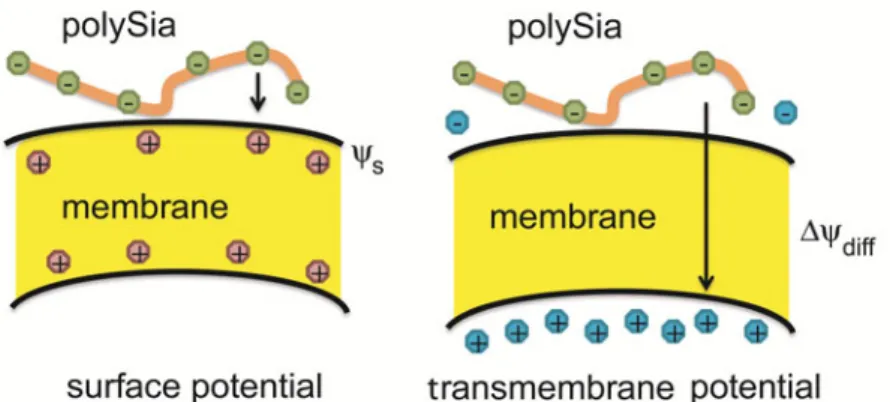
In addition, the presented data indicate that both surface potential and transmembrane potential affect the binding of polySia chains to the surface of a model lipid membrane (Fig. 5). Thus, it seems that there is a continuous interplay between membrane potentials and polySia binding to the membrane surface.
Surface potential can modulate the interaction of other polyanions with lipid bilayers. The thermal phase behavior of multilamellar liposomes containing positively charged sphingolipids was significantly altered by the adsorption of DNA [41]. We observed a similar phenomenon in the case of polySia chains. The fluidity of the positively charged lipid monolayers and bilayers was found to be essential to induce the close packing of the adsorbed DNA [42]. The studies of the adsorption of DNA oligonucleotides on the lipid bilayer showed that the diffusion time of labeled oligonucleotides in the presence of cationic liposomes decreases [43]. The driving force for the formation of charge-neutral complexes between positively charged liposomes and DNA is the release of DNA and lipid counterions [44]. The formation of equilibrium clusters composed of positively charged liposomes stuck together by a polyanionic polyacrylate was demonstrated [45]. The adsorption of tRNA on the surface of lipid bilayers can lead to segregation in the mixed anionic or cationic bilayers, where tRNA is likely excluded from the anionic-rich domains in the first system, and associated with the cationic-rich domains in the second system [46]. Conditions such as the lipid composition of liposomes and salt concentration, under which DNA is still physically associated with positively charged liposomes, were previously studied [47].
bilayers [57] may also be influenced by the changes in the dipole potential caused by the insertion of sterols into the bilayer. Although this possibility was not considered by the authors, they found the outer polar headgroup region as a localization site of AmB. There are no previous reports on the effect of the transmembrane potential on the binding of hydrophilic polyanions or polycations to lipid bilayers.
As follows from Figs 2 through 4, polySia binds to uncharged DOPC and DPPC liposomes and monolayers. This is not surprising since sialic acids can bind to the phospholipid bilayers via hydrogen bonds [58-59], which can occur, e.g. between the amide group of sialic acid and the carbonyl of the phospholipid ester bond. In the case of the polySia molecules binding to phospholipid bilayers, the number of these bonds (per molecule) can be multiplied. It was found that interactions of the amide group in the sialic acid residue play an important role in ganglioside–phosphatidylcholine interactions within the bilayer, the amide being able to act as a donor and as an acceptor of hydrogen bonds [58].
The binding of polySia chains to the membrane was also observed in the case of biological membranes. Cross-linking experiments showed that extracellularly applied polySia or polySia-NCAM and intracellularly expressed MARCKS-ED (a peptide comprising the effector domain of myristoylated alanine-rich C kinase substrate) are in close contact, suggesting insertion of polySia and MARCKS-ED peptide into the cell membrane of hippocampal neurons from opposite sides [60]. From the results of our experiments, we suggest an involvement of membrane potentials in this phenomenon. Since the MARCKS-ED peptide is positively charged (it contains several lysines and an arginine) and the polySia chain is negatively charged, the same intramembrane potential can drive both of them simultaneously in opposite direction within the membrane, thus bringing them closer to each other.
Cell multidimensional solution NMR spectroscopy analysis revealed that free and cell-bound polySia were structurally similar [61]. PolySia free in solution, although predominantly random coil in nature, adopts local, extended helical conformations, like helices [62], and the results were used to suggest that the binding of specific antibodies to polySia arise from the recognition of a high-order local helix. These fleeting helical conformations may also contribute to the binding of polySia to lipid bilayers and monolayers.
Our report shows that both surface potential and transmembrane potential can modulate the binding of polySia chains to model lipid membranes when added free in solution. These observations suggest that these membrane potentials can also affect the polySia-mediated interaction between cells.
REFERENCES
1. Przybyło, M., Borowik, T. and Langner, M. Fluorescence techniques for determination of the membrane potentials in high throughput screening.
J. Fluoresc. 20 (2010) 1139-1157.
2. Rothbard, J.B., Jessop, T.C. and Wender P.A. Adaptive translocation: the role of hydrogen bonding and membrane potential in the uptake of guanidinium-rich transporters into cells. Adv Drug Deliv. Rev. 57 (2005) 495-504.
3. Troy, F.A., Janas, T., Janas, T. and Merker, R.I. Vectorial translocation of polysialic acid chains across the inner membrane of Escherichia coli K1.
FASEB J. 5 (1991) A1548-A1548.
4. Janas, T., Krajiński, H., Timoszyk, A. and Janas, T. Translocation of polysialic acid across model membranes: kinetic analysis and dynamic studies. Acta Biochim. Polon. 48 (2001) 163-173.
5. Sundelacruz, S., Levin, M, and Kaplan, D.L. Role of membrane potential in the regulation of cell proliferation and differentiation. Stem Cell Rev. Rep.
5 (2009) 231-246.
6. Kadenbach, B., Ramzan, R., Moosdorf, R. and Vogt, S. The role of mitochondrial membrane potential in ischemic heart failure. Mitochondrion
11 (2009) 700-706.
7. Brown, G.P. and Douglas, J.G. Influence of transmembrane potential differences of renal tubular epithelial cell on ANG II binding. Am. J. Physiol. 252 (1987) F209-F214.
8. Mahaut-Smith, M.P., Martinez-Pinna, J. and Gurung, I.S. A role for membrane potential in regulating GPCRs? Trends Pharmacol. Sci. 29 (2008) 421-429.
9. Janas, T., Kuczera, J. and Chojnacki, T. Voltage-dependent behaviour of dolichyl phosphate-phosphatidylcholine bilayer lipid membranes. Chem. Phys. Lipids 52 (1990) 151-155.
10. Ferrier, G.R. and Howlett, S.E. Cardiac excitation-contraction coupling: role of membrane potential in regulation of contraction. Am. J. Physiol. Heart Circ. Physiol. 280 (2001) H1928-H1944.
11. Xu, C. and Loew, L.M. The effect of asymmetric surface potentials on the intramembrane electric field measured with voltage-sensitive dyes. Biophys. J.
84 (2003) 2768-2780.
12. Wojtczak, L., Famulski, K.S., Nalecz, M.J. and Zborowski, J. Influence of the surface potential on the Michaelis constant of membrane-bound enzymes: effect of membrane solubilization. FEBS Lett. 139 (1982) 221-224. 13. Yeung, T., Gilbert, G.E., Shi, J., Silvius, J., Kapus, A. and Grinstein, S.
14. Wang, L., Bose, P.S. and Sigworth, F.J. Using cryo-EM to measure the dipole potential of a lipid membrane. Proc. Natl. Ac. Sci. USA 103 (2006) 18528-18533.
15. Troy, F.A. Polysialic acid in molecular medicine. Encyclopedia Biol. Chem. 3 (2004) 407-414.
16. Bonfanti, L. PSA-NCAM in mammalian structural plasticity and neurogenesis. Prog. Neurobiol. 80 (2006) 129-164.
17. Gascon, E., Vutskits, L. and Kiss, J.K. Polysialic acid-neural cell adhesion molecule in brain plasticity: from synapses to integration of new neurons.
Brain Res. Rev. 56 (2007) 101-118.
18. Hildebrandt, H., Muhlenhoff, M., Weinhold, B. and Gerardy-Schahn, R. Dissecting polysialic acid and NCAM functions in brain development.
J. Neurochem. 103 (Suppl 1) (2007) 56-64.
19. Miyata, S., Sato, C. and Kitajima, K. Glycobiology of polysialic acids on sea urchin gametes. Trends Glycosci. Glycotechnol. 19 (2007) 85-98.
20. Rutishauser, U. Polysialic acid in the plasticity of the developing and adult vertebrate nervous system. Nature Rev. Neurosci. 9 (2008) 26-35.
21. Janas, T. and Janas, T. Membrane oligo- and polysialic acids. Biochim. Biophys. Acta - Biomembranes 1808 (2011a) 2923-2932.
22. Janas, T., Janas, T. and Krajiński, H. Membrane transport of polysialic acid chains: modulation of transmembrane potential. Eur. Biophys. J. 29 (2000a) 507-514.
23. Janas, T., Nowotarski, K. and Janas, T. Polysialic acid can mediate membrane interactions by interacting with phospholipids. Chem. Phys. Lipids 163 (2010) 286-291.
24. Hannun, Y.A. and Obeid, L.M. Principles of bioactive lipid signalling: Lessons from sphingolipids. Nat. Rev. Mol. Cell Biol. 9 (2008) 139-150. 25. Hengst, J.A., Guilford, J.M., Fox, T.E., Wang, X., Conroy, E.J. and Yun,
J.K. Sphingosine kinase 1 localized to the plasma membrane lipid raft microdomain overcomes serum deprivation induced growth inhibition.
Arch. Biochem. Biophys. 492 (2009) 62-73.
26. Janas, T., Nowotarski, K. and Janas, T. The effect of long-chain bases on polysialic acid-mediated membrane interactions. Biochim. Biophys. Acta - Biomembranes 1808 (2011b) 2322-2326.
27. Kanato, Y., Kitajima, K. and Sato, C. Direct binding of polysialic acid to a brain-derived neurotrophic factor depends on the degree of polymerization.
Glycobiology 18 (2008) 1044-1053.
28. Janas T., Janas, T. and Yarus, M. Specific RNA binding to ordered phospholipid bilayers. Nucleic Acids Res. 34 (2006) 2128-2136.
29. Janas, T., Janas, T. and Yarus, M. A membrane transporter for tryptophan composed of RNA. RNA 10 (2004) 1541-1549.
31. Negelmann, L., Pisch, S., Bornscheuer, U. and Schmidt, R. Properties of unusual phospholipids. III: Synthesis, monolayer investigations and DSC studies of hydroxyl octadeca(e)noic acids and diacylglycerophosphocholines derived therefrom. Chem. Phys. Lipids 90 (1997) 117-134.
32. Janas, T., Nowotarski, K., Gruszecki, W.I. and Janas, T. The effect of hexadecaprenol on molecular organisation and transport properties of model membranes. Acta Biochim. Polon. 47 (2000c) 661-673.
33. Van Damme, M.P.I., Tiglias, J., Nemet, N. and Preston, B.N. Determination of the charge content at the surface of cells using a colloid titration technique. Anal. Biochem. 223 (1994) 62-70.
34. Yun, S., Ahn, K. and Kim, M.W. Polyelectrolyte flexibility effect on the morphology of charged lipid multilayers. Europhys. Lett. 70 (2005) 555-561. 35. Smith, J.C., Russ, P., Cooperman, S. and Chance, B. Synthesis, structure
determination, spectral properties, and energy-linked spectral responses of the extrinsic probe oxonol V in membranes. Biochemistry 15 (1976) 50794-5105.
36. Bashford, C.L., Chance, B., Smith, J.C. and Yoshida, T. The behavior of Oxonol dyes in phospholipid dispersions. Biophys. J. 25 (1979) 63-85. 37. Clarke, R.J. and Apell, H.J. A stopped-flow kinetic study of the interaction
of potential-sensitive oxonol dyes with lipid vesicles. Biophys. Chem. 34 (1989) 225-237.
38. Holoubek, A., Vecer, J. and Sigler, K. Monitoring of the proton electrochemical gradient in reconstituted vesicles: quantitative measurements of both transmembrane potential and intravesicular pH by ratiometric fluorescent probes. J. Fluoresc. 17 (2007) 201-213.
39. Janas, T. and Janas, T. Polysialic acid: structure and properties. in:
Polysaccharides: Structural Diversity and Functional Versality (Dumitriu, S. Ed.), 2nd edition, Marcel Dekker, New York, NY, 2005a, 707-727.
40. Janas, T., Krajiński, H. and Janas, T. Electromigration of polyion homopolymers across biomembranes: a biophysical model. Biophys. Chem.
87 (2000b) 167-178.
41. Koiv, A., Mustonen, P. and Kinnunen, P.K.J. Differential scanning calorimetry study on the binding of nucleic-acids to dimyristoylphospha-tidylcholine-sphingosine liposomes. Chem. Phys. Lipids 70 (1994) 1-10. 42. Fang, Y. and Yang, J. Two-dimensional condensation of DNA molecules on
cationic lipid membranes. J. Phys. Chem. B 101 (1997) 441-449.
43. Jurkiewicz, P., Okruszek, A., Hof, M. and Langner M. Associating oligonucleotides with positively charged liposomes. Cell. Mol. Biol. Lett. 8 (2003) 77-84.
44. Koltover, I., Salditt, T. and Safinya, C.R. Phase diagram, stability, and overcharging of lamellar cationic lipid-DNA self-assembled complexes.
45. Bordi, F., Cametti, C. and Sennato, S. Does a cluster phase in polyion-liposome colloidal suspensions exist? An integrated experimental overview.
Colloids Surf. A 306 (2007) 102-110.
46. Michanek, A., Kristen, N., Höök, F., Nylander, T. and Sparr, E. RNA and DNA interactions with zwitterionic and charged lipid membranes - a DSC and QCM-D study. Biochim. Biophys. Acta 1798 (2010) 829-838.
47. Even-Chen, S., Cohen, R. and Barenholz, Y. Factors affecting DNA binding and stability of association to cationic liposomes. Chem. Phys. Lipids 165 (2012) 414-423.
48. Diederich, A., Bahr, G. and Winterhalter, M. Influence of polylysine on the rupture of negatively charged membranes. Langmuir 14 (1998) 4597-4605. 49. de Kroon, A.I.P.M., de Gier, J. and de Kruijff, B. Association of synthetic
model peptides with phospholipid vesicles induced by a membrane potential.
Biochim. Biophys. Acta 981 (1989) 371-373.
50. Janas, T., Kotowski, J. and Tien, H.T. Polymer-modified bilayer lipid membranes: the polypyrrole-lecithin system. Bioelectrochem. Bioenerg. 19 (1988) 405-412.
51. Jennings, M.L., Schulz, R.K. and Allen, M. Effects of membrane potential on electrically silent transport. J. Gen. Physiol. 96 (1990) 991-1012.
52. Janas, T. and Janas, T. Involvement of carboxyl groups in chloride transport and reversible DIDS binding to Band 3 protein in human erythrocytes. Cell. Mol. Biol. Lett. 16 (2011) 342-358.
53. Berkovich, A.K., Lukashev, E.P. and Melik-Nubarov, N.S. Dipole potential as a driving force for the membrane insertion of polyacrylic acid in slightly acidic milieu. Biochim. Biophys. Acta 1818 (2012) 375-383.
54. Janas, T. and Yarus, M. Visualization of membrane RNAs. RNA 9 (2003) 1353-1361.
55. Janas, T., Janas, T. and Yarus, M. RNA, lipids and membranes. in: The RNA World III (Gesteland, R., Cech, T.R., Atkins, J., Eds), Cold Spring Harbor Laboratory Press, New York, NY, 2005b, 207-225.
56. Janas, T. and Janas, T. The selection of aptamers specific for membrane molecular targets. Cell. Mol. Biol. Lett. 16 (2011) 25-39.
57. Gabrielska, J., Gagos, M., Gubernator, J. and Gruszecki, W.I. Binding of antibiotic amphotericin B to lipid membranes: A 1H NMR study. FEBS
Lett. 580 (2006) 2677-2685.
58. Müller, E., Giehl, A., Schwarzmann, G., Sandhoff, K. and Blume, A. Oriented I ,2-dimyristoyl-sn-glycero-3-phosphorylcholine/ganglioside membranes: a Fourier transform infrared attenuated total reflection spectroscopic study. Band assignments; orientational, hydrational, and phase behavior; and effects of Ca2+ binding. Biophys. J. 71 (1996) 1400-1421. 59. Khalil, M.B., Kates, M. and Carrier, D. FTIR study of the
a significant interaction between Ca2+ ions and the sialic acid moiety of GM1. Biochemistry 39 (2000) 2980-2988.
60. Theis, T., Mishra, B., von der Ohe, M., Loers, G., Prondzynski, M., Pless, O., Blackshear, P.J., Schachner, M. and Kleene, R. Functional role of the interaction between polysialic acid and myristoylated alanine-rich C kinase substrate at the plasma membrane. J. Biol. Chem. 288 (2013) 6726-6742. 61. Azurmendi, H.F., Vionnet, J., Wrightson, L., Trinh, L.B., Shiloach, J. and
Freedberg, D.I. Extracellular structure of polysialic acid explored by on cell solution NMR. Proc. Natl. Ac. Sci. USA 104 (2007) 11557-11561.