Address for correspondence Vladimír Bartoš, PhD., MPH.
Department of Pathology, Faculty Hospital in Žilina, V. Spanyola 43, 012 07,
Žilina, Slovakia. Ph: +421908386352
Email: [email protected]
Original Article
Immunohistochemical expression of androgen
receptor in cutaneous basal cell carcinoma
Introduction
Androgen receptor (AR) is a member of the steroid and nuclear receptor superfamily, which mediate the biological actions of androgens. Upon binding to the ligands (i.e. testosterone or 5-α-dihydrotestosterone), AR translocates from the cytosol into the nucleus and serves as a
transcriptional factor to regulate the expression of its target genes.1 Physiologically, AR possess a variety of functions, of which the most important one development and maintenance of the male sexual phenotype.1 AR is expressed in several androgen-related tissues, e.g. prostate, skeletal muscle, liver, adrenal gland, and central nervous system.1 In normal skin, AR may be identified in the nuclei of basal and differentiating sebocytes in sebaceous glands, pilosebaceous duct keratinocytes, basal keratinocytes of the interfollicular epidermis, dermal fibroblasts, and in certain secretory cells of eccrine sweat glands.2 As for epithelial skin Vladimír Bartoš, Milada Kullová*
Department of Pathology, Faculty Hospital in Žilina, Slovakia.
* Department of Dermatovenerology, Faculty Hospital in Žilina, Slovakia.
Abstract
Background Androgen receptor (AR) is known to be expressed in a variety of skin tumors, including basal cell carcinoma (BCC). The aim of this study was to investigate an expression status of AR by immunohistochemistry in a set of cutaneous BCC.Material and Methods The study group consisted of 54 BCCs from 49 patients. Based on histomorphology, BCC subtypes were merged into non-aggressive (superficial and nodular subtypes) and aggressive (nodular-infiltrative and infiltrative subtypes) subgroups. Specific monoclonal antibody against AR was used for immunostaining.
Results AR was expressed in 32 BCCs (59.3%). In superficial, nodular, nodular-infiltrative and infiltrative BCC subtypes, nuclear reactivity for AR was found in 70% (7/10), 73.1% (19/26), 35.7% (5/14) and 25% (1/4), respectively. A wide quantitative range of AR expression (1–90% of tumor tissue) was seen. There were twenty-seven cases (84.4%) with focal staining and five cases (15.6%) with diffuse staining. There was significantly lower rate of AR expression in BCCs exhibiting aggressive histomorphologic phenotype. No correlation between AR expression and anatomic location and gender was found.
Conclusion Although cutaneous BCC is generally considered to be AR-positive neoplasm, in the present study, immunoreactivity for AR was detected only in about half of the cases. Most AR-positive tumors showed only focal staining. The pathologists should be aware of these findings, which may cause difficulties in distinguishing BCC from related AR-negative epithelial skin tumors.
Key words
tumors, some neoplasms and tumor-related lesions of sebaceous origin (sebaceous adenoma, sebaceous carcinoma, sebaceoma, sebaceous hyperplasia)3, as well as sweat gland neoplasms (eccrine poroma, hidrocystoma, spiradenoma, syringoma, hidradenoma, mixed tumor, microcystic adnexal carcinoma, sweat gland carcinoma)4 express AR. In addition, cutaneous basal cell carcinoma (BCC) is also known to be immunoreactive for AR. Untill now, few studies have been published addressing this issue3,5-12, some of which have provided inconsistent conclusions. Herein, we have investigated the immunohistochemical expression of AR in a set of cutaneous BCCs and confronted our results with eligible literature data.
Material and Methods
Excisional biopsy samples from 54 cases of cutaneous BCCs were entered into this study. They were obtained from 49 patients (27 men, 22 women) in the age range 26 - 83 years (mean age 68.7 years). All patients were treated at the clinical departments of the Faculty Hospital in Zilina (Slovakia) and biopsy specimens were microscopically investigated at the Department of Pathology in this institution. The topographic location of lesions was as follows: face (29 cases), auricle (3 cases), neck (2 cases), back (12 cases), chest (2 cases), upper limbs (4 cases) and lower limbs (2 cases). The tumors included four histomorphological subtypes: superficial (10 cases), nodular (26 cases), nodular-infiltrative (14 cases), and infiltrative (4 cases). Biopsy specimens were routinely processed and
immunohistochemically stained for AR
according to manufacturer's instructions.
Specific monoclonal mouse antibody against AR
Amoli et al.8, we adopted the following
simplified semiquantitative scoring scheme: a) nuclear staining in more than 50% of neoplastic cells was assessed as diffuse positivity, b) nuclear staining between 1 - 50% of neoplastic cells was considered focal positivity, and c) nuclear staining in fewer than 1% of neoplastic cells was considered negative. Based on histomorphology, BCC subtypes were merged into: a) non-aggressive (superficial and nodular), and b) aggressive (nodular-infiltrative and infiltrative) subgroups. The affected topographic sites were merged into: a) chronically sun-exposed (head & neck), and b) sun-protected (trunk & extremities) regions. Data were collected in a databank, using a software SPSS Statistics. Chi-square test was employed and P value < 0.05 was considered significant.
Results
A summary of the basic clinical and histopathological characteristics of our cohort of patients in relation to AR expression status is presented in Table 1 & 2. Overall, AR was expressed in 32 BCCs (59.3%) showing apparent nuclear staining in the neoplastic cells. In superficial, nodular, nodular-infiltrative and infiltrative BCC subtypes, AR reactivity (defined as ≥ 1% of immunolabelled tumor cells) was found in 70% (7/10), 73.1% (19/26), 35.7% (5/14) and 25% (1/4), respectively. Within this immunopositive subgroup, a wide quantitative range of AR expression (cca 1–90% of tumor tissue, mean value 20%) was recognized. There were twenty-seven cases (84.4%) with focal immunostaining and only
five cases (15.6%) with diffuse
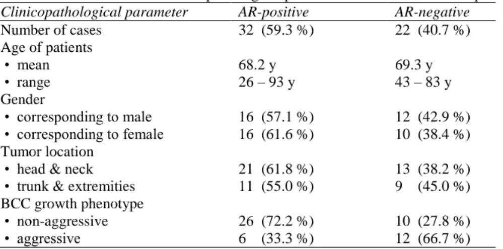
Table 1 An overview of the clinicopathological parameters in relation to AR expression status. Clinicopathological parameter AR-positive AR-negative
Number of cases 32 (59.3 %) 22 (40.7 %)
Age of patients • mean • range
68.2 y 26 – 93 y
69.3 y 43 – 83 y Gender
• corresponding to male • corresponding to female
16 (57.1 %) 16 (61.6 %)
12 (42.9 %) 10 (38.4 %) Tumor location
• head & neck • trunk & extremities
21 (61.8 %) 11 (55.0 %)
13 (38.2 %) 9 (45.0 %) BCC growth phenotype
• non-aggressive • aggressive
26 (72.2 %) 6 (33.3 %)
10 (27.8 %) 12 (66.7 %)
Table 2 A summary of AR expression data in different BCC subtypes.
BCC subtype N AR positive AR negative
Superficial 10 7 cases (70.0%)
- focal expression (7/7; 100%) - diffuse expresion (0/7; 0%)
3 cases (30.0%)
Nodular 26 19 cases (73.1%)
- focal expression (15/19; 78.9%) - diffuse expresion (4/19; 21.1%)
7 cases (26.9%)
Nodular-infiltrative 14 5 cases (35.7%)
- focal expression (4/5; 80%) - diffuse expresion (1/5; 20%)
9 cases (64.3%)
Infiltrative 4 1 case (25.0%)
- focal expression (1/1; 100%) - diffuse expresion (0/0; 0%)
3 cases (75.0%)
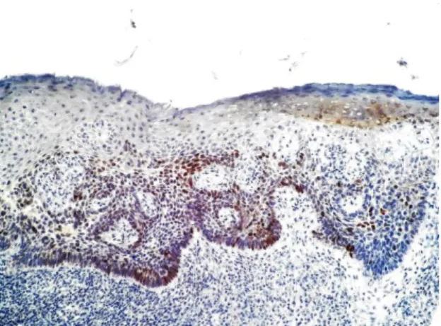
Figure 1 Disperse expression of AR in superficial BCC. (magnification 100x)
Figure 3 Diffuse expression of AR (about 70%) in nodular-infiltrative BCC. Note decreased expression in tumor area (right) manifesting an infiltrative growth pattern. (magnification 40x)
Figure 4 Disperse expresion of AR in infiltrative BCC. (magnification 100x)
(72.2% vs 33.3%, respectively). In the majority of cases, spatial distribution of AR-positive neoplastic cells in tumor tissue was uneven and the cells usually displayed focal or patchy staining pattern. The tumor areas with a high number of cells expressing AR commonly occurred adjacent to the areas with no
AR-labeled cell population (Figures 1–4). Larger
solid nests and nodules of cancer displayed more extensive positivity than smaller aggregations and strands of tumor epithelium in infiltrative BCCs. Although AR-positive BCCs resided on the head and neck region in a slightly larger percentage (61.8%) compared to AR-negative tumors (55.0%), there was no significant association between the expression of AR (present vs absent) and anatomic location (head & neck vs trunk & extremities) (p = 0.6). Nevertheless, all five BCCs exhibiting diffuse immunoreactivity were located on the face. In
the AR-positive and AR-negative BCC
subgroups, the male-to-female ratio was 1.0 and 1.2, respectively, achieving no statistical significance (p = 0.7).
growth with almost total absence of metastases and overall benign clinical course.13 As already mentioned, AR is expressed in a variety of skin tumors, including BCC. In routine biopsy practice, immunohistochemical staining for AR is considered to be helpful tool for differentiation between BCC, which is usually reactive, and benign tumor of follicular origin - trichoepithelioma, which is non reactive.5-12 However, the percentages of AR-positive BCC cases differ among the studies derived from various authors. We have reviewed nine original articles published in English-language literature3,5-12 and found out that the prevalence of AR-positive BCCs ranges between 33-100% (mean 68%) of the cases. The controversial results could be attributed to several factors, such as a total number of lesions investigated, different processing technique and methods used, different cut-off values defining the AR-positivity, and case selection bias in terms of the prevalence of certain histological BCC subtypes.
Many studies3,5,11,12 have shown that nuclear expression of AR in cutaneous BCC is usually focal and conducted in clusters or scattered individual tumor cells. Two decades ago, Bayer-Garner et al.3 found that immunoreactivity for AR was present only in fewer than 5% of tumor cells. Astarci et al.5 revealed that among twenty-three AR-positive BCCs, over half (twelve) manifested a scarce positivity of up to 5% of neoplastic tissue. The remaining lesions displayed a reactivity between 6-50%, whereas none showed an immunostaining in more than half of total tumor tissue. Asadi-Amoli et al.8 observed that immunolabelled neoplastic cells constituted more than 5% of cancer cell population in almost all AR-positive BCCs, but none displayed a diffuse staining pattern. In cases studied by us, only a small proportion of AR-positive lesions showed an immunoreactivity exceeding 50% of cancer tissue.
It remains unclear why some cutaneous BCCs express AR, while the others do not. No unique clinical characteristics of BCC samples manifesting positive staining for AR have been proven so far. This field is poorly elucidate, as most studies done till present time did not specifically focus on this issue. We have tried to reveal a potential relationship between AR expression status and certain clinicopathological variables. Histomorphology of BCC may be directly linked with an expression of AR in cancer cells. In a study of Arits et al.10, AR positivity was found in 22 out of 23 (95.6%) non-aggressive (superficial or nodular) BCC subtypes, while only in 10 out of 15 (66.6%) infiltrative BCCs. Our current analysis has demonstrated a positive association between the expression of AR and non-aggressive BCC subtypes (superficial and nodular), corroborating the finding of previous work.10 It suggests, a loss of AR expression in BCC cells may relate with an acquisition of more aggressive tumor
phenotype. However, the results of Costache et al.9 did not support such a hypothesis. They investigated 18 cases of infiltrative BCC and found, all of them had AR expression.
With regard to gender some papers reported5,11 that an expression of AR in cutaneous BCC occured more commonly in men than women. In a study of Lee et al,11 the AR positivity rates in males and females were 58.8% and 25%, respectively. Even Turkish investigators5 showed a male preponderance in AR-positive subset of BCC. However, both studies did not mention any statistically definite conclusion. In our own analysis, there were almost equal percentages of men and women in both, the AR-positive and AR-negative BCC categories. Provided the AR/ androgen interactions really modify biologic behaviour of cutaneous BCC, then AR-positive BCCs should grow faster in men, who generally have higher concentration of androgens. This area is unexplained and may represent a promising subject for further research.
Conclusion
Although cutaneous BCC is generally considered to be AR-positive neoplasm, in the present study, nuclear immunoreactivity for AR was detected only in about half of the cases. It was less common in BCCs exhibiting aggressive histomorphologic phenotype. Most AR-positive tumors showed only focal staining. The pathologists should be aware of these findings, which may cause difficulties in distinguishing BCC from related AR-negative epithelial skin tumors.
References
1. Gao W, Bohl CE, Dalton JT. Chemistry and structural biology of androgen receptor. Chem Rev 2005; 105: 3352-70.
2. Choudhry R, Hodgins MB, Van der Kwast TH, Brinkmann AO, Boersma WJ. Localization of androgen receptors in human skin by immunohistochemistry: implications for the hormonal regulation of hair growth, sebaceous glands and sweat glands. J Endocrinol 1992; 133: 467-75.
3. Bayer-Garner IB, Givens V, Smoller B. Immunohistochemical staining for androgen receptors: a sensitive marker of sebaceous differentiation. Am J Dermatopathol 1999; 21: 426-31.
4. Kariya Y, Moriya T, Suzuki T, et al. Sex steroid hormone receptors in human skin appendage and its neoplasms. Endocr J 2005; 52: 317-325.
5. Astarci HM, Gurbuz GA, Sengul D et al. Significance of androgen receptor and CD10 expression in cutaneous basal cell carcinoma and trichoepithelioma. Oncol Lett 2015; 10: 3466-3470.
6. Izikson L, Bhan A, Zembowicz A. Androgen receptor expression helps to
differentiate basal cell carcinoma from benign trichoblastic tumors. Am J Dermatopathol 2005; 27: 91–95.
7. Katona TM, Perkins SM, Billings SD. Does the panel of cytokeratin 20 and androgen receptor antibodies differentiate desmoplastic trichoepithelioma from morpheaform/infiltrative basal cell carcinoma? J Cutan Pathol 2008; 35: 174– 79.
8. Asadi-Amoli F, Khoshnevis F, Haeri H, Jahanzad I, Pazira R, Shahsiah R. Comparative examination of androgen receptor reactivity for differential diagnosis of sebaceous carcinoma from squamous cell and basal cell carcinoma. Am J Clin Pathol 2010; 134: 22–26.
9. Costache M, Bresch M, Böer A. Desmoplastic trichoepithelioma versus morphoeic basal cell carcinoma: A critical reappraisal of histomorphological and immunohistochemical criteria for differentiation. Histopathology 2008; 52: 865–76.
10. Arits AH, Van Marion AM, Lohman BG, et al. Differentiation between basal cell carcinoma and trichoepithelioma by immunohistochemical staining of the androgen receptor: an overview. Eur J Dermatol 2011; 21: 870-3.
11. Lee YF, Chang YT, Liu HN. Differentiating basal cell carcinoma from trichoepithelioma by using androgen receptor expression. Dermatol Sinica 2009; 27: 154-60.
12. Ahmed NAA, Ahmed NS. Androgen receptor and CD10 expression in differential diagnosis of trichoepithelioma and basal cell carcinoma. Al-Azhar Assiut Med J 2011; 9: 151-160.