facilitate major physiological processes in humans. GPCRs are targets of more than 40% of therapeutic drugs, which lead to activation of intracellular signal transduction proteins
(transducers). Drugs targeting GPCRs have recently been shown to have preference for certain transduction pathways, a phenomenon termed functional selectivity. Drugs that are biased to exclusively activate one transduction pathway have been found to be more efficacious and produce fewer side effects. Therefore, understanding the nature of a ligand’s preference for a particular transduction pathway may lead to the development of novel drugs with superior therapeutic efficacy. No methods have been developed to specifically characterize the
Introduction
Until recently, pharmacological theory has postulated that a ligand’s ability to occupy a G protein-coupled receptor (GPCR), and therefore activate a response pathway, was solely dependent on the inherent properties of the ligand-receptor pair. This is described as intrinsic efficacy, a phenomenon that has been greatly relied upon for the last half-century (Urban et al., 2007). Intrinsic efficacy depicts a system-independent outcome that is constant for each ligand at a given receptor, regardless of where the receptor is expressed. However, it has been found that some molecules, such as dopamine receptor agonists, do not follow this system-independent pattern for agonist-receptor complexes. These agonists are described as being “biased” toward certain transduction pathways, a phenomenon termed “functional selectivity”.
comprehension and discovery related to agonists’ affinity at GPCRs of interest, such as D2R, will allow for expansion and improvement of drugs and therapies.
Currently, little is known about the relationship between an agonist’s ability to signal at a receptor and the transducers bound to that receptor. In order to provide insight to this unknown, the Roth laboratory is developing a screening platform to characterize ligand bias towards individual transducer-coupled receptors. This platform is an expansion of pilot work done by Dr. Ryan Strachan (Strachan et al., 2014). Using methods from Dr. Strachan’s study, we fused D2R to 17 human transducers and then performed in vitro competitive radioligand binding assays with quinpirole, a D2-selective drug, and dopamine, the endogenous D2R ligand (Figure 1). Drawing upon established knowledge of the dopamine D2 receptor (Montmayeur, Guiramand, & Borrelli, 1993), we predicted that quinpirole and dopamine would exhibit high affinity for D2R bound to its canonical transducer proteins, Gαi (also denoted as Ga(i)), and lack high affinity for D2R bound to non-canonical Gα transducers.
Figure 1. The chemical structures of dopamine and quinpirole. Dopamine, the endogenous ligand for the dopamine D2 receptor, binds to the D2R active state. Quinpirole, a D2-selective drug, also binds strongly to the D2R active state. Quinpirole lacks free rotating bonds and has structural similarity to dopamine (shown in red), making it a high affinity ligand.
Methods
Creating D2R-transducer fusions
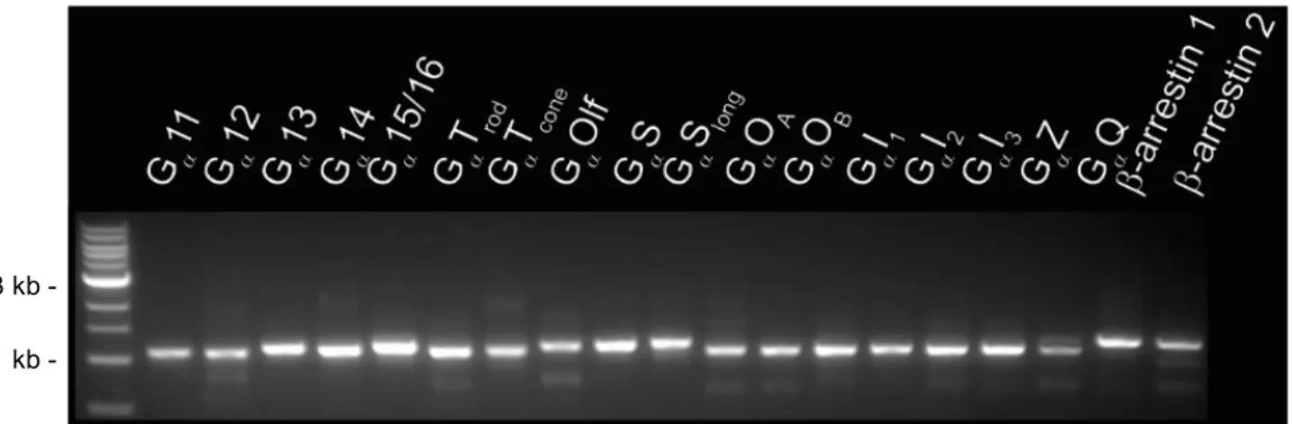
Prior to fusing the 17 transducer elements to the D2 receptor, each transducer component was PCR amplified using Primestar MAX DNA Polymerase according to manufacturer’s
Figure 2. Gel image of amplified human transducer elements. Elements confirmed to be around 1,000 base pairs with 1 kb ladder as reference (left lane).
Figure 3. Example of amplification verification via sequencing, shown for D2-β arrestin 2 fusion with forward primer. Template base code indicated in top row. Clean peaks verify proper
sequence in bottom row. 1 kb -
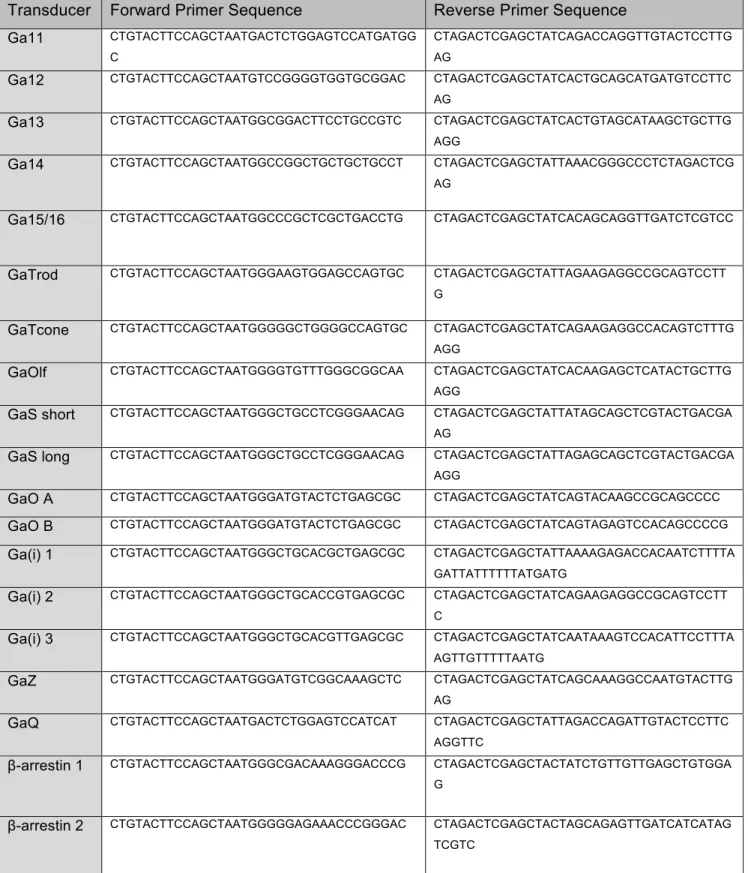
Transducer Forward Primer Sequence Reverse Primer Sequence Ga11 CTGTACTTCCAGCTAATGACTCTGGAGTCCATGATGG
C
CTAGACTCGAGCTATCAGACCAGGTTGTACTCCTTG AG
Ga12 CTGTACTTCCAGCTAATGTCCGGGGTGGTGCGGAC CTAGACTCGAGCTATCACTGCAGCATGATGTCCTTC AG
Ga13 CTGTACTTCCAGCTAATGGCGGACTTCCTGCCGTC CTAGACTCGAGCTATCACTGTAGCATAAGCTGCTTG AGG
Ga14 CTGTACTTCCAGCTAATGGCCGGCTGCTGCTGCCT CTAGACTCGAGCTATTAAACGGGCCCTCTAGACTCG AG
Ga15/16 CTGTACTTCCAGCTAATGGCCCGCTCGCTGACCTG CTAGACTCGAGCTATCACAGCAGGTTGATCTCGTCC
GaTrod CTGTACTTCCAGCTAATGGGAAGTGGAGCCAGTGC CTAGACTCGAGCTATTAGAAGAGGCCGCAGTCCTT G
GaTcone CTGTACTTCCAGCTAATGGGGGCTGGGGCCAGTGC CTAGACTCGAGCTATCAGAAGAGGCCACAGTCTTTG AGG
GaOlf CTGTACTTCCAGCTAATGGGGTGTTTGGGCGGCAA CTAGACTCGAGCTATCACAAGAGCTCATACTGCTTG AGG
GaS short CTGTACTTCCAGCTAATGGGCTGCCTCGGGAACAG CTAGACTCGAGCTATTATAGCAGCTCGTACTGACGA AG
GaS long CTGTACTTCCAGCTAATGGGCTGCCTCGGGAACAG CTAGACTCGAGCTATTAGAGCAGCTCGTACTGACGA AGG
GaO A CTGTACTTCCAGCTAATGGGATGTACTCTGAGCGC CTAGACTCGAGCTATCAGTACAAGCCGCAGCCCC
GaO B CTGTACTTCCAGCTAATGGGATGTACTCTGAGCGC CTAGACTCGAGCTATCAGTAGAGTCCACAGCCCCG
Ga(i) 1 CTGTACTTCCAGCTAATGGGCTGCACGCTGAGCGC CTAGACTCGAGCTATTAAAAGAGACCACAATCTTTTA GATTATTTTTTATGATG
Ga(i) 2 CTGTACTTCCAGCTAATGGGCTGCACCGTGAGCGC CTAGACTCGAGCTATCAGAAGAGGCCGCAGTCCTT C
Ga(i) 3 CTGTACTTCCAGCTAATGGGCTGCACGTTGAGCGC CTAGACTCGAGCTATCAATAAAGTCCACATTCCTTTA AGTTGTTTTTAATG
GaZ CTGTACTTCCAGCTAATGGGATGTCGGCAAAGCTC CTAGACTCGAGCTATCAGCAAAGGCCAATGTACTTG AG
GaQ CTGTACTTCCAGCTAATGACTCTGGAGTCCATCAT CTAGACTCGAGCTATTAGACCAGATTGTACTCCTTC AGGTTC
β-arrestin 1 CTGTACTTCCAGCTAATGGGCGACAAAGGGACCCG CTAGACTCGAGCTACTATCTGTTGTTGAGCTGTGGA G
β-arrestin 2 CTGTACTTCCAGCTAATGGGGGAGAAACCCGGGAC CTAGACTCGAGCTACTAGCAGAGTTGATCATCATAG TCGTC
The D2R GPCR (DRD2-Tango from addgene.org) (Kroeze et al., 2015) was amplified using Primestar MAX DNA according to manufacturer’s instructions. Primers used for
amplification were transfus-tangobbf (TAGCTCGAGTCTAGAGGG) and transfus-tangobbr (TAGCTGGAAGTACAGGTTC).
All DNA amplification was done using 10-beta competent E. coli cells. DNA was purified using Zymo mini-prep kit according to manufacturer’s instructions. Zymo midi-prep kit was used according to manufacturer’s instructions for full DNA preparation for transfection.
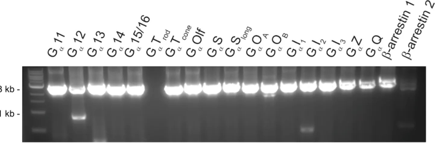
The GPCR-transducer fusions were then generated using homologous recombination cloning with NEBuilder Hi-Fi DNA Assembly Master Mix, according to manufacturer’s instructions. Successful incorporation of the transducer was confirmed via gel imaging (Fig. 4) and sequencing (Genewiz) (Fig. 3). Sequencing confirmation was done with primers BGHR (TAGAAGGCACAGTCGAGG), CMV-Forward (CGCAAATGGGCGGTAGGCGTG),
Figure 4. Gel image of fusion constructs. Successful insertion of transducer into DRD2-Tango backbone is visualized by appropriate size, about 3,000 base pairs. 1 kb ladder used as reference (left lane). Fusions missing in image (D2-GaTrod and β-arrestin 2) were made and confirmed at a later time.
Figure 5. General plasmid design for GPCR-transducer fusion proteins.Arrows indicate primer sites for PCR verification of fusions used to generate Figure 4. Includes signal for
overexpression in membrane, a FLAG affinity tag for visualizing receptor expression,
Vasopressin 2 receptor (V2R) C-terminus as a linker and arrestin recruitment domain, and a TEV protease cleavage site to remove fused transducer for control experiments.
Expression and membrane preparation of DRD2-transducer fusion proteins
HEK293T cell membranes were transfected using a modified calcium phosphate method (Jordan, Schallhorn, & Wurm, 1996). Briefly, cells were transfected with 20 µg of D2 fusion cDNA and allowed to express for at least 48 hours. On the day of membrane preparation, media
was decanted and cells were in 10 mL ice-cold lysis buffer (1 mM HEPES, 2 mM EDTA, pH 7.4) and left at 4°C for at least 10 minutes. Cell lysates were re-suspended, transferred to 30 mL centrifuge tubes, and centrifuged at 30,000xg for 30 minutes at 4°C. Tubes were decanted and membrane pellets were re-suspended in 4 mL/15 cm plate of Standard Binding Buffer (SBB; 50 mM Tris-HCl, 10 mM MgCl2, 0.1 mM EDTA, pH 7.4). Cells were re-suspended using a mechanical homogenizer (Polytron, setting 4) for 5 seconds, and 1 mL aliquots of the re-suspension were transferred to pre-chilled 1.7 mL microcentrifuge tubes. Re-suspended membranes were then centrifuged at 13,000xg for 10 minutes at 4°C. The SBB was decanted and membrane pellets were stored at -80°C until use.
Competitive radioligand binding
Elmer). Data was quantified and graphed using Prism 6.0 software and methods from Strachan et al., 2014.
Figure 6. Chemical structure of radioactive D2R antagonist [3H] N-methyl spiperone.
Results
Figure 7. Binding curves for [3H] N-Methyl spiperone. No significant high affinity shifts for this radioligand at any fusions with canonical transducers of interest (Ga(i)1,2,3). Data is from three (n=3) separate set-ups and runs with [3H] N-Methyl spiperone.
Quinpirole, a D2-selective drug, was tested with twelve of the transducer fusions (Figure 8). Quinpirole showed high-affinity site binding shifts for canonical transducers Ga(i)1, Ga(i)2, and Ga(i)3. The high- and low-affinity half maximal inhibitory concentration (IC50Hi and IC50Lo, respectively) for the fusion curves was used to determine fold shift from the D2-unfused receptor curve. Quinpirole showed a 30-fold shift with Ga(i)1, a 118-fold shift with Ga(i)2, and a 68-fold shift with Ga(i)3 (Figure 9). These shifts demonstrated significant bias for the canonical Ga(i) transducers.
N-Methyl Spiperone
n
= 3
-12 -11 -10 -9 -8 -7 -6 -5 -4
-25 0 25 50 75
100 D2-unfused
D2-Ga(i)1 D2-Ga(i)2 D2-Ga(i)3 D2-Ga(o)A D2-Ga(o)B D2-Barr1 D2-Barr2
log [drug] M
%
Di
sp
la
ce
m
en
Figure 8. Quinpirole binding curves for 12 transducer-fusions. Quinpirole showed high-affinity site binding shifts for canonical transducers Ga(i)1, Ga(i)2, and Ga(i)3, taken from three (n=3) sets of data. Significant shifts were not detected for other transducers, performed with two experimental runs (n=2).
Quinpirole n = 2, 3
-11 -10 -9 -8 -7 -6 -5 -4
-25 0 25 50 75 100
D2-unfused D2-Ga(i)1 D2-Ga(i)2 D2-Ga(i)3
D2-Barr1 D2-Ga(o)A D2-Ga(o)B
D2-Barr2 D2-Ga(s) long D2-Ga(s) short D2-Ga(q) D2-Ga(z) D2-Ga(olf)
log [drug] M
%
D
is
p
la
ce
m
en
-12 -11 -10 -9 -8 -7 -6 -5 -4
0 50 100
D2-unfused D2-Ga(i)1
log [Quinpirole] M
% Di sp la ce m en t IC50Hi IC50Lo D2-unfused (not used) 1.416e-006 D2-Ga(i)1 4.764e-008 1.416e-006 Quinpirole
n = 3
-12 -11 -10 -9 -8 -7 -6 -5 -4 0 50 100 D2-unfused D2-Ga(i)2 IC50Hi IC50Lo D2-unfused (not used) 1.332e-006 D2-Ga(i)2 1.132e-008 1.332e-006 log [Quinpirole] M
% Di sp la ce m en t Quinpirole
n = 3
-12 -11 -10 -9 -8 -7 -6 -5 -4
0 50 100 D2-unfused D2-Ga(i)3 IC50Hi IC50Lo D2-unfused (not used) 1.473e-006 D2-Ga(i)3 2.171e-008 1.473e-006 log [Quinpirole] M
% Di sp la ce m en t
Figure 9. Quinpirole high-affinity site binding shifts at the Ga(i)1, 2, and 3 transducers. D2-Ga(i)1 fusion showed a 30-fold shift in its high affinity IC50 (IC50Hi = 47.6 nM) relative to the low affinity IC50 (IC50Lo =1416 nM) (a). D2-Ga(i)2 fusion showed a 118-fold shift in its high affinity IC50 (IC50Hi =11.3 nM) relative to the low affinity IC50 (IC50Lo =1332 nM) (b). D2-Ga(i)3 fusion showed a 68-fold shift in its high affinity IC50 (IC50Hi =21.7 nM) relative to low affinity IC50
(IC50Lo = 1473 nM) (c). All three fold-shift values for the Ga(i) transducers were significant in
determining high-affinity binding sites for
quinpirole. (n=3) for (a), (b), and (c). b.
Quinpirole showed no significant affinity shifts for non-canonical transducers, including Ga(s) long, Ga(s) short, Ga(q), Ga(z), and Ga(olf) (Figure 10).
a. b. Quinpirole n = 2
-11 -10 -9 -8 -7 -6 -5 -4
-25 0 25 50 75 100 D2-unfused D2-Ga(s) long D2-Ga(s) short
log [Quinpirole] M
% Di sp la ce m en t Quinpirole
n = 2
-11 -10 -9 -8 -7 -6 -5 -4
-25 0 25 50 75 100 D2-unfused D2-Ga(q) D2-Ga(z) D2-Ga(olf)
log [Quinpirole] M
% Di sp la ce m en t
Dopamine, the endogenous D2R ligand, was also found to show high-affinity site binding shifts for canonical Ga(i) transducers (Figure 11). The high- and low-affinity half maximal inhibitory concentration for the fusion curves was used to determine fold shift from the D2-unfused receptor. Dopamine showed a 181-fold shift with Ga(i)1, a 81-fold shift with Ga(i)2, and a 19-fold shift with Ga(i)3 (Figure 12). The fold shifts indicate that dopamine significantly showed preference for Ga(i)1, Ga(i)2, and Ga(i)3.
Figure 11. Dopamine binding curves for canonical Ga(i) transducers. Dopamine showed high-affinity site binding shifts for canonical transducers Ga(i)1, Ga(i)2, and Ga(i)3. Data taken from three (n=3) sets of data.
Dopamine n = 3
-11 -10 -9 -8 -7 -6 -5 -4
-25 0 25 50 75
100 D2-unfused
D2-Ga(i)1 D2-Ga(i)2 D2-Ga(i)3
log [Dopamine] M
%
D
is
p
la
ce
m
en
-12 -11 -10 -9 -8 -7 -6 -5 -4 0 50 100 D2-unfused D2-Ga(i)1 IC50Hi IC50Lo D2-unfused (not used) 4.197e-007 D2-Ga(i)1 2.318e-009 4.197e-007 log [Dopamine] M
% Di sp la ce m en t Dopamine n = 3
-12 -11 -10 -9 -8 -7 -6 -5 -4
0 50 100 D2-unfused D2-Ga(i)2 IC50Hi IC50Lo D2-unfused (not used) 3.683e-007 D2-Ga(i)2 4.522e-009 3.683e-007 log [Dopamine] M
% Di sp la ce m en t Dopamine n = 3
-12 -11 -10 -9 -8 -7 -6 -5 -4 0 50 100 D2-unfused D2-Ga(i)3 IC50Hi IC50Lo D2-unfused (not used) 4.231e-007 D2-Ga(i)3 2.278e-008 4.231e-007 log [Dopamine] M
% Di sp la ce m en t
Figure 12. Dopamine high-affinity site binding shifts at the Ga(i)1, 2, and 3 transducers. D2-Ga(i)1 fusion showed a 181-fold shift in its high affinity IC50 (IC50Hi = 2.3 nM) relative to the low affinity IC50 (IC50Lo =419.7 nM) (a). D2-Ga(i)2 fusion showed a 81-fold shift in its high affinity IC50 (IC50Hi =4.5 nM) relative to the low affinity IC50 (IC50Lo =368.3 nM) (b). D2-Ga(i)3 fusion showed a 19-fold shift in its high affinity IC50 (IC50Hi =22.7 nM) relative to low affinity IC50 (IC50Lo = 423.1 nM) (c). All three fold-shift values for the Ga(i) transducers were significant in determining high-affinity binding sites for dopamine. (n=3) for (a), (b), and (c). a.
b.
Discussion
Our assay provides insight into the functional selectivity of quinpirole and dopamine at the dopamine D2 receptor. Quinpirole, a D2-selective drug, and dopamine, the D2R endogenous ligand, both showed high affinity shifts for D2R fused to canonical Gαi transducers (Figures 9, 11, 12). Quinpirole did not show high affinity shifts for non-canonical Gα transducers (Figure 10). This data is consistent with our predictions and the accepted knowledge of the nature of the dopamine D2 receptor (Lane, Powney, Wise, Rees, & Milligan, 2007) and therefore provides validation for the developing screening platform used in the Roth laboratory.
The fusion constructs created were designed to express D2R fused to one transducer. The subsequent isolation of the complexes allowed for in vitro binding assays that would provide data without in vivo variables, such as the presence of other proteins and transducers in a cell’s internal environment. Therefore, it is understood that the signaling profiles produced provide data that is result of the definitive receptor-transducer pair. The variations in affinity can be attributed to the presence of a transducer at the receptor, as seen with the quinpirole and dopamine data presented here.
This screening platform can be utilized to investigate ligands’ pharmaceutical potential. Expression of transducers is cell- and tissue-type dependent (Sharma et al., 2015). By
understanding a ligand’s varying preference for transducers, this differential transducer
likely be more efficacious and trigger less side-effects than a drug that was not characterized as having high affinity for Ga(i)3.
In addition to facilitating new drug design, our platform can uncover novel biology occurring between current pharmaceutical and their targeted receptors. This information will allow for clearer understanding of a drug’s preference for particular transducers, therefore prompting speculation of the drug’s effects on a body-wide scale. For example, the serotonin 5-HT2A receptor is mostly known for its expression in the central nervous system and is clinically targeted accordingly. However, 5-HT2A is also highly expressed in the intestine, platelets, and endothelial cells (Raote, Bhattacharya, & Panicker, 2007). Using our platform to create affinity profiles for 5-HT2A-targeted drugs may reveal new information about the drug’s behavior at the body’s different cell types, considering that transducer expression is differential between cells and tissues. This insight may then uncover new uses for the drug or allow for modification of the drug to reduce its effects at untargeted sites of the body.
To further broaden and develop this platform, more ligands should be tested against our D2R transducer fusions. This may include characterizing known clinical antipsychotics that are D2R-targeted, such as aripiprazole which is prescribed to treat schizophrenia (Burris et al., 2002) and bipolar disorder (Kanba et al., 2012). Additionally, other GPCRs of pharmacological
importance can be used for fusions in order to create high-affinity profiles for a variety of drugs. For our next step, the Roth lab plans to expand this screening platform by creating fusions with the aforementioned serotonin 5-HT2A receptor, the target of clinical drugs such as mirtazapine which is prescribed to treat depression (Celada, Puig, Amargós-Bosch, Adell, & Artigas, 2004).
Acknowledgements
I would like to thank Dr. Justin English for his mentorship over the past nine months. He has introduced me to the Roth lab and its projects and has trained me in the molecular biology used for this project. Thank you to Dr. John McCorvy for his mentorship in radioligand binding assays and for his assistance with the results figures. I am grateful to both Dr. English and Dr. McCorvy for their advice, conversation, and dedicated support while forming this thesis.
Thank you to Dr. Ryan Strachan for his intellectual piloting and pioneering of this continuing project. Thank you to Dr. Bryan Roth for his support and lab’s resources that have made this project possible. To all the members of the Roth lab, thank you for being inspiring scientific role models and for your readiness to provide assistance and wisdom when needed.
References
Burris, K. D., Molski, T. F., Xu, C., Ryan, E., Tottori, K., Kikuchi, T., … Molinoff, P. B. (2002). Aripiprazole, a novel antipsychotic, is a high-affinity partial agonist at human dopamine D2 receptors. The Journal of Pharmacology and Experimental Therapeutics, 302(1), 381–389. http://doi.org/10.1124/jpet.102.033175.Carson
Celada, P., Puig, M. V., Amargós-Bosch, M., Adell, A., & Artigas, F. (2004). The therapeutic role of 5-HT1A and 5-HT2A receptors in depression. In Journal of Psychiatry and Neuroscience (Vol. 29, pp. 252–265).
Civelli, O., Bunzow, J. R., & Grandy, D. K. (1993). Molecular diversity of the dopamine receptors. Annual Review of …. Retrieved from
http://www.annualreviews.org/doi/pdf/10.1146/annurev.pa.33.040193.001433\npapers2://p ublication/uuid/7E8EE902-9295-49CE-8AA9-1C3CB20C77C6
Filmore, D. (2004). It’s a GPCR world. Modern Drug Discovery, 7(11), 24–27. Retrieved from http://pubs.acs.org/subscribe/journals/mdd/v07/i11/html/1104feature_filmore.html
Jordan, M., Schallhorn, A., & Wurm, F. M. (1996). Transfecting mammalian cells: Optimization of critical parameters affecting calcium-phosphate precipitate formation. Nucleic Acids Research, 24(4), 596–601. http://doi.org/10.1093/nar/24.4.596
Kanba, S., Kawasaki, H., Ishigooka, J., Sakamoto, K., Kinoshita, T., & Kuroki, T. (2012). A placebo-controlled, double-blind study of the efficacy and safety of aripiprazole for the treatment of acute manic or mixed episodes in Asian patients with bipolar I disorder (the AMAZE study). The World Journal of Biological Psychiatry : The Official Journal of the World Federation of Societies of Biological Psychiatry, (February), 1–9.
Kroeze, W. K., Sassano, M. F., Huang, X.-P., Lansu, K., McCorvy, J. D., Giguère, P. M., … Roth, B. L. (2015). PRESTO-Tango as an open-source resource for interrogation of the druggable human GPCRome. Nature Structural & Molecular Biology, 22(5), 362–9. http://doi.org/10.1038/nsmb.3014
Lane, J. R., Powney, B., Wise, A., Rees, S., & Milligan, G. (2007). Protean agonism at the dopamine D2 receptor: (S)-3-(3-hydroxyphenyl)-N-propylpiperidine is an agonist for activation of Go1 but an antagonist/inverse agonist for Gi1,Gi2, and Gi3. Molecular Pharmacology, 71(5), 1349–1359. http://doi.org/10.1124/mol.106.032722.2000
Masri, B., Salahpour, A., Didriksen, M., Ghisi, V., Beaulieu, J.-M., Gainetdinov, R. R., & Caron, M. G. (2008). Antagonism of dopamine D2 receptor/beta-arrestin 2 interaction is a common property of clinically effective antipsychotics. Proceedings of the National Academy of Sciences of the United States of America, 105(36), 13656–61.
http://doi.org/10.1073/pnas.0803522105
Montmayeur, J. P., Guiramand, J., & Borrelli, E. (1993). Preferential coupling between dopamine D2 receptors and G-proteins. Molecular Endocrinology (Baltimore, Md.), 7(March), 161–170. http://doi.org/10.1210/me.7.2.161
Raote, I., Bhattacharya, A., & Panicker, M. M. (2007). Serotonin 2A (5-HT2A) Receptor Function: Ligand-Dependent Mechanisms and Pathways. Serotonin Receptors in Neurobiology, 1–17. http://doi.org/NBK1853 [bookaccession]
Seeman, P. (2006). Targeting the dopamine D2 receptor in schizophrenia. Expert Opinion on Therapeutic Targets, 10(4), 515–531. http://doi.org/10.1517/14728222.10.4.515
Neuroscience, 18(12), 1819–31. http://doi.org/10.1038/nn.4160
Strachan, R. T., Sun, J. P., Rominger, D. H., Violin, J. D., Ahn, S., Bie Thomsen, A. R., … Lefkowitz, R. J. (2014). Divergent transducer-specific molecular efficacies generate biased agonism at a G protein-coupled receptor (GPCR). Journal of Biological Chemistry, 289(20), 14211–14224. http://doi.org/10.1074/jbc.M114.548131
Urban, J. D., Clarke, W. P., von Zastrow, M., Nichols, D. E., Kobilka, B., Weinstein, H., … Mailman, R. B. (2007). Functional selectivity and classical concepts of quantitative pharmacology. J Pharmacol Exp Ther, 320(1), 1–13.
http://doi.org/10.1124/jpet.106.104463

![Figure 6. Chemical structure of radioactive D2R antagonist [3H] N-methyl spiperone.](https://thumb-us.123doks.com/thumbv2/123dok_us/8331688.2210385/10.918.324.593.275.412/figure-chemical-structure-radioactive-d-antagonist-methyl-spiperone.webp)

