Characterization of Dormant Tumor Cells in Pancreatic Ductal
Adenocarcinoma (PDAC) and Therapeutic Strategies
Sonya Anderson, PharmD Candidate1; Limei Shen, PhD2; Leaf Huang, PhD2
1
University of North Carolina in Chapel Hill Eshelman School of Pharmacy
2
University of North Carolina in Chapel Hill Department of Pharmacoengineering and Molecular Pharmaceutics
Itemized Counts: Total words: 3,350
Figures: 3
Tables: 0
References: 26
Characterization of Dormant Tumor Cells in Pancreatic Ductal
Adenocarcinoma (PDAC) and Therapeutic Strategies
Authors: Sonya L. Anderson1*, Limei Shen2, Leaf Huang2
Affiliations: 1
University of North Carolina in Chapel Hill Eshelman School of Pharmacy
2
University of North Carolina in Chapel Hill Department of Pharmacoengineering and Molecular Pharmaceutics
*To whom correspondence should be addressed: Tel: 630-636-0264. Email: [email protected].
One Sentence Summary:
The CXCL13 trap works to decrease tumor burden in pancreatic cancer by reducing the differentiation of B-cells responsible for inhibiting the anti-tumor immune response, shown through the expression levels of cytokines measured through western blot and flow cytometry.
Abstract:
Introduction: Pancreatic cancer represents around 3% of all cancers in the United States. Despite this low incidence, it represents around 7% of all cancer deaths in the United States, making it the fourth leading cause of cancer-related death. While prognosis does depend on histologic grade and extend of spread, pancreatic cancer is often difficult to treat due to lack of effective screening tests, quick spread, recurrence, and low immunogenic nature. Additionally, pancreatic cancer, such as its most common form: Pancreatic Ductal Adenocarcinoma (PDAC), is resistant to most systemic/targeted therapies. This facilitates the need for novel drug delivery. One such novel therapeutic treatment option is traps. Traps are fusion proteins that bind to chemokines in a manner similar to monoclonal antibodies. The traps are then formulated into nanoparticles which can be delivered to the pancreas. The trap can be formulated to target chemokines like CXCL13 which has been implicated in the suppression of the anti-tumor response through B-reg
recruitment. Methods: To analyze the effectiveness of the CXCL13 trap, mouse tumor studies were completed, in which KPC98207 cells were injected into the tail of the pancreas. Mice were randomized into two treatment groups: PBS or CXCL13 trap nanoparticles. Tumor tissues were then harvested to be analyzed through western blot and flow cytometry for the presence of certain cytokines (pSTAT3, IL-35, IL-10) and vimentin. Results: The PDAC tumors treated with CXCL13 trap had significantly reduced presence of pSTAT3, IL-35, IL-10, and vimentin. Conclusion: The CXCL13 trap works to decrease tumor burden in PDAC by reducing differentiation of B-cells into B-reg cells, shown by the reduction of cytokines involved in inhibiting the anti-tumor immune response (pSTAT3, IL-35, IL-10) and vimentin. Thus, the CXCL13 trap presents a new treatment option to be used in conjunction with current
[Main Text: ] Introduction
Epidemiology
Within the United States approximately 53,670 patients are diagnosed with pancreatic cancer each year, representing around 3% of all cancers in the United States.1 Despite this low incidence, approximately 43,090 patients will die from pancreatic cancer each year, representing around 7% of all cancer-related deaths in the United States.1 This makes pancreatic cancer the fourth leading cause of cancer-related deaths in the United States.1 Additionally, pancreatic cancer’s 5-year survival rate is particularly low, with only around 8% of patients expected to survive.1
While the cause of pancreatic cancer is complex and multifactorial, cigarette smoking and family history are among the most common causes. Cigarette smoking has been linked to around 20% of all pancreatic tumors.2 Additionally, cancers in those that smoke harbor more genetic mutations as compared to those that are non-smokers.2 With respect to family history, approximately 7-10% of affected individuals have some form of family history.3 Additionally, first-degree relatives of individuals with familial pancreatic cancer, defined as those with a pair of first-degree relatives with pancreatic cancer, have a nine-fold increased risk as compared to the general population.4 In kindreds, defined as those with three or more first-degree relatives with pancreatic cancer, have a 32-fold increased risk as compared to the general population. Furthermore, patients with familial pancreatic cancer are at a higher risk for developing precancerous lesions and extra-pancreatic cancers as compared to patients with sporadic pancreatic tumors.5 Pancreatic cancer is also a disease that is associated with advancing age. It culminates to a 40-fold increased risk by the age of 80.6 Patients with these high-risk factors could potentially benefit the most from improved treatment modalities.
Although prognosis is dependent on histologic grade and extent of spread, pancreatic cancer often still has poor prognosis due to a number of factors. Most often is that early detection is fairly uncommon.7 Signs and symptoms of pancreatic cancer, such as upper abdominal pain, yellowing of the skin and eyes, and weight loss, do not typically occur until later stages of the disease.7 Furthermore, there are no effective screening tests for pancreatic cancer.8 Pancreatic cancer also tends to spread very quickly as it lies at the junction of a number of structures allowing it to spread easily. It often spreads to nearby organs, such as the liver, gallbladder, and intestines, even in the early stages of the disease.8 Additionally, recurrence is likely. Even after surgical resection, pancreatic cancer reoccurs. There is a high rate of systemic, 80%, and local, 20%, recurrence after surgery alone.9 It is also difficult to achieve a wide resection margin due to proximity to a number of veins and arteries within the pancreas.9 Lastly, the tumors are low or non-immunogenic, and typically lack tumor-infiltrating effector lymphocytes.10
Pathophysiology
BRCA2.13 Other genetic abnormalities include widespread chromosomal losses, gene amplifications, and telomere shortening.14,15
Early ductal lesions typically do not present with any genetic alterations. KRAS
mutations are among the first genetic changes that are detected in the progression series. KRAS mutations are prevalent in 30% of early stage lesions.16 These mutations increase in frequency with disease progression, such that they are present in close to 100% of PDAC tumors.13
Activating mutations in the RAS-family oncogenes result in cellular effects such as proliferation induction, survival, and invasion through effector pathway stimulation.17
With respect to the TP53 tumor-suppressor gene mutation, more than 50% of PDAC tumors have this mutation present.13 It is generally caused by missense alterations and arises in the later stages of pancreatic intraepithelial neoplasias. This typically causes significant
dysplasia, reflecting TP53’s role in preventing malignant progression.16
Loss of TP53 facilitates genetic instability that leads to malignancy.16
The tumor microenvironment plays an important role in modulating immune responses during cancer progression. Tumor associated fibroblasts (TAF) are a major component of the tumor microenvironment and produce a number cytokines that promote initiation, progression, metastasis of solid tumors.18 In addition, TAFs synthesize extracellular matrix, as well as metabolic and immune reprogramming of the tumor microenvironment, which impacts adaptive chemoresistance.19 TAFs also shape immune cell populations within the tumor
microenvironment by secreting chemokines such as CXCL13. CXCL13 recruits B cells into the tumor microenvironment. Regulatory B cells (B-regs) and total B cells promote cancer growth by inhibiting the cytotoxic activity of Th1/CD8+ cells. Additionally, B-regs suppress different cell subtypes, such as CD4+ T cells, through the secretion of suppressive mediators like TGF-β. This facilitates the conversion of CD4+ T cells into regulatory T cells (T-regs) which promote tumor progression.20 As differing B cell subpopulations are recruited to the tumor
microenvironment, they can acquire immunosuppressive properties within the tumor bed and attenuate anti-tumor immune responses by promoting IL-10 and PD-L1 expression.20 B-reg secretion of IL-10 can also convert dendritic cells into a tolerogenic phenotype, thus attenuating anti-tumor immune responses. B cell–macrophage interactions also promote PI3Kγ- and Bruton tyrosine kinase (BTK)–dependent macrophage Th2 polarization, leading to immune suppression and pancreatic cancer progression.21
Current Standards of Treatment
Standards of treatment vary depending on the patient’s extent of disease at presentation. Resectable disease is defined as a pancreatic tumor without involvement of the superior
mesenteric artery or coeliac axis, a patent superior mesenteric-portal venous confluence, and the lack of distant metastatic disease.22 Currently, surgical resection is the only curative option for pancreatic cancer. Unfortunately as the disease frequently presents late, only 15-20% of patients are candidates for pancreatectomy.23 Adjuvant chemotherapy is recommended in all patients with resected pancreatic cancer. Adjuvant treatment includes gemcitabine +/- capecitabine +/-
to thymidylic acid, interfering with DNA synthesis. It again is associated with a number of side effects including hand-foot syndrome and GI toxicities.24
Locally advanced, unresectable pancreatic cancer is defined as a tumor that encases a vascular structure (ie: superior mesenteric artery, coeliac axis, superior mesenteric vein-portal vein confluence) or bulky peripancreatic lymphadenopathy with no evidence of distant
metastatic disease.22 Treatment options include gemcitabine +/- nanoparticle albumin-bound paclitaxel (nabpaclitaxel) or FOLFIRINOX (oxaliplatin, leucovorin, irinotecan, fluorouracil, FU).25 Paclitaxel works by promoting microtubule assembly by enhance the action of tubulin dimers, stabilizing existing microtubules, and inhibiting their disassembly. This interferes with the late G2 mitotic phase and cell replication. However it does have side effects including
myelosuppression, peripheral neuropathies, and hypersensitivities.25 As FOLFIRINOX is a combination of a number of different significant side effects including myelotoxicity, diarrhea, mucositis, hand-foot syndrome, and pulmonary toxicities. It is typically only used in those with a good performance status and normal total bilirubin.25
As for metastatic disease, depending on performance status and bilirubin levels, FOLFIRINOX, FOLFOX, gemcitabine + nabpaclitaxel, gemcitabine + capecitabine, or gemcitabine alone.26 Palliative care alone may be considered in those with too poor of a performance status.26
Traps
Traps are fusion proteins that are designed to bind to chemokines, such as CXCL13 with a high affinity in a manner similar to monoclonal antibodies. Nanoparticles can be used to deliver and express the plasmid DNA encoding the trap.
Importance and Objective
PDAC is characterized by the presence of abundant stroma and is resistant to most systemic and targeted therapies. Even immunotherapies, including checkpoint inhibitors, are not effective against PDAC. Treatment modalities that are effective are not targeted and cause a number of toxicities in the patient. The objective is to evaluate a trap against CXCL13’s in its potential use in PDAC. As CXCL13 recruits B-reg cells into the tumor microenvironment, by blocking CXCL13, B-reg infiltration will be blocked and reduce tumor progression. This targeted therapy represents a novel treatment modality for PDAC. Preliminary data has shown that the CXCL13 trap (OT1) reduces tumor growth; however its mechanism of action has not yet been shown to reduce B-reg differentiation. Thus, the goal is to prove OT1 decreases PDAC tumor growth by reducing B-reg differentiation through known marker of B-reg expression.
Results
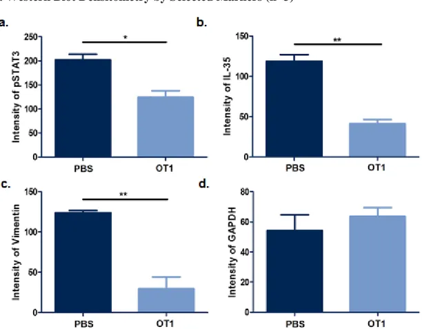
OT1 Decreases pSTAT3, IL-35, and Vimentin Expression
After immunoblotting with the corresponding antibodies, OT1 was shown to reduce expression of pSTAT3, IL-35, and Vimentin (Fig. 1). Each is a known marker of B-reg
differentiation. IL-35 is directly secreted by B-reg cells. pSTAT3 is upregulated by IL-10, which is secreted by B-reg cells. Vimentin is a general marker of accelerated growth, increased
invasion, and poor prognosis. After densitometry analysis, OT1 decreases pSTAT3 expression at a significance level of p<0.05 (Fig. 2). Additionally, OT1 decreases IL-35 and Vimentin
OT1 Decreases IL-10 Expression
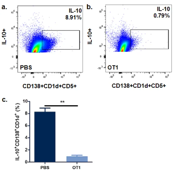
IL-10 expression in B-reg cells was analyzed through the use of flow cytometry. CD138+CD1d+CD5+ is the specific marker for B-reg cells. After specifically targeting these cells in the tumor lysate and utilizing antibodies against IL-10, OT1 was shown to decrease expression of IL-10 as compared to placebo (0.79% vs. 8.91% respectively) (Fig. 3). Through statistical analysis, OT1 significantly reduced expressed of IL-10 at a level of p<0.01.
Discussion
In conclusion, OT1 significantly reduces expression of known markers of B-reg expression: pSTAT3, IL-35, Vimentin, and IL-10. This shows that OT1 does reduce B-reg differentiation, allow the body to product an anti-tumor response, decreasing tumor progression. OT1 offers a new targeted treatment modality to be used alongside current chemotherapy and potentially future immunotherapy. Limitations of this study include that marker confirmation was only assessed through two methods: western blot and FC, and that other known markers of B-reg expression were not assessed (ie: TGF-β, PD-L1, PD-1, and IL-8). Future research includes assessing cytokine levels through other methods: RT-PCR, ELISA, and
immunofluorescence staining; assessing other known markers; and evaluating OT1’s use in other models (ie: melanoma).
Materials and Methods
Materials
distearoryl-sn-glycero-3-phosphoethanolamine-N-[methoxy(polyethyleneglycol-2000)] ammonium salt (DSPE-PEG) was purchased from NOF (Ebisu Shibuya-ku, Tokyo). 1,2-dioleoyl-3-trimethylammonium-propane chloride salt (DOTAP) was purchased from Avanti Polar Lipids (Alabaster, AL, USA). Cholesterol and protamine were purchased from Sigma-Aldrich (St. Louis, MO, USA). DiI for biodistribution studies was purchased from ThermoFisher Scientific. All other chemicals were purchased from Sigma-Aldrich if not specifically
mentioned.
Methods
Construction of CXCL13 Trap Genes
The CXCL13 trap was engineered through a known antibody that binds to human CXCL13 by grafting the CDRs into a murine scFv scaffold. In brief, the codon-optimized DNA sequences code for a signaling peptide, VH domain, a flexible linker, VL domain, E tag, and
His(6×) tag, respectively was synthesized. The resulting cDNA was cloned into pCDNA3.1 between Nhe I and Xho I sites and the accuracy was confirmed by DNA sequencing.
Expression and Purification of Recombinant Trap Proteins
days in a CO2 incubator at 37 C. After protein expression, media was harvested and incubated
with IMAC Nickel-charged resin (BIORAD, Hercules, CA) equilibrated with the binding buffer (25 mL HEPES, 300 mM NaCl, pH 7.4) for 1 h at 4 C. The slurry was packed into a column followed by washing with 20 mM and 50 mM imidazole in the binding buffer sequentially. Bound trap was eluted with 200 mM imidazole in the binding buffer and dialyzed against PBS at 4°C.
Binding Affinity Measurement
The affinity of CXCL13 trap to CXCL13 was accessed with Microscale Thermophoresis (MST). One hundred µL of 200 nM IL-10 trap was first fluorescently labeled by mixing with 100 L of 100 nM RED-tris-NTA dye followed by incubation at room temperature for 30 min. Ten L of the labeled trap was then added to 10 L of serially 2-fold diluted mouse IL-10 or other interleukins (Biolegend, San Diego, CA) at 10 M using a PBST buffer (PBS with 0.05 % Tween 20). The resulting samples were subsequently loaded into capillaries, and the
thermophoresis of each sample was measured using Auto Red laser power and medium MST power on Monolith NT.115 (NanoTemper Technologie, Munich, Germany).
Preparation and Characterization of LPD
LPD NPs were prepared through a stepwise self-assembly process as described
previously. Briefly, DOTAP and cholesterol (1:1, mol/mol) were dissolved in chloroform, and the solvent was removed. The lipid film was then hydrated with distilled water to make the final concentration of 10 mM cholesterol and DOTAP. The hydrated lipid suspension was
sequentially extruded through 200 nm and 100 nm polycarbonate membranes (Millipore, MA) to form 70 – 100 nm unilamellar liposomes. The LPD polyplex cores were formulated by mixing 100 μL of 50 μg protamine in 5 % glucose with equal volume of 50 μg plasmid (plasmids encoding CXCL13 trap) in 5 % glucose. The mixture was incubated at room temperature for 10 min followed by the addition of 60 μL cholesterol/DOTAP liposomes (10 mM each). Post insertion of 15 % DSPE-PEG-AEAA was performed at 60 °C for 15 min. The size and surface charge of the NPs were determined by a Malvern ZetaSizer Nano series (Westborough, MA). TEM images were acquired where NPs were negatively stained with uranyl acetate using a JEOL 100 CX II TEM (JEOL, Japan).
Bio-distribution of LPD NPs
Approximately 0.1 % of hydrophobic dye DiI was incorporated into DOTAP-cholesterol liposomes to formulate the DiI-labeled LPD NPs. Twenty-four hours after intravenous injection of the DiI-labeled LPD NPs, mice were euthanized, major organs and tumors were collected. The distribution of LPD NPs in major organs was quantitatively visualized with IVIS® Kinetics Optical System (Perkin Elmer, CA). Images were captured at an excitation wavelength of 520 nm and emission wavelength of 560 nm.
Cell Lines and Tissue Cultures
(FBS) (Gibco) and 1 % Penicillin/Streptomycin at 37 °C and 5 % CO2 in a humidified
atmosphere.
Orthotopic Allograft KPC Mouse Model
Six-week-old female C57BL/J mice were purchased from Charles River Laboratories and maintained under pathogen-free conditions. All animal handling procedures were approved by the University of North Carolina at Chapel Hill’s Institutional Animal Care and Use Committee. Sub-confluent KPC98207 (with or without RFP/Luc) cells were harvested and washed with cold PBS just prior to implantation. Orthotopic allografting KPC model was established by orthotopic injection of 1x10⁶ cells into the tail of pancreas. In brief, eight-week-old C57BL/6 mice were anesthetized by 2.5 % isoflurane and placed in supine position. A midline incision was made to exteriorize the spleen and pancreas. Using an insulin-gage syringe, 1x10⁶ cells in 50 μL (PBS + Matrigel) were injected into the tail of pancreas. The abdominal wall and skin were closed with 6-0 polyglycolic acid sutures. The injection site was sealed with a tissue adhesive (3M, St. Paul, MN) and sterilized with 70 % alcohol. Tumor growth was monitored by intraperitoneal injection of 100 µL of D-Luciferin (10 mg/ml) followed by bioluminescence analysis using an IVIS® Kinetics Optical System (BD, CA).
Tumor Growth Inhibition and Survival Analysis
Mice bearing KPC98027 RFP/Luc allografts were established as described above. Treatments were initiated on day 18. Mice were then randomized into 4 groups (n = 8 - 10) as follows: Untreated group (PBS), CXCL13 trap NP (encapsulated with pcDNA3.1 containing the trap gene). Intravenous injections were performed every three days for a total of 3 doses of 50 µg plasmid/mice. Tumor growth was monitored using IVIS® Kinetics Optical System (Perkin Elmer, CA) every 3 days. The increases of tumor volumes were calculated as the radiance of the intensities and standardized with the initial tumor volume (Vt/V0).
Western Blot Analysis
Tumor extracts were separated by SDS-PAGE and transferred onto PVDF membranes. These extracts were probed with antibodies against GAPDH, pSTAT3, IL-35, and Vimentin (1:1000, Invitrogen). Proteins of interest were detected with HRP-conjugated goat anti-mouse IgG antibody (1:4000, Invitrogen) and visualized with the Pierce ECL Western blotting substrate (Thermo Scientific), according to the provided protocol.
Flow cytometry assay
Tumor-infiltrating immune cells were analyzed by flow cytometry. In brief, tumor tissues were harvested and digested with collagenase A and DNase at 37 °C for 60 min. After lysis of red blood cells, cells were resuspended in 5 mL of PBS. 2x10⁶ cells were stained with
fluorescently labeled antibodies for surface marker expression analysis. After staining, cells were fixed with 300 μL 4 % PFA and analyzed via FACS (BD LSR II).
References and Notes:
1. Siegel RL, Miller KD, Jemal A. Cancer Statistics, 2018. CA Cancer J Clin. 2018;68(1):7-30. 2. Blackford A, Parmigiani G, Kensler TW, et al. Genetic mutations associated with cigarette
smoking in pancreatic cancer. Cancer Res 2009; 69: 3681–88.
3. Petersen GM, de Andrade M, Goggins M, et al. Pancreatic cancer genetic epidemiology consortium. Cancer Epidemiol Biomarkers Prev 2006; 15: 704–10.
4. Klein AP, Brune KA, Petersen GM, et al. Prospective risk of pancreatic cancer in familial pancreatic cancer kindreds. Cancer Res 2004; 64: 2634–38.
5. Brune KA, Lau B, Palmisano E, et al. Importance of age of onset in pancreatic cancer kindreds. J Natl Cancer Inst 2010; 102: 119–26.
6. Anderson, K. E., Potter, J. D. & Mack, T. M. in Cancer Epidemiology and Prevention (eds Schottenfeld, D. & Fraumeni, J. J.) 725–771 (Oxford University Press, New York, 1996). 7. Chari ST. Detecting Early Pancreatic Cancer – Problems and Prospects. Semin Oncol.
2009;34(4):284-294.
8. Poruk KE, Firpo MA, Adler DG, et al. Screening for Pancreatic Cancer: Why, How, and Who? Ann Surg. 2013;257(1):17-26.
9. Zakharova OP, Karmazanovsky GG, Egorov VI. Pancreatic adenocarcinoma: Outstanding problems. World J Gastrointest Surg. 2012;4(5):104-113.
10.Lutz ER, Wu AA, Bigelow E, et al. Immunotherapy Converts Non-immunogenic Pancreatic Tumors into Immunogenic Foci of Immune Regulation. Cancer Immunol Res.
2014;2(7):616-631.
11.Stewart BW, Wild CP. World Cancer Report 2014. WHO. 2014.
12.Vincent A, Herman J, Schulick R, et al. Pancreatic Cancer. Lancet. 2011;378:607-20. 13.Rozenblum E, Schutte M, Goggins M, et al. Tumor-suppressive pathways in pancreatic
carcinoma. Cancer Res 1997; 57: 1731–34.
14.Jones S, Zhang X, Parsons DW, et al. Core signaling pathways in human pancreatic cancers revealed by global genomic analyses. Science 2008; 321: 1801–06.
15.van Heek NT, Meeker AK, Kern SE, et al. Telomere shortening is nearly universal in pancreatic intraepithelial neoplasia. Am J Pathol 2002; 161: 1541–47.
16.Bardeesy N, DePinho RA. Pancreatic Cancer Biology and Genetics. Nature Rev. 2002;2:897-909.
17.Shields JM, Pruitt K, McFall A, et al. Understanding Ras: ‘it ain’t over ‘til it’s over’. Trends
Cell Biol. 2000;10:147–154.
18.Quail DF, Joyce JA. Microenvironmental regulation of tumor progression and metastasis.
Nature medicine. 2013;19:1423-1437.
19.Kalluri R. The biology and function of fibroblasts in cancer. Nat Rev Cancer. 2016;16:582-598.
20.Roghanian A, Fraser C, Kleyman M, et al. B Cells Promote Pancreatic Tumorigenesis.
Cancer Discov. 2016;6:230-232.
22.Li D, Xie K, Wolff R, et al. Pancreatic cancer. Lancet. 2004;363:1049-57.
23.Sewnath ME, Karsten TM, Prins MH, et al. A meta-analysis on the efficacy of preoperative biliary drainage for tumors causing obstructive jaundice. Ann Surg. 2002; 236: 17–27. 24.Khorana AA, Mangu PB, Berlin J, et al. Potentially Curable Pancreatic Cancer: American
Society of Clinical Oncology Clinical Practice Guideline. J Clin Oncol. 2016;34(21):2541. 25.Balaban EP, Mangu PB, Khorana AA, et al. Locally Advanced, Unresectable Pancreatic
Cancer: American Society of Clinical Oncology Clinical Practice Guideline. J Clin Oncol. 2016;34(22):2654. Epub 2016 May 31.
26.Sohal DP, Mangu PB, Khorana AA, et al. Metastatic Pancreatic Cancer: American Society of Clinical Oncology Clinical Practice Guideline. J Clin Oncol. 2016;34(23):2784. Epub 2016 May 31.
Figures:
Fig. 1. pSTAT3, IL-35, Vimentin, and GAPDH Western Blot (n=3)
Fig. 1: Protein expression of pSTAT3, IL-35, vimentin, and GAPDH were detected by
Fig. 2.Western Blot Densitometry by Selected Markers (n=3)
Fig. 3. IL-10 Flow Cytometry
Fig. 3: a. 8.91% IL-10 expression by B-reg cells in the PBS treatment group. b. 0.79% IL-10 expression by B-reg cells in the OT1 treatment group. c. OT1 significantly reduces IL-10 expression by B-reg cells. **denotes p < 0.01.
Report Addendum
Acknowledgments: None.
Funding Support: The authors have no funding support to disclose.
Conflicts of Interest: Leaf Huang is a co-owner of the company OncoTrap Inc.