STROKE is the 5th leading cause of death in the United States, killing about 130,000 Americans a year1. The prevalence of stroke is roughly 3% of the population per year1. There are two types of stroke: hemorrhagic and ischemic. Hemorrhagic stroke is caused by bleeding into the cranial cavity whileischemic involves occlusion or restriction of blood flow to an area of the brain.1 Potential pathophysiologic mechanisms of ischemic stroke include decreased
perfusion due to a systemic cause or due to stenosis of a vessel that feeds the brain. Decreased perfusion due to a systemic cause, like persistent hypotension, causes global cerebral hypo perfusion. Vascular occlusion can occur due to plaque or embolization and causes decreased perfusion in the associated vascular bed. Cardioembolism is the most common cause
representing about 37% of ischemic strokes2. Carotid artery atherosclerosis is a major risk factor and causes 10-15% of ischemic stroke3. Atherosclerosis can cause a gradual change in the vessel causing a narrowed area that restricts flow and can accumulate platelets which lead to an acute episode4 . Risk of stroke from carotid artery stenosis depends on the severity of the stenosis among other risk factors including diabetes and hypertension.
Carotid endarterectomy (CEA) is an open surgical procedure was established in 1954 as a reliable treatment for carotid stenosis. In approximately 1990, a clinical trial supported the use of endarterectomy over aspirin alone5. Carotid artery stenting is a more recent procedure developed in the 1980s as a less invasive alternative treatment. It is important to compare and contrast each method of treatment to further identify the long-term outcomes associated with each option.
Carotid artery stenting (CAS) is usually completed in the cardiac catheterization lab via percutaneous access of the right radial or femoral artery with a catheter. A wire is used to cross the lesion. A distal protection filter is usually deployed distally to catch any debris and prevent intraoperative stroke. A balloon is used to dilate the lesion and a stent is deployed. Following this the filter is removed. CAS is typically completed using conscious sedation. Risks include stroke, myocardial infarction, access site hematoma.
It is important to mention lifestyle modification, smoking cessation, diet, exercise and medical management of carotid artery stenosis. Many patients will not undergo carotid stenting or carotid endarterectomy. Medical therapy involves correcting or treatment for modifiable risk factors. The medical management typically includes antihypertensive medications, statin therapy, glucose control for diabetic patients, and antiplatelet therapy.
This paper serves to discuss presentation, diagnosis, and treatment options for carotid artery stenosis. The available evidence in systematic reviews will be examined comparing periprocedural risks of CEA versus CAS.
Clinical Presentation
Carotid Artery Stenosis can be symptomatic or asymptomatic which makes it difficult to
identify. Asymptomatic carotid artery stenosis is when a patient is not aware that they have the
disease, and they do not experience any symptoms. Symptomatic carotid artery stenosis is easier
to identify. A patient is symptomatic if they have permanent or transient neurological symptoms
related to the ipsilateral retina or hemisphere of their brain. Symptoms can include contralateral
weakness, numbness of the extremities, loss of vision, dysarthria, aphasia, amaurosis fugax.
Technically symptoms such as dizziness and syncope are not considered symptoms of carotid
Physical Exam
Auscultation of the carotid arteries is a physical exam technique that can be utilized to
identify potential plaque in the carotid arteries. There is a “whoosh” sound created as blood flow
is more turbulent in arteries that have plaque. The presence of a carotid bruit is associated with
increased risk of vascular disease, including stroke, myocardial infarction, and cardiovascular
death8. Bruits can radiate from cardiac murmurs. Bruits that are louder above the clavicle more
often to be a true carotid bruit, whereas bruits heard more intensely below the clavicle are likely
to be cardiac murmurs. Bruits may not be present if there is a total occlusion of the vessel. It is
important to listen to the heart sounds in all positions for multiple cardiac cycles to differentiate
sounds heard.
Diagnostic Testing
Carotid Duplex Ultrasound is a non-invasive, cost effective test used as the first
technique to identify potential plaque in the vessels. This technique uses doppler ultrasound to
document flow in the vessel. This can be completed in the office or hospital setting in a little
amount of time. It does not require contrast dye or radiation. On ultrasound, identifying carotid
artery disease is based on flow velocities within the vessels. Increased velocity of flow may
signify stenosis in a vessel. Completely occluded vessels will not have flow documented.
Computed tomography angiography (CTA)/Magnetic Resonance Angiography (MRA) is
another diagnostic tool used to image patients with suspected carotid artery stenosis. Both the
CTA and MRA require IV contrast although the risk is less with MRA. CTA involves radiation
Carotid angiogram is the definitive way to identify vessel features including tortuosity
and degree of stenosis. This requires catheter placement into either the femoral or radial artery
while the patient is under conscious sedation and involves contrast and radiation exposure. The
potential risks include stroke, MI, bleeding, and infection.
Treatment Options
The cornerstone of treatment is lifestyle modification and medical management. Invasive
treatment options include carotid artery stenting and carotid endarterectomy9. There are many
risks and benefits to consider in the use of either method. The following systematic reviews pool
data to show the efficacy of stenting versus carotid endarterectomy and the risk of peri
procedural and long-term stroke.
Methods: The search databases utilized were the following: Cochrane Database of Systematic Reviews, PubMed, and TRIP database using keywords carotid artery stenosis, carotid artery stenting and carotid endarterectomy. My inclusion criteria consisted of: systematic reviews and meta analyses of randomized controlled trials. I excluded observational studies, clinical review papers, and abstracts. The quality evaluation will be completed through review of each
systematic reviews use of the Cochrane risk of bias tool. Library Search dates include: June 2017 – November 2017
Terms from PubMed
Carotid artery stenting OR endarterectomy AND stroke
Carotid artery stenosis AND stroke
Mesh Terms: Carotid stenosis, treatment, stroke, endarterectomy, stenting
Results: After searching Cochrane Database of Systematic Reviews, PubMed, and TRIP, I
utilized four systematic reviews to compare the rates of periprocedural risks in CEA and CAS. I utilized systematic reviews published after 2015. Included in the four systematic reviews were a total of 17 different clinical trials.
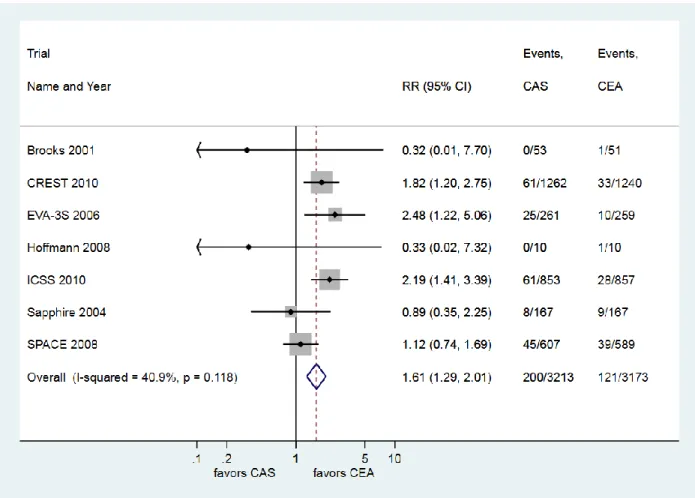
Zhang et al10 compared carotid artery stenting versus endarterectomy in a meta-analysis in 2015.
This meta-analysis compared the effectiveness of stenting versus endarterectomy in studies
between 2006 and 2015 to include primary endpoints of stroke and death in 30 days. The
inclusion criteria included both randomized and non-randomized studies, with at least 20 patients
in the study with at least 10 patients in each group for stenting versus endarterectomy. A total of
35 studies (27,525 patients) were used in the data analysis after excluding 734 studies. The
article excluded systematic reviews, guidelines, case reports, and reviews. The average age of
patients was 70 years old with 68% of the patients being men. They assessed the quality of the
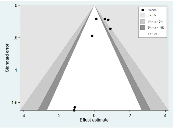
study utilizing the Cochrane risk bias tool with evaluation of publication bias using a funnel plot.
Their results (RR 1.61 95% CI 1.29-2.01) revealed that CEA was superior to CAS in regards to
stroke/death free rates in 30 days post intervention and that CEA was inferior to CAS for
stroke/death at 1 year. The limitations include the inclusion of both RCTs and non RCTS and the
lack of consideration of confounding factors such as age, anesthesia time, and symptomatic
versus asymptomatic status. The results did suggest a difference in the results dependent on age,
anesthesia, time, and symptoms. This can be due to recovery and difference in population. This
review included all studies such as prospective, randomized, and retrospective trials which may
weaken the data and limit the evidence significance. However, presented here in this paper are
factors that are not able to be adjusted for in analyzing this data. Those factors include operator
experience, antiplatelet therapy, symptomatic versus asymptomatic, stent types, and surgical
technique of endarterectomy. These factors are important because they add variables that are
hard to account for in the data analysis.
Figure 1. Forest plot utilizing raw data from RCTs from Zhang et al10 showing stroke or death in
Figure 2. Funnel plot comparing RCTs that evaluated stroke or death in thirty days in stenting
versus endarterectomy
Yang et al11 included eight clinical trials with 7,005 patients to compare the efficacy and safety
of CAS versus CEA. The studies that were included were randomized control trials, involved at
least 20 patients, and were published and peer reviewed. They excluded all retrospective trials,
observational studies, systematic reviews, and meta-analyses. The Cochrane risk bias tool was
used to evaluate the quality of the studies included. There were not any high risk of bias
domains. There was unclear risk of bias for the EVA-3s RCT in allocation concealment. The
and blinding. The average age was 66-70 years old and the percentage of males was up to 80%.
This data included primarily symptomatic patients. The results (RR 1.42 95% CI 1.20-1.67)
reveled that CAS resulted in a significantly higher risk of stroke both in the long-term and
periprocedural timeframe. They did not find heterogeneity that affected the results of the study.
There were however, a small number of patients included in the meta-analysis. The limitations
include an inability to do subgroup data analysis because of the lack of patients. There are
important items to consider in the subgroups including age, symptoms, gender, use of distal
protection devices that could shift the results and recommendations for treatment.
Figure 3. Forest plot utilizing raw data from Yang et al illustrating odds ratios for the cumulative
Sardar et al12 utilized five clinical trials and 6,526 patients to compare the effectiveness of CAS
versus CEA for the prevention of stroke in patients with carotid artery stenosis. Inclusion criteria
included randomized clinic trials with at least 50 patients that used embolic protection. Studies
that were excluded were isolated balloon angioplasty and abstracts only. These studies were
evaluated using the Cochrane risk bias tool. Most of the included RCTs were determined to be
low risk of bias in all categories. There were no designations for high risk of bias. EVA-3S 2006,
did receive unclear bias in the allocation concealment domain. The Sapphire trials from 2008 and
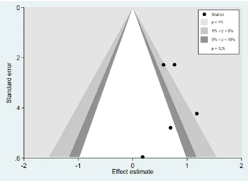
2004 received unclear bias in the selective reporting domain. From review of the funnel plots,
there did not seem to be evidence of publication bias. Of the five trials, two of them included
only symptomatic patients. In this set of studies, the average age was 67-72 years old with
56-78% of participants being male. The results suggested an increased risk of any kind of stroke
from the date of procedure through long term follow up with CAS versus CEA, however they
were mostly non-disabling periprocedural strokes. The limitations of this study include the
inability to adjust for different protocols, characteristics of patients, and definitions of outcomes.
There continues to be a wide variety of lesion, types of stents, and operator experience, which
cannot be accounted for in this research. Operator experience is key because most of the major
clinical trials have been conducted at centers of excellence in these procedures which may not
Figure 4. Forest plot from Sardar et al showing the increased risk of any periprocedural stroke in
comparison of CAS and CEA.
Moresoli et al13 reviewed five randomized control trials for a total of 4,534 patients who were
randomized between CEA and CAS. This systematic review was specifically interested in the
rates of events in asymptomatic patients, which consisted of 3,019 patients in those five
randomized control trials. This review included studies only if they had greater than or equal to
50 asymptomatic patients. They did not include trials with differences in
anticoagulation/antiplatelet pre-and post procedure. However, they did not make exclusion
decisions based on embolic protection device use. Evaluation of the quality of the study was
completed using the Cochrane risk bias tool. In their evaluation of bias, the risk was low or
unclear in all categories except for the Sapphire trial in which it was a high risk due to
incomplete outcome data due to the difference in lost to follow up in each arm: 14.4% in the
CAS arm and 29.9% in the CEA arm. The unclear categories were noted in sequence generation
and funding. In this collection of patients that were asymptomatic, the average age was 67-69
years old, with the majority of patients being male. The results suggested an increased rate of
stroke peri procedurally in asymptomatic patients that underwent CAS (RR of 1.90 and a 95% CI
1.01-3.56). There is heterogeneity in the studies including length of follow up and patient
characteristics, types of devices used, and types of embolic filters used, as well as technical skill
of the operator. Another large limitation includes the few number of RCTs included in this
Figure 6. Forest plot from Moresoli et al showing increased risk of cranial nerve palsy in
Figure 7. Forest plot from Moresoli et al showing increased risk of myocardial infarction in
endarterectomy versus stenting
Figure 8. Forest plot from Moresoli et al showing increased risk of periprocedural stroke in
Discussion
Based on these systematic reviews, there is evidence to suggest the reduced risk of stroke
following CEA compared to CAS.
Carotid artery stenosis can be asymptomatic or symptomatic which makes it difficult to
diagnose. Currently, no screening recommendations exist for asymptomatic disease. While
diagnosis includes an ultrasound and carotid angiogram, different treatment modalities exist and
may vary depending on the individual patient. The mainstay treatment is lifestyle modification
and medical therapy including risk factor modification with statin therapy, blood pressure
management, glucose control and antiplatelet therapy. All patients with carotid artery stenosis
should be receiving medical therapy at minimum in addition to any intervention to reduce the
risk of stroke. Carotid endarterectomy could be considered for any patient without high surgical
risk. High risk surgical features include: severe coronary artery disease, congestive heart failure,
myocardial infarction in past 6 weeks, severe pulmonary disease, renal failure, age >80 and
anatomical difficulties. Carotid artery stenting is an alternative to CEA for selected patients who
need revascularization and are high surgical risk. There are risks and benefits associated with
each procedure and choosing the best treatment option for your patient is largely individualized.
Patient preferences for treatment must be considered as an integral part of the decision. While
there have been an increasing number of studies comparing stenting and endarterectomy, these
studies do not address medical management alone. The CREST 2 randomized control trial is
looking to address this variable. The CREST 2 trial consists of two multicenter RCTs of carotid
revascularization and intensive medical management versus medical management alone in
patients with asymptomatic carotid stenosis14. One trial will randomize patients 1:1 to
stenting with embolic protection versus no stenting. In all of the treatment arms, the medical
management will be identical. This trial is currently enrolling and will address an integral
component of intensive medical management. Intensive medical management should be
implemented regardless of invasive treatment modality. Dividing and investigating treatment
options in asymptomatic versus symptomatic patients must be thoroughly evaluated. Overall, it is
hard to use data from meta analyses because these have limitations including study heterogeneity
and experience of the operators. There is continued need to evaluate the risk and benefits
associated with treatment for carotid artery stenosis. As technology and medical management
excel, there may be more ways to integrate treatment for individual patients that decreases the
Bibliography
1. Benjamin EJ, Blaha MJ, Chiuve SE, et al. Heart Disease and Stroke Statistics-2017
Update: A Report From the American Heart Association. Circulation 2017; 135:e146.
2. Hobson RW, Weiss DG, Fields WS, Goldstone J, Moore WS, Towne JB, et al. Efficacy
of carotid endarterectomy for asymptomatic carotid stenosis. The Veterans Affairs
Cooperative Study Group. N Engl J Med. 1993 Jan 28;328(4):221–227
3. Bonati LH, Dobson J, Featherstone RL, Ederle J, van der Worp HB, de Borst GJ, et al. Long-term outcomes after stenting versus endarterectomy for treatment of symptomatic carotid stenosis: the International Carotid Stenting Study (ICSS) randomised trial. The Lancet. 2015 Feb 7;385(9967):529–538.
4. Caplan LR. Basic pathology, anatomy, and pathophysiology of stroke. In: Caplan's
Stroke: A Clinical Approach, 4th ed, Saunders Elsevier, Philadelphia 2009. p.22.
5. Gahremanpour A, Perin EC, Silva G. Carotid artery stenting versus endarterectomy: a systematic review. Tex Heart Inst J. 2012;39(4):474–487.
6. Brott TG, Hobson RW, Howard G, Roubin GS, Clark WM, Brooks W, et al. Stenting versus endarterectomy for treatment of carotid-artery stenosis. N Engl J Med. 2010 Jul 1;363(1):11–23.
7. De Weerd M, Greving JP, de Jong AW, Buskens E, Bots ML. Prevalence of
asymptomatic carotid artery stenosis according to age and sex: systematic review and metaregression analysis. Stroke. 2009 Apr;40(4):1105–1113.
9. Zhang L, Zhao Z, Ouyang Y, et al. Systematic Review and Meta-Analysis of Carotid
Artery Stenting Versus Endarterectomy for Carotid Stenosis: A Chronological and
Worldwide Study. Wilhelm. M, ed. Medicine. 2015;94(26):e1060.
doi:10.1097/MD.0000000000001060.
10. Li Y, Yang J-J, Zhu S-H, Xu B, Wang L. Long-term efficacy and safety of carotid artery
stenting versus endarterectomy: A meta-analysis of randomized controlled trials. Baron
J-C, ed. PLoS ONE. 2017;12(7):e0180804. doi:10.1371/journal.pone.0180804.
11.Sardar P, Chatterjee S, Aronow HD, et al. Carotid artery stenting versus endarterectomy
for stroke prevention: a meta-analysis of clinical trials. J Am Coll
Cardiol. 2017;69:2266–75.[PubMed]
12.Moresoli P, Habib B, Reynier P, Secrest MH, Eisenberg MJ, Filion KB. Carotid Stenting
Versus Endarterectomy for Asymptomatic Carotid Artery Stenosis: A Systematic Review
and Meta-Analysis. Stroke. 2017 Aug;48(8):2150–2157.
13.Mott M, Koroshetz W, Wright CB. CREST-2: Identifying the Best Method of Stroke
Prevention for Carotid Artery Stenosis: National Institute of Neurological Disorders and