Pharmacosynthetics and the Cell-Type-Specific Control of Neuronal Signaling
Martilias Stephen Farrell
A dissertation submitted to the faculty of the University of North Carolina at Chapel Hill in partial fulfillment of the requirements for the degree of Doctor of Philosophy in the
Department of Pharmacology
Chapel Hill 2013
Approved by:
©2013
ABSTRACT
MARTILIAS STEPHEN FARRELL: Pharmacosynthetics and the Cell-Type-Specific Control of Neuronal Signaling
(Under the direction of Bryan L. Roth)
ACKNOWLEDGEMENTS
First and foremost, I would like to acknowledge Dr. Bryan Roth for providing time, guidance, and resources throughout my dissertation research. Bryan instilled within me scientific faculties that I did not possess prior to joining his lab, and I am grateful for his persistence in shaping me into a young scientist.
My dissertation research was performed with the assistance and guidance of many great scientists, and I wish to acknowledge them here. Members of the Roth Lab, present and past, were invaluable to my dissertation. Prem Yadav, Jon Allen, Atheir Abbas, Ryan Strachan, Sarah Rogan, Sandy Hufeisen, Tom Mangano, Neils Jensen, Xi-Ping Huang, Heong-min Lee, and Vince Setola directly helped me along the way in some fashion, and the other members of the lab were fantastic colleagues. I would also like to thank Tanya Daigle, Marc Caron, Yehong Wan, and Nicole Calakos of Duke University for their collaborations.
I’d like to thank Gary Johnson for shaping an excellent department in which to receive doctoral training. I’d also like to thank members of the administrative staff, particularly Eddie Gill and Chris Turner, for making my NRSA fellowship application process incredibly simple. I’d also like to thank the National Institute of Mental Health for the honor of being awarded the National Research Service Award, an investment that I hope I can return to society through a successful scientific career.
I’d like to specifically thank Dan Urban for his comradery during our graduate school career. When we first rotated in the Roth lab together, we both somehow knew that neither was competing to one-up the other, and our tenure in the lab since then has been one of support, encouragement, and endless scientific conversation. In Dan I have found an amazing colleague and friend, and I wish to acknowledge him for being awesome.
Finally, I would like to thank my family and friends for their love and support over the past 6 years. From the countless pickups and drop offs at the airport or train station, to those of you that made the drive down to visit the Paris of the South, I extend my gratitude.
TABLE OF CONTENTS
TABLE OF CONTENTS ... viii
LIST OF TABLES ... xi
LIST OF FIGURES ... xii
LIST OF ABBREVIATIONS ... xiii
LIST OF SYMBOLS... xvii
CHAPTER 1. BACKGROUND ...1
1.1. INTRODUCTION ...1
1.2. APPLICATION OF PHARMACOSYNTHETICS ...6
1.2.1. Selective mimicry of endogenous receptors ...6
1.2.2. Creating drug-like modulation where none exists ...7
1.2.3. Encoding and modulating diffuse neuronal ensembles ... 10
1.2.4. Long-lasting specific neuronal modulation ... 11
1.3. CONSIDERATIONS AND IMPLICATIONS OF THE PHARMACOSYNTHETIC APPROACH ... 12
1.3.1.2. Genomic Insertion of Transgene ... 13
1.3.1.3. Combinatorial approaches ... 17
1.3.2. CNO Doses and Routes of Administration ... 17
1.4. LIMITATIONS OF CURRENT PHARMACOSYNTHETIC TECHNOLOGY ... 18
1.4.1. Pharmacological vs. Physiological Manipulation ... 18
1.4.2. Limitations of Technology ... 19
1.5. CONCLUSIONS ... 20
1.5.1. Key Differences Between Optogenetics and Pharmacosynthetics ... 20
1.5.2. Concluding remarks ... 22
CHAPTER 2. NEURONAL VALIDATION OF GS COUPLED DREADD (RM3DS)... 23
2.1. INTRODUCTION ... 23
2.2. THE STRIATUM AS AN IDEAL REGION FOR TESTING SELECTIVE CONTROL OF NEURONAL SIGNALING IN DEFINED NEURONAL POPULATIONS. ... 24
2.3. METHODS... 26
2.4. RESULTS ... 39
2.4.1. Generation and characterization of adora2A-rM3Ds mice ... 39
2.4.2. CNO-induced modulation of locomotion in adora2A-rM3Ds mice ... 43
2.4.3. CNO-induced modulation of amphetamine sensitization ... 43
3.1. DISCUSSION ... 46
3.2. IMPLICATIONS ... 48
3.2.1. Regarding Technological Validation ... 48
3.2.2. Regarding Neurobiological Findings ... 49
3.3. FUTURE DIRECTIONS ... 50
3.3.1. Future technological development ... 50
3.3.1.1. A non CNO-based DREADD ... 50
3.3.1.2. Enhanced genetic expression strategies... 51
3.3.1.3. Complete experimental control of signal transduction... 51
3.3.2. Future Applications of Pharmacosynthetic Technology ... 52
3.3.2.1. Non-interfering modulation ... 52
3.3.2.2. Cell-type specific GPCR signaling vs. “activation” and “silencing” ... 54
3.3.3. Future Neurobiological Directions ... 58
3.4. FINAL WORDS: ... 58
APPENDIX A. TABLES ... 60
APPENDIX B. FIGURES ... 64
LIST OF TABLES
LIST OF FIGURES
Figure 1: Properties and Composition of Currently Utilized DREADDs. ... 65
Figure 2: Creation and validation of adora2A-rM3Ds mice. ... 67
Figure 3: rM3Ds is expressed in striatopallidal neurons in adora2A-rM3Ds mice. ... 69
Figure 4: Additional immunohistochemistry images comparing a wild-type mouse with an adora2A-rM3Ds transgenic mouse. ... 70
Figure 5: CNO activates canonical Gαs-type signaling in adora2A-rM3Ds mice. ... 71
Figure 6: pErk1/2 and pAkt308 signaling in adora2A-rM3Ds mice. ... 73
Figure 7: Cocaine-induced signaling in wild-type and adora2A-rM3Ds mice. ... 74
Figure 8: Results of baseline behavior screen... 75
Figure 9: Endogenous Gs signaling is intact in adora2A-rM3Ds mice. ... 77
Figure 10: CNO administration inhibits locomotor activity in adora2A-rM3Ds mice. ... 78
Figure 11: CNO administration modulates amphetamine-induced physiological changes. .... 79
Figure 12: Replication in second founder line. ... 81
Figure 13: Shifting the pharmacological equation. ... 83
LIST OF ABBREVIATIONS
A2A – Adenosine A2A receptor
AAV – adeno-associated virus
adora2A – Adenosine A2A receptor gene
AgRP – agouti-related protein
AKT, pAKT – Protein Kinase B (PKB), phosphorylated
AMPAR – alpha-amino-3-hydroxy-5-methyl-4-isoxazolepropionic acid receptor
AMPH – Amphetamine
ANOVA – Analysis of Variance
ARC – arcuate nucleus
ATP – adenosine triphosphate
BAC – Bacterial Artificial Chromosome
BCA – Bicinchoninic acid assay
bp – base pair
BSA – Bovine Serum Albumin
cAMP: cyclic adenosine monophosphate
CNO – Clozapine N-oxide
D2 – dopamine receptor 2
DARPP-32: Dopamine and cAMP regulated neuronal phosphoprotein
dB – decibels
DMEM – Dulbecco’s Modified Eagle Medium
DMSO – Dimethyl Sulfoxide
DNA – Deoxyribonucleic Acid
Drd1 – Doparmine Receptor D1
Drd2 – Doparmine receptor D2
DREADD – Designer Receptors Exclusively Activated by Designer Drug
EGFP – enhanced green fluorescent protein
EPSCs – Excitatory postsynaptic currents
ERK, pERK – Extracellular signal regulated kinase, phosphorylated
Flp – Flippase
FRT – Flippase recognition target
FW – Forward
GDP – guanosine diphosphate
GENSAT – Gene Expression Atlas of Mouse Central Nervous System
GPCR: G protein-coupled receptor
HBSS – Hank’s Buffered Salt Solution
HEK - human embryonic kidney cells
HEPES - 4-(2-hydroxyethyl)-1-piperazineethanesulfonic acid
hM3Dq – Human Muscarinic Receptor 3 Gq DREADD
hM4Di – Human Muscarinic Receptor 4 Gi DREADD
i.p. – intraperitoneally
IRES – Internal Ribosome Entry Site
kb: kilobase
mEPSC: miniature excitatory post synaptic current
MeSH – Medical Subject Headings
Min – minutes
MSN: medium spiny neuron
NAc – Nucleus accumbens
NIH – National Institutes of Health
NINDS – National Institute of Neurological Disorders and Stroke
NMDAR – N-methyl-D-aspartate receptor
NREM: Non Rapid Eye Movement
P23 – Post Natal Day 23
PBS – Phosphate Buffered saline
PCR – Polymerase Chain Reaction
PDZ – post synaptic density protein (PSD95), Drosophila disc large timor suppressor
(Dlg1), and zonula occludens-1 protein(zo-1) domain
PKA – protein kinase A
PLSD – Fisher’s protected least-significant difference
Rbp4: retinol binding protein 4
REV – Reverse
RFP – Red Fluorescent Protein
rM3Ds – Rat Muscarinic Receptor 3 Gs DREADD
Rpm – revolutions per minute
RT – Room temperature
SDS – Sodium Dodecyl Sulfate
TBST – Tris Buffered Saline with 0.05% Tween 20
TRE – Tetracycline Response Element
LIST OF SYMBOLS
Gα – G protein alpha.
Gβ/γ – G proteins beta and gamma
Gαs – Gs-type G protein alpha – stimulates adenylyl cyclase Gαolf – G alpha olf, a Gs-type G protein
CHAPTER 1.BACKGROUND
1.1.INTRODUCTION
The gap between our understanding of receptor mediated signaling and the ultimate functional output of the brain is shrinking. The past decade has witnessed the advent of multiple technologies that allow the exquisite manipulation of neurons in an intact animal, providing the opportunity to definitively determine the neuronal correlates of complex brain function. The primary technologies are optogenetic – the modulation of transgenic receptors and channels via photons-- and pharmacogenetic – the modulation of transgenic receptors via pharmacologic agents. Here I will focus on pharmacogenetics.
reimagination of pharmacogenetics (the modulation of transgenic receptors via pharmacologic agents) as pharmacosynthetics. This term integrates the true meaning and functional mechanisms of the technology: pharmaco- meaning drug and -synthetic meaning the combination of two or more parts in an artificial manner. Pharmacosynthetics provides a clear distinction from both pharmacogenetics and chemicogenetics and, to date, has not been used to describe any phenomenon or identify any technology.
I present the formal definition of pharmacosynthetics as “a branch of biology which deals with the creation of pharmacological modulation using artificial components”. While it is possible to equate conventional drugs with pharmacosynthetics (or having been developed through pharmacosynthesis), there are distinctions within the semantics that should be explored to provide clarification. A chemical is synthesized to have a particular pharmacology, and this pharmacology is based on the system with which that chemical interacts. On the other hand, a pharmacosynthetic approach creates pharmacological modulation within a system using artificial components. While a pharmacological agent may be synthesized, at no point in this effort is the pharmacology of the agent created – instead, it is measured. In one way of thinking about it, a pharmacology (as defined as the study of drug action) is engineered for an otherwise inert chemical by engineering a receptor and inserting the receptor into a living system. On the other hand, when a novel chemical is synthesized, its pharmacology in a living system is studied to determine whether or not it is a drug or has drug-like properties.
signaling (Conklin et al, 2008). The original DREADDs were human muscarinic acetylcholine receptors engineered to be activated by clozapine N-oxide (CNO), an otherwise inert pharmacological agent. Additionally, DREADDs are insensitive to the endogenous ligand, acetylcholine. There are currently three DREADDs in common use – the hM3Dq that activates Gαq signaling, the hM4Di that activates Gαi signaling, and the rM3Ds that activates Gαs signaling. These three DREADDs share the same point mutations (Figure 1) that simultaneously engender CNO efficacy and acetylcholine inefficacy (Armbruster et al, 2007). The rM3Ds was engineered to couple Gαs by replacing intracellular loops 2 and 3 of the hM3Dq with those from the turkey β1-adrenergic receptor (Guettier et al, 2009). With these three DREADDs, it is possible to control 3 of the G protein signaling cascades found in the mammalian brain.
modulate ion conductance via ligand gated ion channels. Namely, the pharmacologically selective actuatory modules (PSAMs) and their cognate pharmacologically selective effector molecule (PSEM) agonists (Magnus et al, 2011) have been shown to be effective neuronal modulators. This system permits for the direct modulation of ion conductance via pharmacological means using chimeras of ligand-binding domains of the alpha7 nicotinic acetylcholine receptor and ion pore domains of other Cys-loop receptors. Similar to DREADDs, PSAMs were engineered to respond to PSEMs in a two-way selective manner, providing for exclusive control of neuronal signaling. Additionally, ivermectin-gated ion channels provide similar modulation of membrane ion conductance using glutamate-gated chlorid channel receptor (GluClR) activated by ivermectin (Lerchner et al, 2007), though this technology has not been extensively utilized.
Finally, therapeutic efficacy is most often obtained through modulation of diffusely expressed albeit specific drug targets (Roth et al, 2004). These three characteristics can only be mimicked via the systemic injection of drug and the dispersed expression of the DREADD. This similarity to conventional therapeutics may thus facilitate an immediate crossover of insights gleaned from research utilizing DREADDs to the physiological phenomena responsible and necessary for therapeutic efficacy.
1.2.APPLICATION OF PHARMACOSYNTHETICS 1.2.1.Selective mimicry of endogenous receptors
1.2.2.Creating drug-like modulation where none exists
Additionally, the DREADD system can be conceptualized of as a way to pharmacologically modulate spatially defined neuronal populations for which no pharmacological modulatory agents exist. Krashes et al. (2011) used DREADDs in this fashion to study the arcuate nucleus (ARC) of the hypothalamus. This nucleus has been implicated in regulating energy homeostasis and has therefore been a focus of intensive study for the understanding and treatment of obesity, with particular focus on agouti-related protein (AgRP) neurons expressed in this area. To date, investigative efforts into the function of these neurons have been limited to conventional genetic, invasive, and ablative approaches: overexpression of AgRP in transgenic mice, central administration of peptides, and ablation of AgRP neurons. The first approach removes the temporal specificity required for definitive experimentation, whereas the latter approaches introduce confounds associated with non-reversible and invasive administration techniques. Krashes et al. (2011) utilized DREADD technology to study the acute effects of AgRP neuronal activity. The hM3Dq DREADD was targeted to AgRP neurons using a Cre-recombinase dependent adeno-associated virus (AAV) injected into AgRP-Ires-cre mice. Following i.p. administration of CNO (0.3 mg/kg), hM3Dq-expressing mice began feeding and consumed almost four times as much food than control mice in the first half hour. Additionally, Krashes et al. infected the same neurons with the hM4Di to induce neuronal silencing and observed a decrease in food intake. This study demonstrates that DREADDs can be used to introduce pharmacological modulation to nuclei for which drug-like compounds do not exist.
modulation modulated food intake, the fact that the AGRP neurons project to disparate brain nuclei raises the question of which of these downstream nuclei is integral for the ultimate behavioral effect of AGRP activation. This was determined through functional-connectivity mapping utilizing a combination of optogenetics and phamacosynthetic tools. In this study, the hM4Di was used to silence pro-opiomelanocortin (POMC) expressing neurons in the arcuate nucleus, a population of neurons modulated by AGRP neurons from within the same nucleus, to determine whether inhibition of these neurons modulated food intake. Atasoy et al. found that CNO (5.0 mg/kg) administration did not significantly alter food intake over 1 hour, but repeated treatment (5.0 mg/kg, 3 injections every 8 hours) increased food intake over a 24 hour period. The paraventricular hypothalamic (PVH) nucleus also receives AGRP innervation, and administration of CNO to mice expressing the hM4Di in these neurons caused an increase in food intake. Furthermore, these mice displayed an increase in break point during a progressive ratio operant task for food reinforcement, indicating that PVH suppression itself can mimic the food seeking and food consumption effects of whole-circuit AGRP activation. These experiments were complemented by activating all of the AGRP neurons via systemic CNO injection and then selectively silencing the projections to the PVH using GABA or NPY antagonists. Even with activation of brain-wide AGRP neural circuits, selective inactivation of these projections caused signifantly reduced food intake. The use of pharmacological agents provides a physiological context for the circuitry described in this report.
of neuropeptide systems is historically difficult due to issues of invasiveness (local microinjection of purified peptide), lack of temporal control (genetic modulation) or off-target effects (physical ablation). Using virally mediated gene transfer, Sasaki et al. (2011) was able to express the hM3Dq and hM4Di in the orexin neurons of the lateral hypothalamic area. Following intraperitoneal administration of CNO (5.0 mg/kg) during the light phase (when mice typically sleep), the percent of wakefulness during the following hour was significantly greater and the NREM time was significantly shorter. Similarly, administration of CNO during the dark phase (when mice are typically awake), caused a significant increase in wakefulness. Conversely, administration of CNO to mice expressing the hM4Di (the inhibitory DREADD) in the orexin neurons decreased wakefulness during the dark phase and the light phase.
1.2.3.Encoding and modulating diffuse neuronal ensembles
1.2.4.Long-lasting specific neuronal modulation
adulthood. This study demonstrates the ability of the pharmacosynthetic approach to provide chronic neuronal modulation.
1.3.CONSIDERATIONS AND IMPLICATIONS OF THE PHARMACOSYNTHETIC APPROACH
Pharmacosynthesis requires a consideration of many factors to be effectively utilized. The key elements to be considered are the expression of DREADD and the dose of CNO required for experimental manipulation. Here I provide a primer on the consideration of these elements.
1.3.1.Expression systems
The primary challenge in pharmacosynthetics is inserting the DREADD receptor into the desired tissue of the model organism. To date, this has been achieved using virally mediated gene transfer and the genomic insertion of a transgene. Although a full review of each approach is beyond the scope of this dissertation, the benefits and complications of each approach will be briefly discussed.
1.3.1.1.Virally Mediated Gene Transfer
Finally, the viral approach permits utilization of the DREADD in model organisms for which transgenic approaches are not available or widespread (e.g., rats, monkeys). The drawbacks of the viral approach arise from the nature of local microinjections and the size limitations of viral packaging. This vector delivery method is invasive, potentially inducing an immune response and causing damage to tissue, including cell populations either directly or indirectly involved in the scrutinized output. Additionally, the spatial resolution provided can also be a limitation, in that DREADD expression is limited to the number of microinjection sites and the spread of viral particles. This latter point, however, can be a benefit depending on the goals of the study. Furthermore, the expression pattern of DREADDs between animal subjects will not be precisely identical due to differential stereotactic coordinate alignment, inconsistent viral diffusion, and experimental variation. Finally, viruses are only capable of carrying a certain quantity of DNA, potentially limiting the addition of desirable vector traits including targeting information (promoter sequences) and cell-type markers (fluorescent proteins).
1.3.1.2.Genomic Insertion of Transgene
only provide expression of the DREADD in the area of viral diffusion, the transgene inserted into the genome is present in all cells. Expression of the DREADD is dependent upon the information contained in the transgene, and while the genetic sequences that confer cell-type specificity of expression are still a matter of research, certain promoter sequences have been determined. Thus, while the noninvasive component is definitively beneficial, the utility of dispersed expression patterns is dependent upon the research goals.
DREADD sequence in addition to the endogenous gene product. To avoid interfering with a particular genomic locus, an alternative transgenic strategy is to create a transgene containing the entirety of the genetic information associated with a particular cell-type specific protein. This approach can be achieved by using bacterial artificial chromosomes, which are capable of carrying 200-300 kb of genetic information, a drastically larger amount than other transgenic approaches utilize (Heintz, 2001). For instance, the camKII-alpha promoter sequence is 8.5 kb (Tsien et al, 1996), whereas the adora2A BAC is 175 kb. Finally, creating a transgenic mouse is both resource intensive and the effort has no guaranteed yield, depending on the strategy.
the Flp excision site (Flpe). Thus, in the cell population that expresses Cre or Flp, the stop cassette is removed from the genetic sequence. The nuclear expression machinery can then translate the DREADD sequence into protein in that cell population. In the cells that do not express Cre or Flp, the DREADD transgene remains silent due to the presence of the stop cassette. The Cre / Flp systems can also take advantage of a second phenomenon of the recombinatorial proteins in that they can reverse the direction of the sequence between the excision sequences. Dependent upon the orientation of the excision sequences, the Cre and Flp can either excise the bookended DNA or flip the direction (Atasoy et al, 2008). The “reversal” approach is less leaky than the excision method; i.e., the intended specificity of expression is more likely to occur.
1.3.1.3.Combinatorial approaches
Already the world of neuroscience research is seeing the full implementation of these technologies and the benefits of combining them. For example, the Krashes et al. (2011) and Sasaki et al. (2011) studies combined the specificity of expression provided by the genomic transgene approach with the spatial resolution and quick turnaround of the viral approach to achieve cell-type specific neuronal modulation. The Ray et al. (2011) study used intersectional genetics, combining Cre and Flp recombination to increase the specificity of DREADD expression with minimal invasiveness. At this point, the ability to target DREADD expression to specific tissue populations depends on the transgenic state of the art. 1.3.2. CNO Doses and Routes of Administration
c-fos promoter to drive expression of the hM3Dq, so the DREADD was expressed at lower levels and higher doses of CNO were apparently necessary. Furthermore, Krashes (2011) used a viral approach to express the hM3Dq in a small nucleus and administered 5.0 mg/kg CNO to elicit a response in these mice. From the body of work performed with DREADDs to date, it can be seen that the dose of CNO is variable and dependent on the type of DREADD and the expression system used.
To date, a majority of studies performed have used the intraperitoneal route of administration, though other routes of administration are possible. Our lab has demonstrated that CNO can be administered through the drinking water to create chronic administration conditions (10 mg/kg/day, unpublished observations).
1.4.LIMITATIONS OF CURRENT PHARMACOSYNTHETIC TECHNOLOGY 1.4.1.Pharmacological vs. Physiological Manipulation
modulation due to synaptic leakage to potentiall influence volume transmission (Goto et al, 2007). This phasic nature of ligand-induced signaling can not be replicated using pharmacosynthetics due to the uniform distribution of a pharmacological agent. Furthermore, the site of action of a neurotransmitter is mostly restricted to the synapse, though extra-synaptic receptors are present. This differential localization of endogenous receptor creates the possibility for differential response to neurotransmitter presence, based on the “leakage” from the synapse. This differential response will not be observed in a pharmacosynthetic system due to the uniform distribution of ligand. For these reasons, pharmacosynthetic tools are best utilized to study a pharmacological response of a system as opposed to the physiology of the system itself.
1.4.2.Limitations of Technology
research (rM3Ds) is influenced by the presence of the turkey beta1 adrenergic receptor loops. Therefore, the targeting of the rM3Ds could be influenced by the trafficking information of both the muscarinic M3 receptor and the beta1 adrenergic receptor. The current targeting and trafficking information encoded by the available DREADDs represents the state-of-the art and is an area requiring further development to enhance the capabilities of pharmacosynthetic technology.
1.5.CONCLUSIONS
1.5.1.Key Differences Between Optogenetics and Pharmacosynthetics
family of light activated ion channels that directly modulate the ion conductance of neuronal membranes and either hyperpolarize or depolarize neurons. In contrast, DREADDs modulate G-protein mediated signaling – signaling cascades for which neuronal hyperpolarization or depolarization are only one outcome. However, there are optogenetic tools available – the OptoXRs - that modulate G-protein mediated signaling using light, though these have not gained widespread use.
1.5.2.Concluding remarks
CHAPTER 2.NEURONAL VALIDATION OF GS COUPLED DREADD (RM3DS)
2.1.INTRODUCTION
tests the utility of DREADDs in interrogating the relationship between intracellular signaling in specific neuronal circuits and behavior. This enabling technology has the potential to provide neuroscientists an innovative research strategy (pharmacological control of cell type-specific signaling with unprecedented precision) to advance towards Strategic Objective 1 of the National Institute of Mental Health (promote discovery in the brain and behavioral sciences to fuel research on the causes of mental disorders). Ultimately, the knowledge obtained could yield new insights into basal ganglia function.
2.2.THE STRIATUM AS AN IDEAL REGION FOR TESTING SELECTIVE CONTROL OF NEURONAL SIGNALING IN DEFINED NEURONAL POPULATIONS.
Emilien et al, 1999). The distinctive functional and biochemical composition of these neurons present a suitable region to validate the ability of the DREADDs to selectively control cell type specific neuronal signaling.
The Gαs- and Gαi- G protein signaling cascades, modulated by D1- and D2-receptors, respectively, are implicated in both the short-term excitability and the long-term plasticity of MSNs (Centonze et al, 2001; Surmeier et al, 2007). Striatal G protein signaling cascades have primarily been studied as a consequence of activating dopamine receptors, but activation of Gαs signaling downstream of other GPCRs also has significant effects. For example, striatopallidal Gαs signaling modulated by the A2A-adenosine receptor influences psychostimulant activity (Brown and Short, 2008). To create a mouse model in which the cellular and behavioral consequences of striatopallidal-specific Gs-type signaling (G protein signaling that increases cAMP production) can be studied, we took advantage of technology we developed whereby evolved GPCRs (DREADDs, or Designer Receptor Exclusively Activated by Designer Drug) are expressed in a cell-type-specific fashion to remotely control cellular signaling (Armbruster et al, 2007).
2007)). Herein, I use this novel transgenic line to validate the rM3Ds by measuring the biochemical, electrophysiological, and behavioral consequences of CNO administration to adora2A-rM3Ds transgenic mice.
2.3.METHODS
Plasmids: The plasmid map of the p-rM3Ds-IRESmCherry construct is detailed in Figure 2.
Drugs: Clozapine N-oxide (CNO) was obtained from the NIH as part of a Rapid Access to Investigative Drug Program funded by the National Institute of Neurological Disorders and Stroke (NINDS). D-amphetamine (AMPH) and isoproterenol were purchased from Sigma (St. Louis, MO). For experiments in mice, CNO was first dissolved in DMSO then brought to final concentration with 0.9% saline and a final concentration of DMSO of 0.5%. Amphetamine was dissolved directly into 0.9% saline. For all experiments, the appropriate (e.g., 0.9% saline for amphetamine experiments and 0.5% DMSO in saline for CNO experiments) vehicle controls were utilized. Unless otherwise noted, the dose of CNO was 1.0 mg/kg and the dose of amphetamine was 2.0 mg/kg. Drugs were injected intraperitoneally (i.p.) at a volume 100 ul /10 g body weight. For in vitro studies, drugs were dissolved in DMSO at 10 mM as stocks and then diluted into sample buffer.
Lentiviral studies were done as previously described with modification (Abbas et al, 2009; Alexander et al, 2009) by Ying Pei. To generate a lentiviral construct, the coding region for rM3Ds-IRESmCherry was subcloned into the lentiviral expression vector FUGW (Lois et al, 2002), a gift from Dr. Guoping Feng (Duke University). Fugene6 (Roche Applied Science, Indianapolis, IN) was used to co-transfect seven 150 cm2 dishes of HEK293T cells with the FUGW plasmid and two viral packaging constructs (Δ8.9 HIV-1 and VSVG) in a ratio of 3.3:2.5:1. Lentivirus-containing media was collected 48 hours post-transfection. Virus was concentrated by centrifugation and Amicon ultra-15 centrifugal filter devices (Millipore, St. Louis, MO), aliquoted, and frozen at -80°C until use. Rat cortical neurons were infected with FUGW-rM3Ds-IRES-mCherry as previously described (Alexander et al, 2009). Two days following infection, cells were exposed to increasing concentrations of CNO, and cAMP accumulation was quantified using the Catchpoint assay per manufacturer’s instructions (Molecular Devices).
with dilute trypsin, resuspended in 1X HBSS (with calcium and magnesium) (Invitrogen) supplemented with 20 mM HEPES, pH 7.4 (drug buffer), counted, and diluted to 15,000 cells/20 microliters. The cell suspension was added to white 384-well plates (Greiner) (20 microliters/well). After a 1-2 hr incubation, the cells were challenged with 10 microliters/well of 3X working dilutions of CNO (for concentration-dependent activation of rM3Ds) or isoproterenol (for concentration-dependent activation of endogenously expressed beta2AR). The 3X working dilutions were prepared in drug buffer containing 6% (i.e., 3X) GloSensor reagent (Promega). Ten minutes after agonist challenge, the luminescence was counted (1 s/well) on a TriLux (Perkin Elmer) microbeta/luminescence plate reader. For each transfection condition (rM3Ds +/- Gα), the luminescence per well was expressed as a function of the log [agonist], and the data were fit using a three-parameter logistic equation as described previously (Alexander et al, 2009). Best-fit pEC50 and Emax values +/- SE were compared across transfection conditions and between agonists.
Animal Subjects: Behavioral, biochemical, and electrophysiology experiments were performed at the University of North Carolina and Duke University in accordance with the National Institutes of Health’s guidelines for the care and use of animals and with approved animal protocols from the Institutional Animal Care and Use Committees of the aforementioned institutions.
was then injected into the pronucleus of B6SJLF1/J mouse oocytes. Genotyping was performed by PCR of genomic DNA extracted from tail clips using the following primers for
mCherry: FW 5´-GTGAGCAAGGGCGAGGAGG-3´ REV:
5´-GTCGGCGGGGTGCTTCAC-3´ using the following cycle: Initial denaturation: 94 – 4 minutes, followed by 30 cycles of 94°C 30s / 65°C 30s / 72°C 30s, followed by 72°C 4 minute final extension. PCR products were analyzed using gel electrophoresis (1%, Aqua Por LE, National Diagnostic, Atlanta, GA), and rM3Ds positive mouse samples present a clear band at 200 bp (Figure 2d). From this screen, 9 genotype-positive mice were found. These mice were bred to wild-type (C57BL/6J) mice, and their offspring were screened for mCherry expression via immunohistochemistry for mCherry following the methods detailed below. Three mCherry-positive founder lines were identified and named AD6, AD8, and AD10. The AD6 line was used in the present study. Following initial screening, AD6 mice were bred onto the C57BL/6J background. The breeding strategy had consistent pairing with an AD6 mouse always paired with a C57BL/6J mouse; thus, all mice used in these studies were hemizygous for the adora2A-rM3Ds gene. The AD6 line of mice is referred to as adora2A-rM3Ds mice in this manuscript. Mice used for behavioral studies were bred in large cohorts using a harem-breeding strategy, and littermate pairings between conditions were used. All behavioral studies were performed on mice of the F3 generation or later. The amphetamine behavioral sensitization studies and behavior core screen were performed on the F3 generation. Electrophysiology studies were performed on the F6 generation or later.
D2-expressing neurons. C57BL/6J mice were obtained from Jackson laboratories (Bar Harbor, ME).
A region of interest in the body of the striatum was selected for N=3 mice, and the number of EGFP positive and mCherry positive cell bodies was quantified.
DARPP-32 study: Mice were injected i.p. with CNO (5.0 mg/kg), cocaine (20.0 mg/kg) or vehicle and then sacrificed 15 min later by cervical dislocation. The ventral striatum was isolated using a rapid head-freeze dissection technique as described previously (Beaulieu et al, 2004). Frozen tissue samples were probe sonicated in 95°C 1% SDS buffer containing 1X Halt phosphatase inhibitor (Halt, Pierce, 87786) and 1X protease inhibitor (Roche Diagnostics, Complete, no. 11697498001). Protein concentration of sample was determined using the BCA method (Pierce). Samples were boiled in Laemmli buffer, and 25 ug of protein were loaded into 10% SDS-PAGE gels, transferred onto nitrocellulose membranes, and incubated with antibodies to pT34 DARPP-32 (Phosphosolutions, Aurora, CO, p1025-34, 1:300, TBST / 3% BSA), total DARPP-32 (BD Transduction, San Diego, CA, 611520, 1:1500, TBST / 5% BSA), pERK1/2 (Cell Signaling, Danvers, MA, 9101, 1:500, TBST / 5% milk), total ERK1/2 (9107, 1:500, TBST / 5% milk), pAKT308 (2965, 1:100, TBST / 5% BSA), or total AKT (2920, 1:1500, TBST / 5% BSA). Blots were imaged on the LI-COR Odyssey instrument (LI-COR Biosciences, Lincoln, NE). The phospho-specific probe band intensity was measured using NIH ImageJ software and was normalized to total probe band intensity. The fold stimulation was determined by normalizing these values to the average of the vehicle treatment group.
genotype. Data were analyzed using one-way or repeated measures Analysis of Variance (ANOVA) to determine effects of genotype. Fisher's protected least-significant difference (PLSD) tests were used for comparing group means only when a significant F value was determined in the overall ANOVA. Within-genotype comparisons were conducted to determine side preference in the social approach test, and quadrant preference in the water maze. For all comparisons, significance was set at p < 0.05.
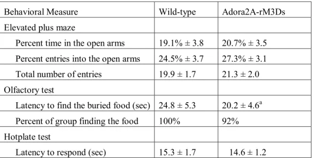
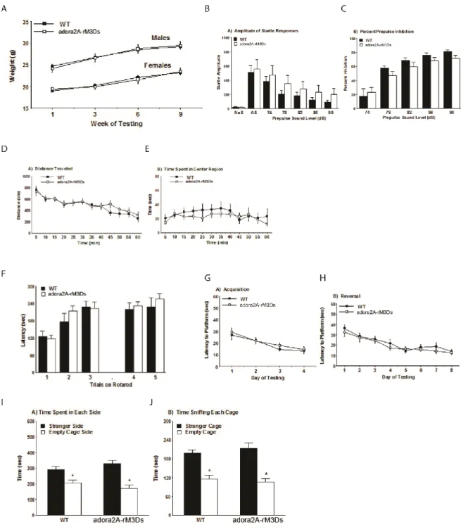
Testing Regimen: Mice were tested in the following procedures, with at least one or two days between each assay: elevated plus maze test for anxiety-like behavior, neurobehavioral screen, activity in an open field, accelerating rotarod (2 tests, 48 hours apart), social approach test, acoustic startle test, buried food test for olfactory ability, visual cue test in the Morris water maze, hidden platform test for spatial learning, reversal learning in the Morris water maze, hotplate test for thermal sensitivity.
Elevated plus-maze test: Mice were given one 5-min trial on a metal plus-maze, which had two closed arms, with walls 20 cm in height, and two open arms. The maze was elevated 50 cm from the floor, and the arms were 30 cm long. Animals were placed on the center section (8 cm x 8 cm), and allowed to freely explore the maze. Arm entries were defined as all four paws entering an arm. Entries and time in each arm were recorded during the trial by a human observer via computer coding. Percent open arm time was calculated as 100 x (time spent on the open arms/time in the open arms + time in the closed arms). Percent open arm entries was calculated using the same formula.
posture, and normality of gait. Normal reflexive reactions to a gentle touch from a cotton-tipped swab to the whiskers on each side of the face, and the approach of the swab to the eyes, were assessed. Each subject was placed in a small, empty plastic cage, and ability to remain upright when the cage was moved from side-to-side or up-and-down was noted. Locomotor coordination was assayed by allowing the mouse to walk across an elevated dowel (wrapped in nylon rope to facilitate grasping) and to climb down a similar pole. Each subject was also placed on a wire grid and allowed to hang for one minute. Reaction to 20 seconds of tail-suspension was recorded.
Buried food test for olfactory function: Several days before the olfactory test, an unfamiliar food (Froot Loops, Kellogg Co., Battle Creek, MI) was placed overnight in the home cages of the subject mice. Observations of consumption were taken to ensure that the novel food was palatable to the mice. Sixteen to twenty hours before the test, all food was removed from the home cage. On the day of the test, each mouse was placed in a large, clean tub cage (46 cm L x 23.5 cm W x 20 cm H), containing paper chip bedding (3 cm deep), and allowed to explore for five minutes. The animal was removed from the cage, and one Froot Loop was buried in the cage bedding. The animal was then returned to the cage and given fifteen minutes to locate the buried food. Measures were taken of latency to find the food reward and whether it was consumed.
Activity in an open field: Exploratory activity in a novel environment was assessed by a one-hour trial in an open field chamber (40 cm x 40 cm x 30 cm) crossed by a grid of photobeams (VersaMax system, AccuScan Instruments). Counts were taken of the number of photobeams broken during the trial in five-minute intervals, with separate measures for ambulation (total distance traveled), fine movements (repeated breaking of the same set of photobeams), and rearing movements. Time spent in the center region of the activity chamber was used as a measure of anxiety-like behavior in a novel environment.
Rotarod: Subjects were tested for motor coordination and learning on an accelerating rotarod (Ugo Basile, Stoelting Co., Wood Dale, IL). For the first test session, animals were given three trials, with 45 seconds between each trial. Two additional trials were given 48 hours later. Rpm (revolutions per minute) was set at an initial value of 3, with a progressive increase to a maximum of 30 rpm across five minutes (the maximum trial length). Measures were taken for latency to fall from the top of the rotating barrel.
Sociability: The three-chamber social approach test was designed to assess whether mice will approach or avoid an unfamiliar stranger mouse. Each session consisted of two ten-minute phases: a habituation period and a test for sociability. For the sociability assay, mice were given a choice between being in the proximity of an unfamiliar conspecific (stranger 1), versus being alone.
The test mouse was first placed in the middle chamber and allowed to explore for ten minutes, with the doorways into the two side chambers open. After the habituation period, the test mouse was enclosed in the center compartment of the social test box, and an unfamiliar C57BL/6J male (stranger 1) was placed in one of the side chambers. The stranger mouse was enclosed in a small wire cage, which allowed nose contact between the bars, but prevented fighting. An identical empty wire cage was placed in the opposite side of the chamber. Following placement of the stranger and the empty wire cage, the doors were re-opened, and the subject was allowed to explore the entire social test box for a ten-minute session. Measures were taken of the amount of time spent in each chamber and the number of entries into each chamber by the automated testing system.
Acoustic startle: The acoustic startle test can be used to assess auditory function and sensorimotor gating. The test is based on the measurement of the reflexive whole-body flinch, or startle response, that follows exposure to a sudden noise. Assessments can be made of startle magnitude and of prepulse inhibition, which occurs when a weak prestimulus leads to a reduced startle in response to a subsequent louder noise. For this study, animals were tested with a San Diego Instruments SR-Lab system. Briefly, mice were placed in a small Plexiglas cylinder within a larger, sound-attenuating chamber. The cylinder was seated upon a piezoelectric transducer, which allowed vibrations to be quantified and displayed on a computer. The chamber included a house light, fan, and a loudspeaker for the acoustic stimuli. Background sound levels (70 dB) and calibration of the acoustic stimuli were confirmed with a digital sound level meter (San Diego Instruments).
stimulus (40 msec; 120 dB) alone, and trials in which a prepulse stimulus (20 msec; either 74, 78, 82, 86, or 90 dB) occurred 100 ms before the onset of the startle stimulus. Measures were taken of the startle amplitude for each trial across a 65-msec sampling window, and an overall analysis was performed for each subject's data for levels of prepulse inhibition at each prepulse sound level (calculated as 100 - [(response amplitude for prepulse stimulus and startle stimulus together / response amplitude for startle stimulus alone) x 100].
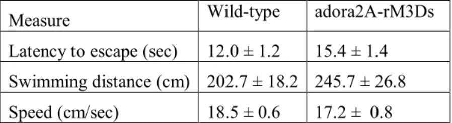
Morris water maze - Visible platform test: The Morris water maze task was used to assess spatial learning in the mice. The water maze consisted of a large circular pool (diameter = 122 cm) partially filled with water (45 cm deep, 24-26o C), located in a room with numerous visual cues. Mice were first tested using a visible platform. In this case, each animal was given four trials on one day to swim to an escape platform cued by a patterned cylinder extending above the surface of the water. For each trial, the mouse was placed in the pool at one of four possible locations (randomly ordered), and then given 60 seconds to find the visible platform. If the mouse found the platform, the trial ended, and the animal was allowed to remain 10 seconds on the platform before the next trial began. If the platform was not found, the mouse was placed on the platform for 10 seconds, and then given the next trial. Measures were taken of latency to find the platform, swimming distance, and swimming speed, via an automated tracking system (Noldus Ethovision).
criterion was reached, with a maximum of nine days of testing. When criterion was reached, mice were given a one-minute probe trial in the pool with the platform removed. In this case, selective target search was evaluated by measuring percent time spent in each quadrant, and the number of crossings for the target location where the platform had previously been located versus corresponding locations in each quadrant of the pool.
Reversal learning: Following the acquisition phase, mice were tested for reversal learning, using the same procedure as described above. In this phase, the hidden platform was located in a different quadrant in the pool, diagonal to its previous location. On the eighth day of testing, the platform was removed from the pool, and the group was given a probe trial to evaluate reversal learning.
Locomotor behavior studies: Locomotor activity was assessed in photocell-based activity chambers under standardized environmental conditions using an AccuScan activity monitor (AccuScan Instruments, Columbus, OH) with a 41 cm x 41 cm x 30 cm Plexiglas chamber and a beam spacing of 1.52 cm as described (Abbas et al, 2009). Horizontal activity was measured as the total distance covered in centimeters as the total of all vectored X-Y coordinate changes and recorded in 5 minute bins.
mg/kg) and placed in activity boxes 20 min. later. Locomotor activity was recorded for 1 hour.
Amphetamine sensitization study
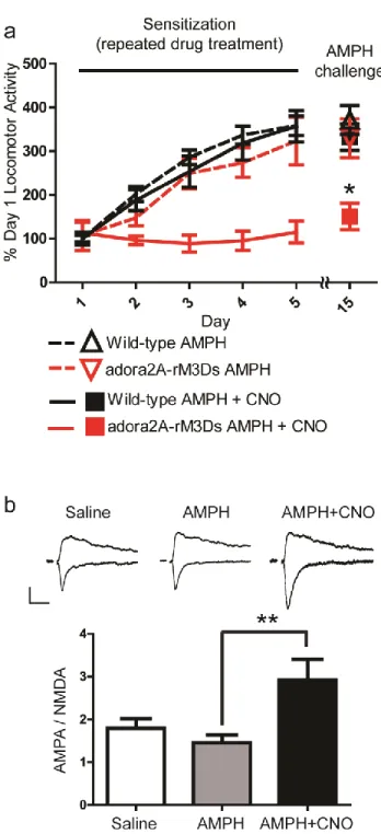
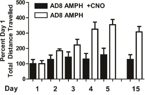
Development Phase: Mice were placed in locomotor activity boxes for 1 hour to acclimate. Mice were then injected (i.p.) with drug(s) and/or vehicle and then returned to the activity chamber for 2 hours. This was repeated once daily for 5 days. Drug doses were 2.0 mg/kg amphetamine and 1.0 mg/kg CNO. The following conditions were tested in separate cohorts: Cohort 1 (all adora2A-rM3Ds mice): amphetamine + CNO vs. amphetamine + vehicle; Cohort 2 (all wild-type mice): amphetamine + CNO vs. amphetamine + vehicle. Incubation phase: On days 6 – 14, mice were left in their home cages with no drug treatment. Expression phase: On day 15, mice were placed in locomotor chambers. One hour later, mice were injected with amphetamine (2.0 mg/kg) and returned to the activity chamber for 2 hours. Locomotor activity chambers were located in a room separate from the mouse colony. All behavioral sensitization sessions were conducted from 1 pm to 5 pm. Data Analysis: For each individual mouse, total distance travelled during the hour post injection was summed. Day 1 distance of a cohort was averaged, and this value was used to calculate each mouse’s percentage of Day 1 distance travelled. Data are presented as the average of these percentages. Significance was determined using a Student’s t -test on Day 15 data between the two conditions for each cohort.
accumbens shell. After at least one hour recovery, slices were transferred to a recording chamber where they were continuously perfused with oxygenated artificial cerebrospinal fluid containing (in mM) 124 NaCl, 2.5 KCl, 1.2 NaH2PO4, 1 MgCl2, 2.0 CaCl2, 26 NaHCO3, and 10 glucose. Whole-cell recordings were made from rM3Ds-containing cells (identified by their mCherry fluorescent signals) at room temperature (24-25° C) in the presence of 50 uM picrotoxin and 1 uM glycine. Recording pipette resistances were 2.5–3.5 MΩ with internal solution containing (in mM) 103 cesium gluconate, 2.8 NaCl, 5 TEA-Cl, 20 HEPES, 0.2 EGTA, 5 lidocaine N-ethyl chloride, 4 Mg-ATP, 0.3 GTP, 10 Na-phosphocreatine and pH 7.2–7.3. Experiments were discarded if series resistance (typically 15-20 MΩ) changed by more than 20%. Signals were low-pass filtered at 2 kHz and sampled at 10–20 kHz with an Axopatch 200B amplifier and a Digidata 1440A (Axon Instruments) for subsequent off-line analysis. To evoke excitatory postsynaptic currents (EPSCs), tungsten bipolar electrodes were placed at the prelimbic cortex-NAc border to stimulate afferents preferentially from prelimbic cortex. Stimuli with 150 μs duration were delivered at 0.05 Hz. AMPAR/NMDAR ratio is the ratio of the peak of the EPSC at -70 mV to the magnitude of the EPSC at +40 mV at 60 ms following stimulation.
2.4.RESULTS
2.4.1.Generation and characterization of adora2A-rM3Ds mice
measured in cultured neurons infected with a lentivirus expressing rM3Ds by Ying Pei (Figure 2b). CNO did not cause cAMP accumulation in uninfected, wild-type neurons as measured by Xi-Ping Huang (Figure 2e,f). Because striatal neurons express Gαolf, a Gαs-like G-protein enriched in striatum (Corvol et al, 2001; Drinnan et al, 1991; Zhuang et al, 2000), HEK-T cells were transfected with a 1:0, 1:1, and 1:3 ratio of rM3Ds to Gαolf and their cAMP accumulation in response to CNO measured by Vincent Setola. In Figure 2c, it can be seen that rM3Ds induces cAMP accumulation through the endogenous Gαs present in HEK-T cells in the 1:0 condition, and this accumulation is increased when Gαolf is co-transfected at ratios of 1:1 and 1:3. These experiments verify the functionality of rM3Ds in neurons and demonstrate that it can couples to both Gαs and Gαolf.
To create striatopallidal-targeted rM3Ds transgenic mice, the adora2A BAC (GENSAT1-BX868) - a gene preferentially expressed in striatopallidal MSNs (Brown et al, 2008; Chen et al, 2001) - was used to create a transgene carrying the rM3Ds construct. The adora2A BAC was recombineered to include an rM3Ds – IRES – mCherry construct (Figure 2a) downstream of the adora2A start site, and mouse oocyte pronuclei were injected with the recombineered and purified BAC to create mice expressing rM3Ds under control of the adora2A BAC by Bernd Gloss at the Duke University neurotransgenic core. Three founder lines had detectable and essentially identical patterns of mCherry fluorescence, and the line denoted “AD6” was used for subsequent studies, referred to as “adora2A-rM3Ds mice”.
adora2A-rM3Ds double transgenic mice displayed 82.15% (± 5.14, n=695 D2 cells in 3 mice) colocalization between adora2A-rM3Ds-mCherry cells and Drd2-EGFP cells (Figure 3b), while Drd1a-EGFP / adora2A-rM3Ds double transgenic mice show a 2.51% (±1.262, n=671 D1 cells in 3 mice) colocalization between adora2A-rM3Ds-mCherry cells and Drd1a-EGFP cells (Figure 3c). Images for the above studies were obtained by Noah Sciaky. As expected, there was no colocalization between parvalbumin- containing interneurons and mCherry (Figure 3d). These data demonstrate that the adora2A-rM3Ds mice express rM3Ds selectively in striatopallidal D2-dopamine receptor-expressing MSNs.
adora2A-rM3Ds and wild-type mice was measured. No difference in cocaine-induced DARPP-32 Thr34 levels between wild-type and adora2A-rM3Ds mice (Figure 7) was observed. These findings demonstrate that rM3Ds activates canonical Gαs-type signaling pathways in vivo and indicate that the endogenous signaling mechanisms are not disturbed.
2.4.2.CNO-induced modulation of locomotion in adora2A-rM3Ds mice
Striatopallidal medium spiny neurons of the indirect pathway are thought to exert an inhibitory effect on locomotor behavior when activated (Albin et al, 1989; Alexander and Crutcher, 1990; DeLong, 1990; Kravitz et al, 2010). To determine whether rM3Ds activation in striatopallidal MSNs inhibits locomotion, adora2A-rM3Ds mice were injected with CNO (1.0 mg/kg) and placed into a novel open field testing chamber 20 minutes later. CNO treatment of transgenic, but not wildtype, mice robustly decreased locomotion (Figure 10a). Similar effects and a dose dependency were demonstrated by testing spontaneous locomotion during the active period (dark phase) of the diurnal cycle (Figure 10b,c). These two data sets indicate that Gαs/olf-activation in striatopallidal neurons of adora2A-rM3Ds mice is sufficient to inhibit locomotion.
2.4.3.CNO-induced modulation of amphetamine sensitization
sensitization significantly altered behavioral sensitization. Mice were administered amphetamine (2.0 mg/kg) with or without CNO (1.0 mg/kg) for five days and their locomotor activity recorded. Mice were then given a 10 day “break” period and the expression of sensitization was tested on day 15 by the administration of amphetamine (2.0 mg/kg) and vehicle. When CNO was co-administered with amphetamine in adora2A-rM3Ds mice during development, behavioral sensitization was inhibited (Figure 11a, red line and box). In contrast, wild-type mice treated with amphetamine, wild-type mice treated with amphetamine and CNO, and adora2A-rM3Ds mice treated with amphetamine alone all had normal sensitization (Figure 11a). These data indicate that (1) rM3Ds activation blocks development of amphetamine sensitization, (2) in the absence of CNO adora2A-rM3Ds mice sensitize normally to amphetamine, and (3) that CNO does not have off-target effects. Lastly, these findings were replicated in an independent founder line of adora2A-rM3Ds mice (Figure 12).
CHAPTER 3.GENERAL DISCUSSION, IMPLICATIONS, AND FUTURE DIRECTIONS
3.1.DISCUSSION
Although striatal GPCR signaling has been studied for decades, the role of whole-striatum, cell type-specific, GPCR-mediated signaling on behavior remains unknown and has been heretofore unknowable. Using a newly developed mouse model in which I selectively and stably expressed a Gs-DREADD (rM3Ds) in striatopallidal neurons, I have validated the use of the DREADD technology to non-invasively control Gs-type signaling in a neuronal context in vivo. Because of the non-invasive spatio-temporal control afforded, DREADD technology has far-reaching applicability to test the role of GPCR activity in a broad array of neuronal and non-neuronal contexts. In this report, I further demonstrate that Gs-DREADD is well-tolerated when expressed long-term in a transgenic context and this construct can be used in future studies to reveal new insights on the cellular and behavioral significance of Gs signaling in defined cellular populations.
behavior. My evidence for this is as follows: adora2A-M3Ds mice, when administered CNO, (1) show increased DARPP-32 phosphorylation indicative of MSN Gαs/olf activity; (2) show a dose-dependent decrease in locomotor activity; and (3) show a blunted behavioral sensitization response to amphetamine indicative of long-term Gαs-induced modulatory effects in striatopallidal MSNs. Additionally, these findings complement recent evidence that selective perturbation of D1 MSN activity influences behavioral sensitization (Pascoli et al, 2012). In this dissertation, I found that manipulations specifically targeting A2AR MSNs are also sufficient. Moreover, the findings of the two studies are parsimonious with the idea of opposing effects of D1R and A2AR MSNs on motor activity. D1 MSN synaptic weakening inhibited expression of behavioral sensitization (Pascoli et al, 2012), and we find that A2AR MSN synaptic strengthening is sufficient to block behavioral sensitization.
genomic transgene expression closely resembles pharmacotherapeutic drug action. Potential applications of this aspect can be further appreciated by the fact that DREADDs are engineered GPCRs, and GPCRs are the target for more than 50% of currently prescribed psychiatric therapeutics (Roth et al, 2004). In this regard, the differences between the two technologies can be observed as differences in scientific objectives: pharmacosynthetics is best suited for the study of how drugs modulate the function of the brain (neuropharmacology), whereas optogenetics is best suited for the study of brain function itself (neurophysiology).
In conclusion, I here provide the first evidence that the Gs-DREADD technology can afford modulation of in vivo neuronal populations in a reproducible and non-invasive manner, and thereby providing the neuroscience community with a new tool to selectively and non-invasively modulate Gαs signaling in a neuronal cell type -specific manner.
3.2.IMPLICATIONS
3.2.1.Regarding Technological Validation
3.2.2.Regarding Neurobiological Findings
phenomena, these findings could lead to the development of analgesic adjuvants that prevent the development of prescription opiate addiction.
In the literature, the leading hypothesis of adenosine A2A receptor modulation on addiction-type behaviors is centered on a proposed dopamine D2 receptor / adenosine A2A receptor heterodimer (Ferre et al, 2008). The findings of the amphetamine sensitization studies suggest that striatopallidal Gs-type signaling is sufficient to block amphetamine behavioral sensitization independent of a heterodimer. Although I did not directly test this hypothesis, for example, by determining whether the rM3Ds forms heterodimers with dopamine D2 receptors, the nature of the DREADD manipulation implies that the effects on amphetamine sensitization were due to downstream biochemical signaling events as opposed to steric receptor influences. This avenue of inquiry could be a fruitful direction of future research DREADDs in general, and the adora2A-rM3Ds mice in particular.
3.3.FUTURE DIRECTIONS
3.3.1.Future technological development
My dissertation research was focused on the validation of a new technology. Therefore, I believe that one of the most relevant future directions is that of further development of the pharmacosynthetic approach. These future developments will permit this technology to have broader and greater impacts in the field of neuropharmacology.
3.3.1.1.A non CNO-based DREADD
neuronal circuits, for example, by placing an excitatory non CNO-based DREADD upstream of a nuclei modulated by the hM4Di. In this fashion, one could determine the functional involvement of a series of nuclei posited to be integral for a given neuronal circuit. Indeed, the Roth laboratory has recently created a new Gi-biased DREADD to this end (Vardy et al, in preparation).
3.3.1.2.Enhanced genetic expression strategies
The cell-type specificity afforded by the pharmacosynthetic approach is dependent upon the available genetic targeting approaches. As mentioned above, virally mediated gene transfer provides for the most effective targeting of small nuclei, whereas genomic modification approaches are the most non-invasive means to obtain cell-type specific DREADD expression, though the expression patterns obtained can be off-target (due to the nature of the gene promoter used). Furthermore, existing technologies have been designed to be versatile as opposed to specific, creating scenarios in which multiple transgenes must be present in a mouse to confer DREADD expression or generations of germline recombination must be undertaken to obtain a useable mouse. Ideally, a DREADD could be expressed in the intended neuronal population using as few transgenes as possible. This both simplifies mouse breeding requirements and would “future proof” a given mouse line. E.g., if a single transgene mouse was created that expressed a DREADD in a specific population, a second transgenic mouse carrying a non CNO-based DREADD could be crossed with this mouse. Thus, one could have two different DREADDs expressed using only two transgenes.
3.3.1.3.Complete experimental control of signal transduction
rightward shift in our control and understanding of the pharmacological equation. Whereas the pharmacosynthetic state of the art currently provides for control of the ligand-receptor pair, the physiological response is still dependent on the effectors present in a given cell type. In the future, it may be possible to control the receptor-effector triplet (or the ligand-receptor-effector-effector quartet, etc), providing unprecedented depth of pharmacological manipulation as was recently described by Yagi et al, (2011). For instance, one can imagine creating polycistronic transgenes that contain custom effectors designed to solely interact with designer receptors (Figure 13c). In this manner, one could truly synthesize signaling states in specific cell populations to create an end-goal for pharmacotherapeutic development. These custom signaling cascades will provide an unprecedented level of signaling control and definitively determine the type of signaling required for a particular physiological response.
3.3.2.Future Applications of Pharmacosynthetic Technology
Pharmacosynthetics has untapped potential. The utilities not yet applied are inherent to the nature of GPCR signaling in general and that of the pharmacosynthetic approach itself. With the advent of more specific cell-type expression and measurement systems, DREADD technology can be utilized to probe the mechanisms of pharmacotherapeutic efficacy and the nature of GPCR-induced neuronal modulation. Here I will discuss currently underutilized aspects of pharmacosynthetics.
3.3.2.1.Non-interfering modulation
benign approaches of DREADD expression (transgenic mice), this lack of interference relates to the nature of the experimental manipulation.
It has been posited elsewhere that the ultimate function of the brain arises from the collection, transmission, and integration of information (deCharms and Zador, 2000; Rolls and Treves, 2011). The brain encodes this information in the biochemical and electrochemical phenomena of neurons, with the transmission and integration occurring through the function of action potentials, neurotransmitters, and receptors. Various nuclei in the brain have been implicated in the etiology of disease and the mechanism of action of therapeutics. Due to the limitations of conventional approaches, it is difficult to separate the role of a nuclei’s transmission, integration, or generation of information in the etiology of the associated diseases. For example, in a standard pharmacological approach, a small molecule ligand would be used to modulate a particular receptor. In addition to inherent off-target confounds of this approach, receptor theory posits that any small molecule will compete with the endogenous ligand for that receptor, ultimately functioning as an antagonist of the endogenous tone. This confound applies to allosteric modulation as well. Whereas measurements from such a study would implicate the role of receptor-mediated changes in the postsynaptic neuron, the phenomena observed may be due, in part, to interference with the endogenous tone of ligand-receptor signaling. Thus, the interpretation of such a study would not be able to resolve whether the experimental manipulation modulated the integrator and transmitter of information (the post-synaptic cell receiving input) or the information itself (the endogenous tone).
where the hM3Dq (with or without CNO) had minimal or no effect (dependent on measurement system used) on the quaternary organization of wild-type and DREADD variants (hM3 and hM3Dq) of the human muscarinic receptor (Alvarez-Curto et al, 2010). Furthermore, the inert clozapine N-oxide does not interfere with endogenous receptor signaling. In this manner, the experimenter can specifically modulate the neuronal nuclei in question, independent of the information transfer. This non-interference of experimental manipulation has yet to be explicitly utilized or considered in experimental design using the DREADDs.
3.3.2.2.Cell-type specific GPCR signaling vs. “activation” and “silencing”
A majority of the studies to date have been designed and the data interpreted in the context of DREADD-induced activation or silencing of neuronal activity. Whereas a result of Gαq-coupled GPCR activation is depolarization and a result of Gαi-coupled GPCR activation is hyperpolarization, these electrophysiological endpoints are only one result of GPCR signaling pathways (Allen and Roth, 2011; Beaulieu et al, 2011). In the pharmacosynthetic field, other physiological endpoints have heretofore been, for the most part, overlooked. G-protein pathways are involved in a myriad of neuronal functions, including gene regulation (West et al, 2002). Indeed, whereas the straightforward interpretation and design of Gαq-mediated depolarization and Gαi-Gαq-mediated hyperpolarization is pragmatic for studies to date, the inherently metabotropic nature of GPCR signaling needs to both be utilized and taken into account when considering pharmacosynthetics for experimental manipulation.
that the simple distinction of ligands as agonists, antagonists, and inverse agonists no longer exists (Allen et al, 2011; Urban et al, 2007). Instead, it is now appreciated that a particular small molecule can impart intracellular signaling entirely dependent on the signaling machinery present in a given cell type. Thus, a given “agonist” to a receptor in cell population A can induce receptor-mediated signaling, whereas the same “agonist” at the same receptor in cell population B can have no effect or a different effect entirely. Furthermore, the observed phenomena to date suggest that the signaling induced upon ligand binding is dependent on the small molecule - a particular “agonist” X to a receptor can induce a particular GPCR-mediated signaling phenomenon (such as cAMP accumulation), whereas another “agonist” Y at the same receptor can cause entirely different GPCR-mediated signaling phenomenon (such as beta-arrestin signaling). This “functional selectivity” of small molecule ligands opens a new chapter in small molecule drug discovery for G-protein coupled receptors, an already well-validated drug targeted.
and the whole organism. While reverse pharmacology has created a wealth of information regarding the relationships between the structure of a small molecule ligand and the response of the receptor, the translation of these findings to the whole organism, in terms of therapeutic efficacy, has been less fruitful. A well-known contributor to inverse pharmacology’s lack of success is that these chemicals can have off-target effects when reintroduced to the whole organism. While this confound is measurable and perhaps rectified with further compound development, a second unmeasurable confound is the differential receptor function in the native cellular environment compared to the cultured cell. Whereas a chemical may induce a unique signaling state when it is bound to a receptor in the model cell culture system, the native neuronal environment of the receptor may not have cellular factors capable of recognizing the signal being transduced by the chemical-receptor complex (for review see Allen and Roth 2011).