SHAPE ANALYSIS OF TELOMERASE RNA FORM AND FUNCTION
Daud Imhotep Cole
A dissertation submitted to the faculty at the University of North Carolina at Chapel Hill in partial fulfillment of the requirements for the degree of Doctor of
Philosophy in the Department of Biochemistry and Biophysics in the School of Medicine.
Chapel Hill
2013
Approved by:
Nikolay Dokholyan Howard Fried Michael Jarstfer Barry Lentz Kevin Weeks
© 2013 Daud Imhotep Cole ALL RIGHTS RESERVED
ABSTRACT
Daud Imhotep Cole: SHAPE analysis of telomerase RNA form and function (Under the direction of Michael Jarstfer)
The conformation of telomerase changes as the multicomponent
ribonucleoprotein complex is assembled and reverse transcription proceeds. The conformational changes associated with the RNA, DNA, and protein components of telomerase assembly and activity are poorly understood. Previously, even the secondary structure and base pairing configuration of individual nucleotides within the telomerase RNA subunit were not accurately identified. To overcome this limitation, I have used a novel combination of results from several low-‐resolution structural techniques to generate models of telomerase during assemblage and catalysis. I have combined secondary structural constraints determined by SHAPE and tertiary constraints generated from reported FRET experiments of telomerase RNA with homology modeling of the reverse transcriptase. This combination of low-‐resolution structural data enabled observation of major telomerase RNA structural changes associated with binding an accessory protein, p65, and the catalytic reverse transcriptase. It appears that binding of p65 does not change the secondary structure of telomerase RNA but it does increase the dynamism of key residues and this facilitates assembly into the active complex. The resulting models explain how the assemblage of the telomerase ribonucleoprotein allows this
TABLE OF CONTENTS
LIST OF TABLES ... x
LIST OF FIGURES ... xi
LIST OF ABBREVIATIONS ... xv
CHAPTER I: LITERATURE REVIEW ... 1
A. Telomeres in medicine ... 1
1. Eukaryotic Cellular Division Faces the End-‐replication Problem ... 1
2. Telomeres Contribute to Cellular Mortality ... 3
3. Telomeres Overcome the End-‐replication Problem ... 4
4. Telomerase Activity is a Potential Therapeutic Target for Cancer ... 7
5. Short Telomeres Have a Role in Genetic Diseases ... 8
B. Telomerase ribonucleoproteins ... 12
1. Telomere Structure is Conserved Across Most Eukaryotes ... 12
2. Telomerase Maintains Telomeres ... 13
4. Telomerase RNA Structures Display More Structural Variety
Than TERT Structures ... 16
5. Structure-‐Function Relationships in Tetrahymena Telomerase RNA ... 20
6. p65 Facilitates Telomerase Assemblage by Increasing Affinity of tTERT to tTER ... 21
CHAPTER II: MODELING TERT INDUCED TELOMERASE RIBONUCLEOPROTEIN CONFORMATIONAL TRANSITIONS ... 34
A. Introduction ... 34
B. Materials and Methods ... 35
1. Preparation of tTER and pFLAG-‐tTERT ... 35
2. Reconstitution and Affinity Purification of Tetrahymena Telomerase ... 35
3. SHAPE Analysis of tTER-‐3-‐Ext ... 37
4. SHAPE Analysis of tTER in Complex with tTERT ... 37
5. Structural Models of tTER ... 38
6. Model of tTERT ... 38
7. Discrete Molecular Dynamics Modeling of tTER ... 39
8. Data Analysis ... 41
9. Predicted Secondary Structure Models of tTER ... 43
C. Results ... 44
2. In Vitro Transcribed Tetrahymena Telomerase RNA Forms an Extended Stem III Instead of a Stem IIIa/IIIb
Pseudoknot ... 46
3. tTERT Induces a Conformational Change in tTER ... 48
4. Predictions of tTER Base Pairing ... 50
5. Test of Predicted Stem IIIa Base Pairs by SHAPE and Activity Analysis of tTER Mutants ... 51
6. DMD Analysis of tTER ... 54
D. Discussion ... 57
1. Binding to tTER Causes a Major Conformational Change in tTER ... 57
2. A Three-‐Dimensional Model of tTER ... 61
3. A Biological Model for the tTER Structural Rearrangement ... 62
E. Conclusions ... 63
CHAPTER III: MODELING P65-‐INDUCED TELOMERASE RIBONUCLEOPROTEIN CONFORMATIONAL TRANSITIONS ... 64
A. Introduction ... 64
B. Methods and Materials ... 70
1. p65 Expression and Purification ... 70
2. RNA Preparation ... 71
3. Electrophoretic Mobility Shift Assay ... 72
4. 1M7 Acylation of tTER ... 73
6. Sequencing Gel Electrophoresis ... 74
7. SHAPE Data Standardization ... 75
8. SHAPE Data Normalization ... 75
9. Secondary Structural Predictions ... 76
C. Results ... 80
1. EMSA ... 80
2. SHAPE of tTER Bound to p65 ... 81
3. SHAPE Reactivity Profile of p65 Bound Wildtype tTER ... 84
a. Helix I ... 85
b. Helix II ... 88
c. Template Boundary Element ... 89
d. Template Region ... 90
e. Template Recognition Element ... 91
f. Helix IV ... 93
g. Helix III ... 95
4. Pseudoknot Component Helix Energy Predictions ... 98
5. SHAPE-‐directed RNAstructure Predictions ... 102
D. Discussion ... 106
E. Conclusions ... 109
CHAPTER IV: MODELING P65-‐INDUCED CONFORMATIONAL TRANSITIONS IN THE TELOMERASE PSEUDOKNOT ... 111
B. Methods and Materials ... 112
1. RNA Preparation ... 113
2. Non-‐Denaturing Gel Electrophoresis Sample Preparation ... 113
C. Results ... 114
1. Native Gel Electrophoresis ... 114
2. EMSA ... 115
3. MS1 SHAPE Reactivities ... 116
a. Helix I ... 116
b. Helix II ... 119
c. Template Boundary Element ... 119
d. Template Region ... 120
e. Template Recognition Element ... 122
f. Helix IV ... 124
g. Helix III ... 125
4. MS2 SHAPE Reactivities ... 127
a. Helix I ... 128
b. Helix II ... 131
c. Template Boundary Element ... 132
d. Template Region ... 132
e. Template Recognition Element ... 134
f. Helix IV ... 135
g. Helix III ... 136
a. Helix II ... 140
b. Template Boundary Element ... 141
c. Template Region ... 142
d. Template Recognition Element ... 143
e. Helix III ... 145
6. Mutant SHAPE Reactivity Comparison ... 147
a. SHAPE Reactivities of Free in Solution Mutant RNAs ... 147
b. SHAPE Reactivities of p65 Bound Mutant RNAs ... 148
7. Secondary Structural Predictions ... 149
a. MS1 ... 149
b. MS2 ... 151
c. MS1 / MS2 ... 152
D. Discussion ... 153
E. Conclusions ... 154
LIST OF TABLES Table 1.1 Telomere dysfunction contributes to a
broad range of diseases ... 11
Table 1.2 In Solution tTER Structural Probing ... 25
Table 1.3 tTER in Assembled Telomerase Structural Probing ... 30
Table S4 RNA primers for SHAPE of wildtype tTER ... 35
Table 3.1 Accuracy of RNAstructure predictions ... 105
LIST OF FIGURES
Figure 1.1 Telomeres solve the end replication problem ... 3 Figure 1.2 Tetrahymena thermophila is a
model organism for telomere research ... 5 Figure 1.3 Telomere structure is conserved throughout
most eukaryotes and cancer cells ... 13 Figure 1.4 Telomerase catalyzes the formation of
extension products with characteristic
banding patterns ... 15 Figure 1.5 Telomerase RNA secondary structural
elements are conserved throughout eukaryotes ... 17 Figure 1.6 Ciliate telomerase ribonucleoprotein
resembles that of vertebrates ... 19 Figure 1.7 p65 changes tTER conformation during
telomerase assemblage ... 24 Figure 2.1 A tTER construct for SHAPE experiments ... 45 Figure 2.2 tTER drastically transitions from helical to
pseudoknotted state upon binding tTERT ... 47 Figure 2.3 Mutational analysis of tTER provides
evidence for base-‐pairing interactions
in the stem III pseudoknot ... 53 Figure 2.4 tTER mutants that disrupt base pairing
in the pseudoknot prevent reconstitution
of robust telomerase activity ... 54 Figure 2.5 Conformations of tTER free in solution
and bound to tTERT ... 57 Figure 3.1 Ciliate telomerase RNA is predicted
to contain a pseudoknot ... 65 Figure 3.2 tTER drastically transitions from helical to
Figure 3.3 p65 changes tTER conformation
during telomerase assemblage ... 69 Figure 3.4 p65 directly binds tTER ... 81 Figure 3.5 Sequencing gel electrophoresis permits
extent of SHAPE adduct formation
to be quantified ... 82 Figure 3.6 Removal of outlier data does not distort
p65 bound tTER SHAPE intensities ... 84 Figure 3.7 Wildtype tTER changes conformation
but not secondary structure upon
binding p65 ... 85 Figure 3.8 Helix I nucleotides do not change
upon binding p65 ... 88 Figure 3.9 Stem loop II nucleotides are stabilized
by p65 binding ... 89 Figure 3.10 p65 alters the template region of tTER
upon binding ... 91 Figure 3.11 TRE nucleotides near the pseudoknot
region are altered upon binding p65 ... 92 Figure 3.12 p65 drastically alters Helix IV structure
upon binding p65 ... 95 Figure 3.13 Pseudoknot region changes conformation
upon binding p65 ... 98 Figure 3.14 Component pseudoknot helices may form
competing basins of attraction ... 100 Figure 3.15 SHAPE-‐directed secondary structural
prediction of wildtype p65 tTER
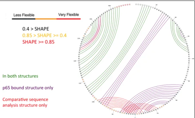
compared to comparative sequence analysis
derived prediction ... 103 Figure 4.1 Mutating tTER sequence
Figure 4.2 MS1 changes conformation but not
secondary structure ... 116 Figure 4.3 MS1 helix I nucleotides do not change
upon binding p65 ... 119 Figure 4.4 p65 alters the template region of
MS1 tTER upon binding ... 122 Figure 4.5 TRE nucleotides near the template
and pseudoknot regions are altered
upon binding p65 ... 124 Figure 4.6 p65 drastically alters MS1 Helix IV
structure upon binding p65 ... 125 Figure 4.7 MS2 pseudoknot region changes
conformation upon binding p65 ... 127 Figure 4.8 MS2 changes conformation but not
secondary structure ... 128 Figure 4.9 MS2 helix I nucleotides do not change
upon binding p65 ... 131 Figure 4.10 MS2 stem loop II nucleotides are stabilized
by p65 binding ... 132 Figure 4.11 p65 alters the template region of
MS2 tTER upon binding ... 134 Figure 4.12 MS2 TRE nucleotides near the template
and pseudoknot regions are altered
upon binding p65 ... 135 Figure 4.13 p65 does not drastically alter MS2 Helix IV
structure upon binding p65 ... 136 Figure 4.14 MS2 pseudoknot region changes conformation
upon binding p65 ... 139 Figure 4.15 MS1 / MS2 changes conformation
but not secondary structure ... 140 Figure 4.16 MS1 / MS2 stem loop II nucleotides
Figure 4.17 p65 alters the template region of
MS1 / MS2 tTER upon binding ... 143 Figure 4.18 MS1 / MS2 TRE nucleotides near the template
and the pseudoknot region are altered
upon binding p65 ... 145 Figure 4.19 MS1 / MS2 pseudoknot region changes
conformation upon binding p65 ... 147 Figure 4.20 Mutating tTER IIIa residues alters
3’ half of tTER when free in solution ... 148 Figure 4.21 Mutating tTER IIIa residues alters helix III
conformation only in the presence of p65 ... 149 Figure 4.22 MS1 mutant prevents helix IIIa
pseudoknot formation ... 151 Figure 4.23 MS2 mutant prevents helix IIIa
and IIIb formation ... 152 Figure 4.24 MS1 / MS2 mutant allows helix IIIa
but not IIIb pseudoknot formation ... 153 Figure 4.25 Model of telomerase assembly ... 155
LIST OF ABBREVIATIONS snoRNP Small nucleolar ribonucleoprotein TER Telomerase RNA
TERT Telomerase reverse transcriptase
tTER Tetrahymena thermophila telomerase RNA
tTERT Tetrahymena thermophila telomerase reverse transcriptase SAFA Semi-‐automated footprint analysis software
SHAPE Selective 2’-‐hydroxyl acylation analyzed by primer extension RNA Ribonucleic acid
p65 Tetrahymena thermophila telomerase holoenzyme protein (65 kDa) RNase Ribonuclease
EDTA Ethylenediaminetetraacetic acid EMSA Electrophoretic mobility shift assay HCl Hydrochloric acid
NaCl Sodium chloride DTT Dithiothreitol
MgCl2 Magnesium chloride
1M7 1-‐methyl-‐7-‐nitroisatoic anhydride BSA Bovine serum albumin
ng Nanogram
U·μL-‐1 Units per microliter DMSO Dimethyl sulfoxide ng/mL-‐1 Nanograms per milliliter ug/mL-‐1 Micrograms per milliliter ssRNA Single strand RNA
IPTG Isopropyl β-‐D-‐1-‐thiogalactopyranoside
Chapter I. Literature Review
A. Telomeres in Medicine
The DNA repair enzymes that fix double-‐stranded breaks within chromosomes must not recognize chromosome ends as breaks, or unwanted nucleolytic degradation and chromosome-‐end fusion can occur. Telomeres are repeating structures flanking repetitive nuclear DNA that help eukaryotes discern the ends of linear chromosomes from double-‐stranded breaks. Telomeres were recognized as a fundamental aspect of cellular biology with the awarding of the 2009 Nobel Prize in Physiology or Medicine to Elizabeth Blackburn, Carol Greider, and Jack Szostak for the discovery of how chromosomes are protected by telomeres and the enzyme telomerase. The essential role telomeres play in the maintenance of genomic integrity and cellular ageing is supported by several observations.
1. Eukaryotic Cellular Division Faces the End-‐replication Problem
The earliest functional description of telomeres was by geneticist Barbara McClintock when she observed in 1931 that chromosome ends never attached to one another after the chromosome was fragmented by x-‐ray irradiation.1
Herman Muller termed these natural ends of linear chromosomes “telomere” to describe the lack of fusing between the natural ends of chromosomes when irradiated by x-‐rays in Drosophila. Muller showed that the ends of the irradiated chromosomes, different from the other genomic regions, did not present alterations such as deletions or inversions, thanks to the presence of a protective cap. Muller named this cap the “terminal gene” and afterwards the “telomere” from the Greek terms -‐telos for “end” and –meros for “part”.2
Although McClintock and Muller monitored differences between natural linear chromosome ends and broken ends, they did not fully understand the molecular nature of the chromosome ends they observed, because several key pieces of information were missing. First, in 1944, Avery, MacLeod, and McCarty identified DNA as the material that chromosomes are made of.3 Next, the double helical structure of DNA was detected in 1953.4 And finally, Arthur Kornberg described the mechanism of DNA polymerase I activity in the 1960s and 70s to explain how DNA is replicated.5
2. Telomeres Contribute to Cellular Mortality
allowed limit of replication and remained active metabolically but that no longer divided were termed “senescent.”
Hayflick’s seminal observations led to the hypothesis that cells possess a counting mechanism capable of both tracking the number of cellular divisions and halting any further division after a predetermined number of cell divisions (known as the “Hayflick limit”) was reached. The same Alexei Olovnikov who had predicted the end replication problem during the replication of linear chromosomes provided the link between the fields of replicative senescence and telomeres when he
suggested that the gradual erosion of the ends of chromosomal DNA could cause the limited proliferative potential observed in Hayflick’s senescent fibroblasts.9 In the mid-‐1970s, Hayflick hypothesized that limited proliferative potential plays an important role in tumor suppression, after which researchers began to question whether the ends of linear chromosomes were important to tumor formation.10
3. Telomeres Overcome the End-‐replication Problem
Elizabeth Blackburn determined the DNA sequence of the ends of the ciliate Tetrahymena thermophila chromosomes in the mid 1970s and noted guanosine rich
sequences at the 3’ end of the lagging strand.11 Tetrahymena was chosen as a model organism because of its unique biology that confers an abundance of telomeres (Figure 1.2). Blackburn went on to show that the same terminal, heterogeneous array of CCCCAA repeats on the leading strand, occurred at the ends of the other chromosomal DNA molecules of the somatic nucleus (the macronucleus) in
the germline nucleus (the micronucleus) from which the somatic nucleus is
generated.12,13 Once Blackburn found that new telomere sequences were added to the ends of the linear rDNA mini-‐chromosomes by an unknown mechanism, the question then became, “How is the telomere repeat added”.14
envisaged: Telomeric sequences are transposed or recombined onto the developing macronuclear DNA termini, or the simple, repeating telomeric sequences are
synthesized de novo onto these termini by specific synthetic machinery.”14 As Blackburn predicted, one model proposed that an unknown enzyme synthesized the ends,15 while a second model proposed that the addition of telomere repeats
occurred through a recombination-‐mediated process.16,17
The debate was settled in 1985 when Carol Greider provided direct
experimental evidence of yeast telomere DNA synthesis18 in Tetrahymena cellular extracts by a specific telomere terminal transferase. Observation of the requirement that the terminal transferase extend yeast telomeric DNA by one guanosine before the characteristic TTGGGG pattern endemic to Tetrahymena was repeatedly synthesized demonstrated compelling evidence of the terminal transferase’s activity.19 The name of the transferase was shortened to “telomerase” in a subsequent paper.20
Greider cloned and separated the RNAs that copurified with telomerase activity to eventually locate a nine-‐nucleotide sequence within one of the purified RNAs that could serve as a template for reverse transcription. After accumulating evidence that oligonucleotides blocking the CAACCCCAA proposed template region also blocked telomerase activity, the implied reverse transcriptase mechanism of elongating telomeric DNA was proposed in 1989. 21 The impact of our
4. Telomerase Activity is a Potential Therapeutic Target for Cancer
The discovery of the human telomerase ribonucleoprotein was announced in 1989 shortly after Greider proposed the mechanism by which Tetrahymena
telomerase reverse transcribes an RNA template into a repeating sequence of DNA i.22 In 1990, Greider confirmed Olovnikov’s prediction that telomere shortening does indeed occur in cultured human cells due to a lack of telomerase expression and a consequent inability to maintain normal telomeres.23 A critical link was made between cancer, the second leading cause of death in the United States, and
telomeres when the authors showed that short telomeres can limit the ability of cells to divide. The link was made when the authors showed that inhibition of telomerase activity could potentially limit the growth of cancer cells.
telomerase maintained telomeres and did not allow them to shorten. Essentially, telomerase activity was shown to prevent senescence.27
These initial findings stimulated the cancer community to begin to study telomerase activity in a variety of tumor types. Attention quickly led to a many hundred-‐fold increase in the number of publications on telomerase at the turn of the 21st century.28 Work in the mid to late 1990s that combined cultured human cells and a novel telomerase knockout mouse model confirmed that inhibition of telomerase expression could indeed limit cancer cell division and tumor production.29
However, the details of the pathways that determine whether telomerase inhibition will be effective in fighting particular types of cancer have still not been fully worked out. It was observed that the loss of telomere function in some situations may lead to chromosome rearrangements that can surprisingly fuel tumor progression instead of arresting it.30 Even though research into telomerase’s role in tumorigenesis continues to be robust, the plethora of biochemical
information produced by the cancer community began to generate unforeseen links between telomeres and human disease.
5. Short Telomeres Have a Role in Genetic Diseases
telomerase activity is tightly regulated in mammals. After initially focusing on fundamental cellular mechanisms in simple model organisms, human genetic studies in the first decade of the 21st century established a clear role for telomere dysfunction in diverse degenerative disease states.
One key biochemical observation in the early 2000s was that human
telomerase RNA is an RNA polymerase II transcript, whereas ciliate telomerase RNA is transcribed by RNA polymerase III.32 Like other small nucleolar RNAs (snoRNAs) that are Pol II transcripts, human telomerase RNA contains a consensus H box sequence (5’ – ANANNA – 3’) and an ACA trinucleotide located three nucleotides upstream of the mature 3’ end. The box H/ACA motif within telomerase RNA serves as a pseudouridylation modification site by a group of four small nucleolar
ribonucleoproteins. (snoRNPs). Specifically, the dyskerin snoRNP is as a pseudouridine synthase that aids in the post-‐transcriptional maturation of telomerase RNA, and as a result, the entire telomerase ribonucleoprotein.33 Alterations in dyskerin were shown to reduce telomerase RNA concentration and shorten telomeres.34,35
Later, mutations in three of the four mammalian snoRNPs were shown to lead to dyskeratosis congenita, a rare congenital disorder characterized by disruptions in the integumentary organ system, such as abnormal skin and bone marrow failure.36-‐38 Though the exact pathology of the disease is not yet fully
understood, it was the initial association of dyskerin to telomeres that eventually led to most evidence pointing to dyskeratosis congenita being a disorder of poor
component either directly or indirectly. Mutations in the human telomerase RNA cause the autosomal dominant form of the disease, while mutations in the gene encoding dyskerin result in the X chromosome linked form, and both display shortened telomeres.38,39
It was not long before the progressive telomere shortening observed over many generations caused by mutations in both the RNA and protein components of the telomerase ribonucleoprotein was linked to limited tissue renewal capacity.40-‐42 Limited tissue renewal capacity due to telomere shortening manifests itself
clinically as dyskeratosis congenita or as other syndromes, depending on the tissue most affected (Table 1.1). The spectrum of telomere length disequilibrium diseases has been greatly extended and now encompasses prevalent disorders that have been poorly understood, such as aplastic anemia.43 The most common manifestation of telomere-‐mediated disease is idiopathic pulmonary fibrosis. 44 However,
cumulative evidence is mounting to suggest that a group of disorders may be the spectrum of the same disease, as they share the molecular defect of short telomere length. Coats plus syndrome, Hoyeraal Hreidarsson syndrome, and Revesz
syndrome have been proposed to be considered as a single syndrome spectrum because of their overlapping clinical features.45 Interestingly, retinopathy,
Biochemical observation of telomeres has yielded insights into the
B. Telomerase ribonucleoproteins
The evolution of linear chromosomes in the common ancestor of eukaryotes is remarkable because it occurred despite the many challenges posed by linear chromosomes. First, conventional primer-‐requiring DNA polymerases cannot copy the end of a DNA template, leading to the progressive loss of end sequence with every round of DNA replication. Second, chromosome ends resemble double-‐ stranded DNA breaks whose recognition and repair can result in chromosome fusions. Finally, DNA ends are vulnerable to the destructive action of exonucleases. A solution to these challenges was to form the protective telomere structure. 46 Although telomere structure is conserved across almost all eukaryotes, telomere structure is dynamic, involves a number of different components, and changes as the cell progresses through its life cycle.47
1. Telomere Structure is Conserved Across Most Eukaryotes
2. Telomerase Maintains Telomeres
Telomere maintenance in most eukaryotes and cancer cells depends on new repeat synthesis of tandem GT-‐rich repeats of DNA. Alternative mechanisms include recombination-‐mediated or transposition-‐mediated elongation, such as that
observed in fruit flies.52,53 Telomerase, the unique reverse transcriptase that
In vitro, telomerase is capable of synthesizing stretches of DNA often
3. Telomerase Reverse Transcriptase Structure is Conserved Across
Eukaryotes
ions necessary for catalysis of deoxynucleotide triphosphate (dNTP) addition. The active site is formed using motifs conserved across all reverse transcriptases,
including the closely related retrotransposons.61,62 The RT domain also positions the template and aligns the substrate 3’ end.63,64
The interaction between the TEN and the RT domains simultaneously alters conformational equilibriums of both the single-‐stranded telomeric DNA substrate and the nascent single-‐stranded product.65 The altering traps the old and new DNA strands in states that convert telomerases only capable of single-‐repeat synthesis into highly processive enzymes.60 The CTE may enhance nucleic acid association and/or otherwise contribute to RT domain function by promoting telomerase processivity and regulating telomerase localization, but does not appear to be essential to catalytic function.66,67
4. Telomerase RNA Structures Display More Structural Variety
Than TERT Structures
Sequences and sizes of telomerase RNAs range from about 150 nucleotides in ciliates to more than 1200 nucleotides in yeast. Even so, functional assays suggest that four TER elements may be considered functionally homologous in ciliates, yeast, and vertebrate TERs, despite the disparities in sequence and size across eukaryotes.68-‐72 Figure 1.5 illustrates the disparities in telomerase RNA sequence and size across eukaryotes by showing specific base pairs as schematic
throughout eukaryotes are maintained in each of the three telomerase RNAs represented in Figure 1.5.
The 3’ region of the template domain helps align the telomeric DNA substrate in the TERT active site while the 5’ portion is copied. The length of the single-‐
stranded template is typically about 1.5 repeats of the telomeric DNA sequence, except in yeasts with especially long and/or degenerate telomeric repeat
A structurally conserved, triple-‐helix stabilized pseudoknot is also adjacent to the template domain. A specific function for the pseudoknot domain has not yet been determined, although the conservation of the structure across eukaryotes is assumed to be important for global TER folding. One plausible model is that the pseudoknot aids in positioning the template relative to the active site and other TER and TERT domains.46 In any event, the pseudoknot domain is typically flanked by long-‐range base pairing at or near the 5’ end of TER. This pseudoknot flanking helix acts as the TBE in human TER, but not in ciliate or yeast TERs.
There is at least one place other than the active site where TER directly interacts with TERT. The highly conserved stem terminus element (STE) stimulates telomerase activity at least in part through its direct interaction with the TRBD of the reverse transcriptase.56,77 The STE is comprised of either a terminal hairpin, a hairpin from a three-‐way junction, or a three-‐way junction.
Only TERT and TER are required to reconstitute telomerase catalytic activity in vitro (although other rabbit reticulocyte lysate components like Heat Shock
Protein 90 are required to properly assemble telomerase).78 In vivo however, cellular telomerase holoenzymes are multiunit complexes that fractionate by gel filtration with an apparent mass of 500 kDa or more.69 Apparently, telomerase holoenzyme proteins other than TERT direct and regulate physical interactions with telomeric chromatin and functional engagement with the chromosome 3’ end.
The identities of telomerase holoenzyme proteins and their exact
telomerase isolated from Tetrahymena thermophila, Saccharomyces cerevisiae, Schizosaccharomyces pombe, mice, or cultured human cells. Less intensively studied, but still important additional model systems include other ciliates, yeasts, vertebrates, and plants.79-‐83 Figure 1.6 summarizes some of the structural and functional similarities between vertebrate and Tetrahymena thermophila telomerase ribonucleoprotein complexes. This thesis focuses on the study of
Tetrahymena thermophila telomerase, which is a valuable model system to study the
mechanism and structure of all telomerases because of the abundance of telomeres and telomerase in organisms’ macronucleus.
5. Structure-‐Function Relationships in Tetrahymena Telomerase
RNA
The model laboratory ciliate possesses a 159 nucleotide TER that includes all four of the conserved motifs summarized in the previous section. The nine-‐
nucleotide template is flanked at its 5’ end by a TBE and at its 3’ end by a
pseudoknot. In addition to defining the template boundary, the TBE and its adjacent single-‐stranded regions also provide the high-‐affinity TRBD-‐binding site.74,84
The pseudoknot sequence does not assemble into a pseudoknot structure in protein-‐free, full-‐length Tetrahymena TER, despite assembling into a stable
pseudoknot conformation when separated from the rest of TER. It is only the assemblage with holoenzyme proteins p65 and TERT that promotes proper folding of the pseudoknot.85,86 The pseudoknot remains stably folded throughout the telomerase catalytic cycle and can form even when some of the base pairing is disrupted.85 These observations, coupled with the fact that the only active telomerase complexes contain a properly folded pseudoknot, suggest that the pseudoknot topology, not the pseudoknot sequence, is critical for an active telomerase ribonucleoprotein.87,88
The template, TBE, and the pseudoknot domains are enclosed by long-‐range base pairing of stem I, while the STE is formed by terminal stem loop IV in
ribonucleoprotein reconstituted in rabbit reticulocyte lysate by TERT and TER. 56,77,88-‐91
Tetrahymena also has an additional motif immediately 3’ of the template termed the template recognition element (TRE). The TRE contributes to efficient copying through the nucleotides in the middle of the template positions as
evidenced by the curiously longer than normal telomeres exhibited by Tetrahymena cells expressing a TER with TRE sequence substitutions.88 The TRE may contribute to increased telomere length by adjusting the conformational dynamics of the template within the active site to alter the time the telomerase enzyme pauses during repeat synthesis equilibrium. TRE also improves the in vitro use of
oligonucleotide templates physically separate from the rest of TER, strengthening the hypothesis that TRE alters the conformational states available to TER.92
6. p65 Facilitates Telomerase Assemblage by Increasing Affinity of
tTERT to tTER
The conformation of telomerase changes as the multicomponent
the polyuridine tail, stem I, and stem IV.93,94 The interaction of p65 with TER
stabilizes a kink in stem IV necessary for tight TERT binding and induces additional conformational changes in stem–loop IV and potentially elsewhere that promote TERT assembly and catalytic activity.95-‐100
p65 is an RNA-‐binding protein in the La-‐family. La-‐family proteins are involved in many aspects of RNA metabolism, including binding and protecting 3’ polyuridine tracts of newly RNA polymerase III-‐transcribed RNA, processing 5’ and 3’ ends of pre-‐tRNA precursors, and acting as an RNA chaperone.101,102 p65 appears to energetically stabilize telomerase RNP and help fold TER into a structure that is more favorable for TERT-‐TER binding and enzyme function than in its
absence.94,96,97,103 These studies provide direct evidence of p65-‐mediated conformational transitions within the telomerase RNP.
quantities at high purity. The ability to rationally target telomerase activity would be dramatically enhanced by a more detailed understanding of telomerase structure and function. However, the problem is that the structure-‐function relationships of telomeres and their components are not fully understood. For example, the
secondary structure of the RNA component of the telomerase ribonucleoprotein during each of its various stages of assembly and reverse transcriptase activity has not been determined.
This dissertation uses telomerase derived from the well-‐established model organism Tetrahymena thermophila to claim that binding of the accessory protein p65 does not alter the secondary structure of ciliate telomerase RNA. Instead, p65 increases the dynamics of the three dimensional conformation of the telomerase RNA nucleotides to facilitate assembly into the active telomerase complex. This claim is supported by evidence from several biochemical and biophysical
experiments, including a novel combination of SHAPE structure-‐probing chemistry and discrete molecular dynamics (DMD) modeling with mutations of the
pseudoknot region of telomerase RNA and previously determined FRET-‐derived tertiary constraints. Chapter II first identifies the secondary structural differences between protein free tTER and reverse transcriptase bound tTER. Chapter III then describes how the subtle differences among pseudoknot region SHAPE intensities in protein free tTER and p65 bound tTER do not translate into secondary structural differences. Finally, Chapter IV focuses on how mutating nucleotides in the
to be differentiated by SHAPE. Our hypothesized model is described in Figure 1.7 as the progression from panels A to C to D rather than from A to B to D.
Table 1.2
In Solution tTER Structural Probing
tTER position
NMIA RN
ONEa
DMSb DEPc RN
T1d RN T1e RN T1f RN V1g RN V1h RN V1i
A1 CBD CBD CBD CBD CBD CBD CBD CBD CBD CBD
U2 CBD CBD CBD CBD CBD CBD CBD CBD CBD CBD
A3 CBD CBD CBD CBD CBD CBD CBD CBD CBD CBD
C4 CBD CBD CBD CBD CBD CBD CBD CBD CBD
C5 CBD CBD CBD CBD CBD CBD CBD CBD CBD
C6 CBD CBD CBD CBD CBD CBD CBD CBD CBD
G7 CBD CBD CBD CBD + CBD +++ CBD
C8 CBD CBD CBD CBD CBD + CBD
U9 CBD CBD CBD CBD CBD CBD
U10 + CBD CBD CBD CBD CBD
A11 ++ CBD +++ CBD CBD CBD
A12 ++ CBD +++ CBD CBD CBD
U13 + CBD CBD CBD CBD
U14 ++ CBD CBD CBD CBD
C15 CBD CBD +++ CBD CBD CBD
A16 + CBD +++ CBD CBD CBD
U17 + CBD CBD CBD CBD
U18 - CBD CBD CBD CBD
C19 - CBD CBD CBD + CBD
A20 - CBD CBD CBD ++ CBD
G21 - CBD CBD +++ CBD +++ +++ CBD
A22 - CBD + CBD CBD + ++ CBD
U23 - CBD CBD CBD + CBD
C24 - CBD CBD CBD + + CBD
U25 - CBD CBD CBD + CBD
G26 + CBD CBD ++ CBD CBD
U27 +++ CBD CBD CBD
A28 +++ CBD +++ +++ CBD CBD
A29 ++ CBD ++ + CBD CBD
U30 +++ CBD
A31 + CBD
G32 - +++ ++ +
A33 -
A34 - + +
C35 - +
U36 -
G37 - ++
U38 ++ +++
C39 ++ +++ +++ ++
A40 +++ +++ +
U41 +++ +++
U42 +++ +++
C43 ++ +++ +++ ++
A44 +++ +++ +
A45 + + +
C46 -
C47 -
C48 - +
tTER position
NMIA RN
ONEa
DMSb DEPc RN
T1d RN T1e RN T1f RN V1g RN V1h RN V1i
A50 +++ ++ +++ + ++
A51 + ++ +++ + +
A52 +++ ++ +++ +
A53 + ++ + +
A54 - ++ +
U55 - ++ +
C56 - ++ ++
U57 + ++ ++ ++
A58 + ++ + ++
G59 + ++ + +++
U60 + ++
G61 - ++ +++ +++
C62 + ++ +++ +
U63 - ++ + ++
G64 - ++ +++ +++ + ++
A65 ++ ++ ++
U66 ++ ++
A67 +++ ++ + + + ++
U68 +++ ++ +
A69 +++ ++ +++ +++ ++
A70 +++ ++ +++ +++ ++
C71 - +++ ++
C72 ++ +++ + ++
U73 + + ++
U74 + + +
C75 + +
A76 +
C77 - ++
C78 + + ++
A79 +++ + + +++ ++
A80 +++ + +++ +
U81 +++ +
U82 + +
A83 - ++
G84 - + + +
G85 - + + + +
U86 - +
U87 + ++
C88 + +++ +++
A89 ++ +++ +++ +++
A90 ++ +++ +++ +++
A91 ++ +++ ++ +++
U92 ++ +++
A93 ++ +++ +++
A94 ++ + +++
G95 + + +++ + +
U96 +++ +
G97 - + +++
G98 - + +++ +
U99 + +
A100 ++ + + +
A101 ++ + +++ +
U102 ++
tTER position
NMIA RN
ONEa
DMSb DEPc RN
T1d RN T1e RN T1f RN V1g RN V1h RN V1i
C104 - + +++
G105 - + +++
G106 - + +++
G107 - +++ +++ +
A108 ++ +++ +++
C109 CBD +++ +++
A110 +++ +++ +++ + +
A111 +++ +++ +
A112 ++ ++
A113 + +
G114 - + +++
A115 - +
C116 -
U117 +++ ++
A118 ++ + +
U119 + +
C120 - +++
G121 +++ +++ ++ +++
A122 +++ ++ +++ +++
C123 - +
A124 + +
U125 ++
U126 ++
U127 ++ +++ +
G128 - +++ +
A129 - CBD CBD +++ CBD CBD
U130 - CBD CBD + CBD CBD
A131 +++ CBD CBD CBD CBD
C132 - CBD CBD CBD CBD CBD
A133 +++ +++ CBD +++ CBD CBD CBD CBD
C134 ++ +++ CBD CBD CBD CBD CBD
U135 +++ +++ CBD CBD CBD CBD CBD
A136 +++ +++ CBD +++ CBD CBD CBD CBD
U137 +++ +++ CBD CBD CBD CBD CBD
U138 +++ +++ CBD CBD CBD CBD CBD
U139 + + CBD CBD CBD CBD CBD
A140 - CBD CBD CBD CBD CBD CBD
U141 - CBD CBD CBD CBD CBD CBD
C142 - CBD CBD CBD CBD CBD CBD
A143 - CBD CBD CBD CBD CBD CBD
A144 + CBD CBD CBD CBD CBD CBD
U145 + CBD CBD CBD CBD CBD CBD
G146 ++ CBD CBD + CBD CBD CBD CBD
G147 + CBD CBD + CBD CBD CBD CBD
A148 + CBD CBD CBD CBD CBD CBD
U149 + CBD CBD CBD CBD CBD CBD
G150 - CBD CBD + CBD CBD CBD CBD
U151 - CBD CBD CBD CBD + CBD CBD
C152 - CBD CBD CBD CBD +++ CBD CBD
U153 + CBD CBD CBD CBD + CBD CBD
U154 +++ CBD CBD CBD CBD +++ CBD CBD
A155 +++ CBD CBD CBD CBD CBD CBD
U156 +++ CBD CBD CBD CBD CBD CBD
tTER position
NMIA RN
ONEa
DMSb DEPc RN
T1d RN T1e RN T1f RN V1g RN V1h RN V1i
U158 +++ CBD CBD CBD CBD CBD CBD
U159 +++ CBD CBD CBD CBD CBD CBD
Lack of a symbol is used for nucleotide positions that were unreactive to the reagent used.
CBD is used for nucleotide positions whose result cannot be determined.
- is used for nucleotide positions with no reactivity to the NMIA reagent.
+ is used for nucleotide positions with low reactivity to the reagent used.
++ is used for nucleotide positions with moderate reactivity to the reagent used.
+++ is used for nucleotide positions with high reactivity to the reagent used.
aRibonuclease ONE digestion of telomerase RNA in the absence of p65 and in the absence of the amino terminal half of TERT (amino acids 1-516) under several buffer conditions. Data is from the minus p65, minus TERT-N lane of Figure 4B in Berman et al104.
bT7 transcripts of telomerase RNA treated with dimethyl sulfate for thirty minutes were subtracted from untreated RNA data from in Figure 3A of Zaug and Cech 105.
cDiethylpyrocarbonate modification of tTER folded at 30°C lanes from Figure 1 in Bhattacharyya and Blackburn 106.
dRibonuclease T1 digestion of telomerase RNA in Tris folding buffer for 10 minutes from Figure 2C of Bhattacharyya and Blackburn 106.
eRibonuclease T1 digestion of telomerase RNA in rabbit reticulocyte lysate with no pET28a-TERT plasmid included from lane 5 of Figure 2A in Sperger and Cech 91.
fRibonuclease T1 digestion of telomerase RNA in the absence of p65 and in the absence of the RNA binding domain of TERT (amino acids 216-516) under several buffer conditions. Data is from the minus p65, minus RBD lane of Figure 4C in Berman et al 99.
hRibonuclease V1 digestion of telomerase RNA in rabbit reticulocyte lysate with no pET28a-TERT plasmid included from lane 8 of Figure 2A in Sperger and Cech 91.
iRibonuclease V1 digestion of telomerase RNA in the absence of p65 and in the absence of the amino terminal half of TERT that contains the TEN and RBD domains. Data is from the minus p65, minus TERT-N lane of Figure 4A in Berman et al 99.
Table 1.3
tTER in Assembled Telomerase Structural
Probing
tTER position
NMIA RN ONEa DMSb RN T1c RN T1d RN V1e RN V1f
A1 CBD CBD CBD CBD CBD CBD CBD
U2 CBD CBD CBD CBD CBD CBD CBD
A3 CBD CBD CBD CBD CBD CBD CBD
C4 CBD CBD CBD CBD CBD CBD
C5 CBD CBD CBD CBD CBD CBD
C6 CBD CBD CBD CBD CBD CBD
G7 CBD CBD CBD CBD
C8 CBD CBD + CBD CBD
U9 CBD CBD CBD CBD
U10 ++ CBD CBD CBD
A11 ++ CBD + CBD CBD
A12 ++ CBD + CBD CBD
U13 ++ CBD CBD CBD
U14 ++ CBD CBD CBD
C15 - CBD CBD CBD
A16 - CBD CBD CBD
U17 - CBD CBD CBD
U18 - CBD CBD CBD
C19 - CBD CBD + CBD
A20 - CBD CBD ++ CBD
G21 - CBD + CBD +++ CBD
A22 - CBD + CBD ++ CBD
U23 - CBD CBD ++ CBD
C24 - CBD CBD ++ CBD
U25 - CBD CBD ++ CBD
G26 - CBD ++ CBD CBD
U27 ++ CBD CBD CBD
A28 +++ CBD + CBD CBD
A29 + CBD CBD CBD
U30 +++ CBD
A31 - CBD
G32 - + +
A33 -
A34 - +
C35 - + +
U36 - +
G37 - + + +
U38 - ++
C39 + ++ ++
A40 +++ ++ +
U41 +++ ++
U42 +++ ++ +++
C43 CBD ++ +++ ++
A44 ++ +++
A45 + +++
C46 - +++ +
tTER position
NMIA RN ONEa DMSb RN T1c RN T1d RN V1e RN V1f
C48 - +++ + ++
C49 - ++ +++ ++ ++
A50 +++ ++ +++ ++ ++
A51 + ++ +++ ++ +
A52 ++ ++ +++ +
A53 ++ ++ +++
A54 ++ ++ +
U55 ++ ++ + ++
C56 + ++ + ++ ++
U57 ++ ++ ++ ++
A58 +++ ++ + + +
G59 +++ ++ +++ + +
U60 +++ ++ +
G61 ++ ++ +++ + ++
C62 + ++ ++
U63 ++ ++ ++ ++
G64 ++ ++ +++ + ++
A65 +++ ++ + +
U66 CBD ++ ++
A67 +++ ++ ++ + +
U68 +++ ++ +
A69 ++ ++ ++
A70 + ++ ++
C71 - ++
C72 - ++
U73 - ++
U74 + + +
C75 + +
A76 + +
C77 - +
C78 - + +
A79 ++ + +
A80 ++ + +
U81 ++ + +
U82 + + +
A83 - + +++
G84 - + + ++
G85 - + +++
U86 + ++
U87 ++
C88 CBD +++
A89 ++ +++
A90 + +++
A91 ++ +++
U92 ++ +++
A93 ++
A94 ++
G95 + + + + +
U96 ++ +
G97 - + +
G98 - + + +
U99 + +
A100 +++ + ++
tTER position
NMIA RN ONEa DMSb RN T1c RN T1d RN V1e RN V1f
U102 +++
G103 - ++
C104 - +++ ++
G105 - +++ +
G106 - ++
G107 - +
A108 CBD ++
C109 CBD +++ ++
A110 +++ +++ ++ +
A111 +++ ++
A112 + +
A113 - +
G114 - + + +
A115 - + +
C116 - +
U117 ++ ++ +
A118 - + +
U119 CBD +
C120 - +
G121 +++ ++ +
A122 +++ ++
C123 - +
A124 + +
U125 CBD
U126 +
U127 ++ +
G128 CBD +
A129 - CBD CBD
U130 - CBD CBD
A131 + CBD CBD
C132 - CBD CBD CBD
A133 ++ +++ CBD CBD CBD
C134 + ++ CBD CBD CBD
U135 ++ ++ CBD CBD CBD
A136 +++ ++ CBD CBD CBD
U137 +++ +++ CBD CBD CBD
U138 +++ +++ CBD CBD CBD
U139 - + CBD CBD CBD
A140 - CBD CBD CBD CBD
U141 - CBD CBD CBD CBD
C142 - CBD CBD CBD CBD
A143 - CBD CBD CBD CBD
A144 - CBD CBD CBD CBD
U145 CBD CBD CBD CBD CBD
G146 CBD CBD CBD CBD CBD
G147 - CBD CBD CBD CBD CBD CBD
A148 - CBD CBD CBD CBD CBD CBD
U149 - CBD CBD CBD CBD CBD CBD
G150 CBD CBD CBD CBD CBD CBD CBD
U151 CBD CBD CBD CBD CBD CBD CBD
C152 - CBD CBD CBD CBD CBD CBD
U153 + CBD CBD CBD CBD CBD CBD
U154 CBD CBD CBD CBD CBD CBD CBD
tTER position
NMIA RN ONEa DMSb RN T1c RN T1d RN V1e RN V1f
U156 +++ CBD CBD CBD CBD CBD CBD
U157 +++ CBD CBD CBD CBD CBD CBD
U158 +++ CBD CBD CBD CBD CBD CBD
U159 +++ CBD CBD CBD CBD CBD CBD
Lack of a symbol is used for nucleotide positions that were unreactive to the reagent used.
CBD is used for nucleotide positions whose result cannot be determined. - is used for nucleotide positions with no reactivity to the NMIA reagent. + is used for nucleotide positions with low reactivity to the reagent used.
++ is used for nucleotide positions with moderate reactivity to the reagent used. +++ is used for nucleotide positions with high reactivity to the reagent used.
aRibonuclease ONE digestion of telomerase RNA in the absence of p65 but in the presence of the amino terminal half of TERT (amino acids 1-516) under several buffer conditions. The amino terminal half of TERT contains the amino terminal and RNA binding domains but does not include the active site. Data is from the minus p65, plus TERT-N lane of Figure 4B in Berman et al104.
bDimethyl sulfate treatment of living Tetrahymena thermophila cells for two minutes were subtracted from cells where β-mercaptoethanol quench solution was added prior to dimethyl sulfate treatment. The data from Figure 3A and isolation of the nuclear RNA can be found in Zaug and Cech 105.
cRibonuclease T1 digestion of telomerase RNA assembled in telomerase that was translated in rabbit reticulocyte lysate using the pET28a-TERT plasmid from lane 4 of Figure 2A in Sperger and Cech 91.
dRibonuclease T1 digestion of telomerase RNA in the absence of p65 but in the presence of the RNA binding domain of TERT (amino acids 216-516) under several buffer conditions. Again, the reverse transcriptase active site is not included in the TERT portion tested. Data is from the minus p65, plus RBD lane of Figure 4C in Berman et al 99.
eRibonuclease V1 digestion of telomerase RNA assembled in telomerase that was translated in rabbit reticulocyte lysate using the pET28a-TERT plasmid from lane 8 of Figure 2A in Sperger and Cech 91.
Chapter II. Modeling TERT induced telomerase ribonucleoprotein conformational transitions 1
INTRODUCTION
We sought to better understand the three-‐dimensional structure and conformational changes associated with tTER function within the telomerase RNP. Therefore, we combined secondary structural constraints of tTER obtained using the high-‐resolution footprinting technique selective 2’-‐hydroxyl acylation analyzed by primer extension (SHAPE),107 distance constraints obtained from single-‐molecule FRET data,95 and biochemical inference gleaned from previous biochemical
experiments to generate constraints. We then modeled the structure of tTER in the minimal complex using discrete molecular dynamics (DMD) that allows facile incorporation of experimental information.108 In addition, we docked the resulting model with a homology model of tTERT based on the crystal structure of the T. Castaneum TERT59 and the tTERT RNA binding domain76 to generate a three-‐ dimensional model of tTER in the minimal telomerase complex. The results reveal conformational changes that occur during telomerase assembly and suggest a model for stem IV binding to tTERT.108
MATERIALS AND METHODS
1. Preparation of tTER and pFLAG-‐tTERT.
RNA was transcribed in vitro using Ampliscribe T7 Transcription Kit (Epicenter Technologies). Templates were generated by PCR using the plasmid pTET-‐telo, a pUC19-‐based plasmid containing the tTER gene, a T7 RNA polymerase promoter, and a self-‐cleaving hammerhead ribozyme that processes the 5′-‐end of the RNA. Primers are listed in Table S4. PCR products were gel purified using Wizard PCR Prep Kits and RNAs gel purified and stored in TE (pH 7.5) at −80 °C.
A sequence encoding the FLAG epitope was ligated into a pET-‐28a plasmid containing tTERT cloned into the BamH1 and Xho1 sites. Oligonucleotides were gel purified and annealed before ligation into the Nco1 and BamH1 sites in pET-‐28a-‐ tTERT. This removed the Nco1 site and an Nde1 site, allowing for easy screening of positive clones, and removed the N-‐terminal His-‐ and T7-‐tags.