METFORMIN EFFICACY AGAINST BREAST CANCER DEPENDS ON ITS CELLULAR UPTAKE VIA CATION TRANSPORTERS AND MODULATION OF INSULIN/IGF1 PATHWAY
Hao Cai
A dissertation submitted to the faculty at the University of North Carolina at Chapel Hill in partial fulfillment of the requirements for the degree of Doctor of Philosophy in the Eshelman
School of Pharmacy.
Chapel Hill 2016
Approved by: Dhiren R. Thakker
Kim L.R. Brouwer
William Zamboni Roy L. Hawke Cary Anders
© 2016 Hao Cai
ABSTRACT
Hao Cai: Metformin Efficacy Against Breast Cancer Depends on Its Cellular Uptake via Cation Transporters and Modulation of Insulin/IGF1 Pathway
(Under the direction of Dhiren R. Thakker)
Clinical evidence suggests that metformin is efficacious as an anticancer agent in diabetic patients; however, results about its efficacy are mixed, especially in non-diabetic patients. The goal of this dissertation project was to improve metformin treatment for breast cancer by elucidating molecular and cellular mechanisms that play an important role in its efficacy. Research conducted in this project showed that breast cancer cells exhibit wide
variability in the expression of cation transporters, which are required for intracellular uptake and accumulation of metformin. Further, metformin requires a functional intracellular adenosine monophosphate-activated protein kinase (AMPK) pathway to exert its anticancer activity. Interestingly, cancer stem cells, which are more sensitive to the antiproliferative effect of metformin, express higher levels of cation-selective transporters than non-stem cancer cells.
Preclinical dose-response studies showed that estrogen receptor positive breast tumors with low expression of cation transporters required a minimum metformin dose (in combination with 30 mg/kg/day paclitaxel) that is equivalent to the highest current anti-diabetic dose of 2,550 mg/day, suggesting that an even higher metformin dose is needed to optimally treat these patients. The minimum efficacious metformin dose (in combination with 50 mg/kg/day
Studies in mice showed that attenuation of the insulin/IGF1 pathway sensitized breast cancer cells to the antiproliferative efficacy of metformin exerted via modulation of the AMPK pathway. These results provide a rationale for lower efficacy of metformin in non-diabetic patients, and suggest that co-administration of metformin with an insulin/IGF1 pathway inhibitor may improve metformin efficacy in non-diabetic breast cancer patients.
In summary, the dissertation research provides valuable insights into cellular and molecular factors that contribute to the variable responses of diabetic and non-diabetic breast cancer patients to metformin therapy. The findings of this research will contribute to
To my parents, for their unending love, support, and understanding.
ACKNOWLEDGEMENTS
First, I would like to express my deepest gratitude to my advisor Dr. Dhiren R. Thakker for his support and mentorship. I still remember the afternoon we first met in 2011. At that time, I was confused about whether I should pursue a Ph.D. degree. It was Dr. Thakker who patiently listened to my concerns and encouraged me to follow my interest in research. During my four years in graduate school, he instructed and guided me not only in scientific thinking and experimental techniques, but also in communication, professional development, and personal life. As an international student who is weak in written and oral communication in English, I was under a lot of pressure due to Dr. Thakker’s high expectations. However, when I look back on
this difficult experience, it was a period in my life when I was able to make the most progress. I would also like to thank my dissertation committee. As the chair of my committee, Dr. Brouwer not only gave me suggestions on pharmacokinetics (PK) and drug transporters, but also helped me examine the feasibility of my dissertation project. As an expert in the field of cancer PK and preclinical models, Dr. Zamboni provided numerous suggestions on using preclinical models to evaluate the distribution of drugs. Drs. Bae-Jump and Anders provided several valuable suggestions from a clinical aspect. My discussions with them always made me feel that my research could make a real contribution to breast cancer patients. I would also like to thank Dr. Lola Reid, who is the stem cell expert on my committee, and who assisted me in designing studies on cancer stem cells. As a student working on drug transport and PK, these “fancy” cancer stem cells assays really intrigued me. I would also like to thank Dr. Roy Hawke,
I would like to acknowledge current and former members of the Thakker Laboratory. I want to show my deep appreciation to Dr. Ruth Everett, who is always willing to help me when I am facing difficulties, and spent a lot of time helping to revise and refine my writing. Dr. Everett is my writing mentor, no doubt. Without her patient guidance with my writing skills, I would not have made a lot of progress. I would like to thank Drs. Kevin Han, Chester Costales, Yunhui Zhang, Ravindra Alluri, Nicole Zane, Muhammad Wahajuddin, Arti Thakkar and Yufeng Xia for their help with experimental design and techniques. Besides, I want to thank Christine Lee, Dr. Lawrence Ku, Bryan Mackowiak, Dr. Kelsey Browder, and Dr. Frank Fanizza for their friendship.
I also want to thank my colleagues at the University of North Carolina, as well as Zawadi Walker, Arlo Brown, Anna Crollman, Kathy Maboll, and Lauren McQuillan for their assistance with scheduling committee meetings and my defense. I would like to extend my appreciation to Charlene Santos for helping me with animal studies, and to Dr. Alan Forrest and Jingxian Chen for assistance with modeling.
TABLE OF CONTENTS
LIST OF TABLES……….………..ix LIST OF FIGURES……….……….x LIST OF ABBREVIATIONS……….………xiii
CHAPTER 1 REPURPOSING METFORMIN FOR BREAST CANCER THERAPY: FROM CLINICAL OBSERVATIONS TO
MOLECULAR MECHANISMS TO DRUG OPTIMIZATION ……….……….1
CHAPTER 2 VARIABILITY IN CATION-SELECTIVE TRANSPORTER
EXPRESSION IN HUMAN BREAST CANCER CELL LINES AND BREAST TUMOR TISSUES RESULTED IN VARIABILITY IN THE
ANTIPROLIFERATIVE EFFICACY OF METFORMIN……….……26
CHAPTER 3 CANCER STEM CELLS ARE MORE SUSCEPTIBLE TO
METFORMIN DUE TO ENHANCED TRANSPORTER-MEDIATED
METFORMIN UPTAKE……….………52
CHAPTER 4 CATION-SELECTIVE TRANSPORTERS IN BREAST
TUMORS ENHANCE THE INTRATUMORAL CONCENTRATION OF METFORMIN AND METFORMIN-MEDIATED ACTIVATION OF
THE AMPK PATHWAY AND ANTITUMOR EFFICACY.………,………66
CHAPTER 5 RELATIONSHIP BETWEEN METFORMIN DOSE AND
EFFICACY AGAINST ESTROGEN RECEPTOR-POSITIVE AND
TRIPLE-NEGATIVE BREAST CANCER………...93
CHAPTER 6 ANTIPROLIFERATIVE EFFICACY OF METFORMIN
AGAINST BREAST CANCER IS ENHANCED BY INHIBITION OF
LIST OF TABLES
Table 1.1 Retrospective Studies on the Effect of Metformin against
Breast Cancer Risk……….………17
Table 1.2 Ongoing Clinical Studies Using Metformin for Breast
Cancer Treatment………..….18
STable 4.1 The Effect of Metformin Plus DOX on Tumor Volumes and
Tumor Weights of OCT3-MCF7 Tumors and MCF-7 Tumors ………..………88 STable 4.2 AUCplasma, Cmax and Cl of Metformin in Xenograft Mice-bearing
OCT3-MCF7 and MCF-7 Tumor………..……….89
Table 5.1 Estimates of the PK Parameters Calculated Using
the Model Described in Figure 5.1 for Mice Bearing MDA-MB-468
Tumors and MCF-7 Tumors………....……114
Table 5.2 Summary of the Simulated Intratumoral Exposures of Metformin
in Tumor-bearing Mice Treated with Varying Doses of Metformin…………..…...115 STable 5.3 Systemic Exposures of Metformin Tumor-bearing Mice Treated
with 360 mg/kg/day Metformin versus 360 mg/kg/day Metformin and
Carboplatin or Paclitaxel……….…….…116 .
LIST OF FIGURES
Figure 1.1 Interactions between AMPK Pathway and Insulin Pathway………...14
Figure 1.2 Metformin Effect on Cell Cycle Check Point Genes………15 Figure 1.3 Metformin Effect on the Generation of CSCs………...16 Figure 2.1 Expression of Cation-selective Transporters OCT1-3, PMAT and
MATE1-2 in Human Breast Cancer Cell Lines…………..……….42 Figure 2.2 Expression of Cation-Selective Transporter Genes in Normal
Breast Tissues, Breast Tumor Tissues and Their Adjacent
Non-Malignant Tissues……….43 Figure 2.3 Metformin Uptake in Human Breast Cancer Cell Lines with High
and Low Cation-Selective Transporter Expression………..44 Figure 2.4 Antiproliferative Effects of Metformin in Human Breast Cancer
Cell Lines with Varying Transporter Expression Profiles……….………45 Figure 2.5 Contribution of Cation-selective Transporters to Metformin-mediated
AMPK and P70S6K Phosphorylation in Human Breast Cancer
Cell Lines………46 SFigure 2.1 The Interaction of Cation-selective Transporters and the AMPK
Signaling Cascade in the Antiproliferative Efficacy of Metformin………..47 Figure 3.1 Expression of Cation-selective Transporter Genes in BT-549
CSCs and NSCCs……….60 Figure 3.2 Expression of MATE1 Protein in BT-549 CSCs and NSCCs………...…….61 Figure 3.3 Metformin Uptake in BT-549 CSCs and NSCCs………....………….……..62
SFigure 3.1 Expression of Cation-selective Transporter Genes in HMLER
CSCs and NSCCs………...……….63 Figure 4.1 Generation and Characterization of OCT3-MCF7 Cells…….………....…….81 Figure 4.3 IHC Staining of Tissue Sections from OCT3-MCF7 Tumors and
MCF-7 Tumors to Evaluate OCT3 Expression and the
Antiproliferative Efficacy of Metformin………82 Figure 4.3 The Effects of Saline, DOX Alone, Metformin Alone and Metformin
Plus DOX on Breast Tumors………...…….…………...83 Figure 4.4 Western Blot Analyses Showing the Effect of Metformin Treatment
on AMPK Phosphorylation and P70S6K Phosphorylation in
Figure 4.5 Plasma and Tissue Concentrations of Metformin in Xenograft
Mice Bearing OCV3-MCF7 Tumors and MCF-7 Tumors………85 SFigure 4.1 The Antiproliferative Efficacy of Metformin in OCT3-MCF7 Cells
and MCF-7 Cells……….………..86 Sfigure 4.2 Metformin-induced Decrease in the Proportion of CSC in
OCT3-MCF7 Tumors and MCF-7 Tumors……….………..………..….…87 Figure 5.1 Design of the In Vivo Study to Evaluate Anticancer Efficacy of
Metformin as a Monotherapy and in Combination with
Chemotherapeutic Agents Paclitaxel (for MCF-7 Tumors) and
Carboplatin (for MDA-MB-468 Tumors)………...….…..108 Figure 5.2 Antitumor Efficacy of Metformin as a Monotherapy and in
Combination with Carboplatin (for MDA-MB-468 Tumors) and
Paclitaxel (for MCF-7 Tumors)……….………..….…..109 Figure 5.3 Kaplan–Meier Survival Curves Showing the Effect of Metformin
Treatment as a Monotherapy and in Combination with (A) Carboplatin or (B) Paclitaxel on the Survival of Mice Bearing
MDA-MB-468 or MCF-7 Tumors, Respectively……….………..110 Figure 5.4 Activation of AMPK Caused by Metformin and/or Paclitaxel and
Carboplatin in MDA-MB-468 Tumors (A) and MCF-7 Tumors (B)..…….………..111 Figure 5.5 Plasma and Intratumoral Concentrations of Metformin in Mice
Treated with Different Doses of Metformin………...……….…….112 Figure 5.6 Visual Predictive Checks of the PK Model to Predict
Plasma and Tumor Metformin Concentrations as a Function of
Time in Tumor-bearing Mice……….……….113 Figure 6.1 Generation of MCF-7
T-CTRL and MCF-7IRS-1 KD Cell Lines……….………..131
Figure 6.2 Comparison of the Antiproliferative Efficacy of Metformin against MCF7
T-CTRL Cells and MCF-7IRS-1 KD Cells.……….………..132
Figure 6.3 Impact of Insulin/IGF-1 on Metformin-mediated Modulation of the AMPK Pathway in MCF-7
T-CTRL and MCF-7IRS1 KD Cell………133
Figure 6.4 Effect of Insulin/IGF1 Treatment on the Expression of PMAT and MATE1 Genes in MCF-7 Breast Cancer Cells with Attenuated
Insulin/IGF1 Signaling Pathway……….………...…134 Figure 6.5 Effect of Insulin/IFG1 Treatment on Intracellular Uptake of Metformin
in Intact and Attenuated Insulin/IGF1 Signaling Pathway.….………..135 SFigure 6.1 Endogenous Growth Rates of Wildtype MCF-7 Tumors,
MCF-7
SFigure 6.2 Cell Proliferation (A), and Metformin Intracellular Uptake (B) in Wildtype MCF-7 Cells and MCF-7
T-CTRL Cells………..……...….137
SFigure 6.3 The Effects of Metformin Treatment (360 mg/kg/day) in Relation to Saline Treatment (Control) on Wildtype MCF-7 Tumors and MCF-7
IRS-1 KD Tumors……….……….……138
LIST OF ABBREVIATIONS ADME AKT AMPK ATM AUC CDC CHT CSC CV DOX EMT ER+ HMLER IGF IGF1R IR IRS-1 LKB-1 MATE MCF-7T-CTRL MCF-7IRS-1 KD NSCC OCT OCT3-BT20
Absorption, distribution, metabolism, and excretion Protein kinase B (PKB)
Adenosine monophosphate-activated protein kinase
Ataxia telangiectasia mutated Area under the curve
Cyclin-dependent kinase Choline transporter Cancer stem cell Coefficient of variation Doxorubicin
Epithelial-mesenchymal transition Estrogen receptor-positive
Oncogenic transformed immortalized human mammary epithelial Insulin-like growth factor
Insulin-like growth factor receptor Insulin receptor
Insulin receptor substrate 1 Liver kinase B1
Multidrug and toxin extrusion protein MCF-7 cells used as transfection control
MCF-7 cells in which IRS-1 expression is knocked down Non-stem cancer cell
Organic cation transporter
OCT3-MCF7 PD
PI3K PK PMAT P70S6K SD ST
TGF TN VPC
OCT3-overexpressing MCF-7 cells Pharmacodynamics
Phosphoinositide 3-kinase Pharmacokinetics
Plasma membrane monoamine transporter Ribosomal protein S6 kinase
Standard deviation Serotonin transporter Transforming growth factor Triple-negative
CHAPTER 1
Repurposing Metformin for Breast Cancer Therapy: from Clinical Observations to Molecular Mechanisms to Drug Optimization
1.1 Metformin is A Leading Drug for Type 2 Diabetes
Metformin, also known as 1,1-dimethylbiguanide, is currently the first-line therapeutic agent for type 2 diabetes. Metformin is a type of biguanide compound which originates from goat's rue (Galega officinalis), a herbal medicine used for the treatment of diabetes for centuries.
Metformin was synthesized in 1922 by Emil Werner and James Bell (1). Compared to other
biguanide compounds such as phenformin, metformin is much safer, but also has a relatively
lower anti-glycemic efficacy. As a result, metformin was not used in the clinic for treatment of
diabetes until the 1970s when other biguanide drugs were withdrawn from the market due to
toxicity.
Metformin was approved by the U.S. Food and Drug Administration (FDA) for type 2 diabetes in 1994. Today, it has become the most widely prescribed anti-diabetic agent in the
U.S. with 59.2 million prescriptions in 2014 (2). For the treatment of type 2 diabetes, metformin is orally administered as either immediate-release GLUCOPHAGE® Tablets or extended-release
GLUCOPHAGE®Tablets (Bristol-Myers-Squibb Inc.). The dose of metformin generally used for anti-diabetic treatment ranges from 500 to 1000 mg, with a maximum recommended daily dose of 2550 mg (for immediate-release formulation) or 2000 mg (for extended-release formulation).
Metformin exerts its anti-glycemic effects in diabetic patients by increasing insulin sensitivity
subsequent downregulation of the genes that drive gluconeogenesis (3). Metformin treatment has also been reported to activate AMPK in the skeletal muscle and recruit glucose transporters to increase glucose uptake into skeletal muscle (4). Studies also suggested that metformin reduces circulating glucose levels by decreasing intestinal glucose absorption, demonstrated by impaired anti-glycemic efficacy of metformin when the drug was delivered through the portal vein (5). The anti-diabetic effect of metformin (i.e. its glucose-lowering effect) in the non-diabetic people has been reported to be insignificant (6). Since metformin has been shown to impact circulating fatty acids (7), growth hormone (8), and transforming growth factor beta (TGFβ) (9),
etc., it is also used for the treatment of polycystic ovary syndrome (10), excessive weight (11), insulin resistance (12), and arterial hypertension (13).
1.2 Metformin Pharmacokinetics (PK): The Role of Transporters in Metformin Disposition
and Efficacy against Type 2 Diabetes
Metformin is a highly hydrophilic small molecule (logD of -6.13 at pH 6.0) that is positively charged (pka 12.4) at all physiological pH values (structure shown as the insert of
Figure 1.1). Due to the physicochemical properties of metformin, its intracellular uptake through passive diffusion is very limited (14). Instead, studies have shown that the cellular transport of metformin is primarily mediated by cation-selective transporters (15, 16), as evidenced by observations showing significantly higher permeability of metformin in Caco-2 cells compared to neutral molecules with similar physicochemical properties, such as mannitol (16-18).
expressed in human intestinal tissues and are responsible for metformin intestinal absorption, (15). As metformin absorption was also shown to be dependent on intestinal length, a “sponge”
mechanism was proposed by Proctor et al., which suggested that the intestinal absorption of metformin is enhanced by an uptake-efflux-reuptake process mediated by cation-selective transporters (16).
Upon being taken up into the intestine, metformin accumulates in the liver and small intestine (17), with a volume of distribution in humans ranging from 60 to 280 L. The absorption of metformin in its primary target organ, the liver, is mediated by OCT1. In the liver, metformin remains unmetabolized (20) and a small proportion of it (<20%)) is secreted into bile by the multidrug and toxin extrusion protein (MATE)1 transporter. The elimination of metformin from the body is achieved primarily through renal clearance. In the kidney, metformin is taken up into the proximal tubules via OCT2, and secreted into the urine via MATE1 and MATE2 (21, 22).
Since cation-selective transporters play a critical role in the ADME of metformin, studies have been conducted to evaluate how mutations in these transporters affect the PK and diabetic efficacy of metformin. Becker et al. reported that OCT1 mutations impaired the anti-glycemic efficacy of metformin in diabetic patients by reducing OCT1-mediated intestinal absorption and decreasing the systemic concentrations of the drug (23). Mutations in
transporters, which only reduce accumulation of metformin in its target organ, also affect the efficacy of the drug. A study conducted by Chen, et al. shown that mutations in OCT3, the predominant transporter expressed in skeletal muscles, impaired the uptake and accumulation of metformin in muscle tissues, and led to a reduced anti-diabetic efficacy as
1.3 From Leading Anti-diabetic Agent to Potential Anticancer Drug: the Beneficial Effects
of Metformin against Breast Cancer in Clinical Studies
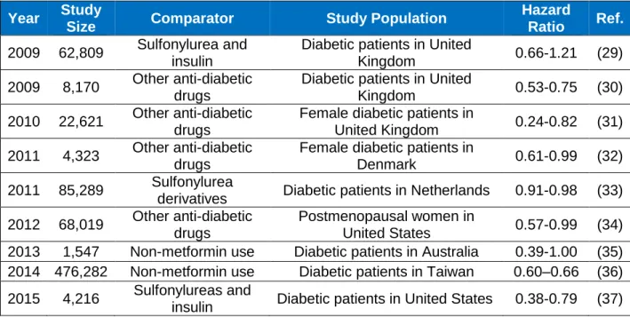
Epidemiological studies have shown that patients with type 2 diabetes exhibited an increased risk of developing breast cancer compared to the non-diabetic population (26-28). These clinical observations led to further retrospective analyses on the potential impact of anti-glycemic agents on the incidence of breast cancer. A majority of retrospective studies have shown that diabetic patients on metformin treatment had a significantly lower risk of developing breast cancer compared to those on other therapeutic agents such as insulin and sulfonylurea, although some studies only observed a trend (Table 1.1) (29-37). This beneficial effect of metformin against breast cancer occurrence aroused further interest in evaluating the possibility of using metformin as an anticancer agent. In a retrospective study by He, et al., it was
concluded that metformin improved the breast cancer-specific survival rate of diabetic patients compared to other anti-glycemic drugs (38). Not only is metformin implicated in cancer
prevention, but evidence is mounting for the inhibitory effects of metformin against cancer cell proliferation and tumor growth. In a study of breast cancer patients on neoadjuvant
chemotherapy, diabetic patients on metformin therapy showed a significantly improved response to chemotherapy (reflected by higher rates of pathologic complete response) compared to diabetic patients on other anti-glycemic drugs (39). Additionally, treatment of presurgery diabetic breast cancer patients with metformin significantly reduced the proportion of proliferating cells (identified by immunohistochemical (IHC) staining of the proliferation
biomarker, Ki-67) in tumor tissues (40).
although the anticancer efficacy of metformin in non-diabetic cancer patients was not as significant as its efficacy in diabetic patients with cancer.
Since breast cancer is the second leading cause of cancer death among women in the U.S (48), and there is currently no efficacious therapeutic agent for some subtypes of breast cancer (e.g. triple-negative breast cancer), repurposing metformin (a highly cost-effective drug with limited toxicity) for breast cancer therapy would be a significant improvement on the current treatment of this disease. However, metformin has not been used in the clinic for the treatment of breast cancer as some clinical trials report that the drug failed to show significant anticancer efficacy (49, 50). Therefore, there have been an increasing number of clinical studies being conducted on the anticancer efficacy of metformin (Table 1.2). At the same time, preclinical studies are also being conducted to identify the anticancer mechanisms of metformin to provide insights into its optimization for future clinical trials.
1.4 Molecular Mechanisms of the Anticancer Effects of Metformin
1.4.1 Reduction of Insulin and Insulin-like Growth Factor (IGF)1
Since metformin has been widely used for the treatment of diabetes, the role of its anti-diabetic effects in its anticancer pharmacology should be evaluated. Studies have shown that the proliferation of cancer cells can be stimulated under hyperglycemic culture conditions versus
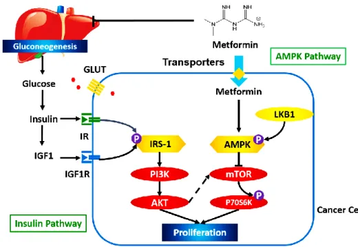
Studies have been conducted to illustrate the role of insulin and IGF1 in breast cancer development (Figure 1.2). Both insulin and IGF1 activate the insulin pathway through binding to their receptors, namely insulin receptor (IR) and IGF1 receptor (IGF1R), on cancer cell
membranes. Activated IR and IGF1R phosphorylate insulin receptor substrate (IRS)-1 and induce a conformational change in IRS-1, which enables downstream molecules to bind to it (55). Phosphoinositide 3-kinase (PI3K) is one of the major downstream targets of IRS-1. Activation of PI3K and its downstream protein kinase B (Akt) induces protein synthesis, enhances glucose uptake, and inhibits cell apoptosis (56). Compared to somatic cells, cancer cells have a hyperfunctional insulin pathway, which is the result of unregulated expression of IR and IGF1R or mutation of PI3K (57, 58). Several inhibitors of the insulin pathway have been approved for cancer therapy. Clinical studies have shown that co-administration of a PI3K inhibitor and trastuzumab significantly improved progression-free survival of breast cancer patients compared to trastuzumab monotherapy (hazard ratio: 0.78, p<0.01) (59), which implies that the insulin pathway plays a critical role in breast cancer development. Besides indirect attenuation of the insulin pathway through modulation of insulin and IGF1 levels, metformin was reported to directly reduce the expression of IR and IGF1R (60). The suppression of the insulin-dependent pathway by metformin was observed only in diabetic patients, which could be the likely cause of a poor response of non-diabetic breast cancer patients to metformin treatment compared to breast cancer patients with type 2 diabetes, as observed in clinical studies.
1.4.2 Activation of the AMPK Pathway
One of the primary roles of AMPK in breast cancer cells is the regulation of energy homeostasis. In somatic cells, energy is produced by two steps: 1) glycolysis in the cytosol to generate pyruvate, and 2) oxidation of pyruvate in mitochondria via Krebs Cycle. In cancer cells, however, pyruvate gets further oxidized into lactate instead of entering into the mitochondria (the Warburg effect) (61). The Warburg effect is critical to cancer cell proliferation as it allows the cancer cells to survive without the need of oxygen, since the microenvironment in tumor tissues is hypoxic. Besides, the Warburg effect not only produces energy but also provides intermediate products that are required for protein and lipid synthesis (61). AMPK is the central regulator of the Warburg effect. Activation of AMPK, on one hand, inhibits multiple enzymes that are involved in Warburg effect, such as phosphofructokinase-1 (62). On the other hand, AMPK activation inhibits the mammalian target of rapamycin (mTOR) and attenuates the
phosphorylation of P70S6 kinase (a downstream molecule of mTOR). Suppression of the mTOR pathway attenuates lipid and protein synthesis, inhibits cell proliferation, and induces apoptosis (63).
Studies have shown that metformin can activate AMPK in cancer cells (Figure 1.1). After being taken up into cancer cells, metformin blocks Mitochondrial Complex I in the electron transport chain and suppresses ATP synthesis. The elevated AMP/ATP ratio induces a
conformational change of AMPK and exposes the site (Thr172) for phosphorylation by liver kinase B1 (LKB-1) (64). The critical role of LKB-1 and AMPK phosphorylation was confirmed in
in vitro studies by Dowling et al., in which the LKB-1 deficient MDA-MB-231 human breast
cancer cell line showed a limited response to the anti-proliferative effects of metformin (65). Studies have implied that the effects of metformin on the AMPK pathway and the insulin pathway are not independent of each other. MCF-7 breast cancer cells cultured in low
insulin/IGF1 media exhibited greater metformin-induced activation of the AMPK pathway
1.4.3 Regulation of the Cell Cycle
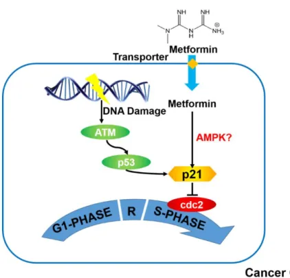
Besides the molecular targets involved in the anti-diabetic effects of metformin, the contributions of other signaling pathways, such as the cell cycle, to the antiproliferative effects of metformin have also been evaluated. Cell proliferation is regulated by the cell cycle regulatory pathway which maintains genomic stability by preventing cells with damaged DNA from proliferating. DNA damage in the cells induces the phosphorylation of Ataxia Telangiectasia Mutated (ATM), which subsequently stabilizes and activates its major downstream modulator, p53 (67). Activation of p53 leads to cell cycle arrest at the G1/S phase or G2/M phase via induction of p21 synthesis, which inhibits a group of cell proliferation initiators including cyclin-dependent kinase 2 (cdc2) (68). Mutations in p53 and ATM are the most frequently observed mutations in tumor tissues, which highlights the critical role of cell cycle regulation in cancer development.
There are some reports to suggest that metformin inhibits cell proliferation through modulation of the cell cycle checkpoint genes (69-72) (Figure 1.2). For example, metformin treatment resulted in an increase in the proportion of breast cancer cells that were arrested in the G1 phase (69). Metformin is believed to regulate the cell cycle by enhancing the synthesis of p21 (69). Although some studies also showed that metformin can induce phosphorylation of the two upstream molecules of p21, namely ATM and p53 (70, 71), a functional p53 is not required for metformin-mediated regulation of the cell cycle in cancer cells. Interestingly, studies have shown that metformin exerts better efficacy against deficient tumors rather than p53-competent tumors (72). This suggests that activation of p21 by metformin is likely to be regulated by other signaling pathways such as the AMPK-dependent pathway (71).
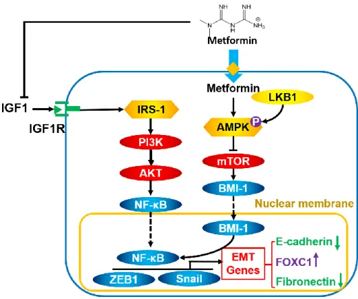
1.4.4 Effect on Cancer Stem Cells (CSCs)
The effect of metformin on cancer relapse and metastasis has also been investigated. According to some reports, the preventive effect of metformin against breast cancer
tumors are believed to be generated from a group of cells named CSCs due to cellular
properties that are similar to embryonic stem cells. Several studies showed that CSCs originate primarily from somatic epithelial cells through epithelial-mesenchymal transition (EMT) (73), a process originally discovered in the early development stage by which epithelial cells lose their cell-cell adhesion, acquire the ability to migrate, and become mesenchymal stem cells.
Compared to non-stem cancer cells (NSCCs), CSCs are more resistant to chemotherapeutic agents due to higher expression of efflux transporters (ATP-binding cassette transporters or ABC transporter), enhanced DNA damage response system, and increased aldehyde
dehydrogenase (ALDH) activity (74, 75). Additionally, CSCs have a lower expression of cell-cell adhesion protein and are more tumorigenic compared to NSCCs (76). It is widely believed that these properties make CSCs major cause of cancer reoccurrence and metastasis.
It has been reported that CSCs are more sensitive to metformin treatment compared to NSCCs (77). Several mechanisms have been proposed to explain this observation (Figure 1.3). First, the intracellular concentration of metformin in CSCs is not affected by the upregulation of efflux transporters because metformin is not an ABC transporter substrate under physiological conditions. Second, the inhibitory effect of metformin on ATP synthesis has greater impact on the proliferation of CSCs versus NSCCs since CSCs heavily rely on mitochondrial ATP production compared to NSCCs (77). Third, metformin inhibits the generation of CSCs by blocking the EMT process. Metformin reduces the secretion of IGF1 and transforming growth factor (TGF)β, both of which are required to maintain the “stemness” of CSCs and initiate EMT
from NSCCs (78, 79). Furthermore, metformin-induced activation of AMPK leads to the
inhibition of mTOR. As mTOR is a regulator of E-cadherin expression, its inhibition by metformin increases E-cadherin expression, improves cell-cell adhesion, and halts the EMT process (80).
1.4.5 Other Mechanisms
expression of human epidermal growth factor receptor (HER)2 (81) which is critical for the growth of HER+ breast cancer cells. The study conducted by Marini et al. revealed that metformin altered glucose metabolism in breast cancer cells by impairing hexokinase activity through steadily binding to its catalytic pocket (82, 83). These studies imply that, unlike other widely used chemotherapeutic agents, metformin inhibits tumor growth by modulating multiple signaling pathways simultaneously.
1.5 The Role of Transporters in the Efficacy of Metformin as An Anticancer Agent
Although multiple molecular mechanisms/targets have been proposed from preclinical studies to explain the anticancer effects of metformin, their clinical relevance remains unclear. Based on the need for intracellular uptake of metformin for activation of these targets, the molecular targets can be categorized into two types: 1) extracellular targets (e.g. insulin and IGF1) and 2) intracellular targets (e.g. AMPK and p21).
1.6 Rationale for the Dissertation Project
The overall goal of this dissertation research is to optimize current treatment of metformin against breast cancer. Based on existing knowledge on metformin PK and its molecular mechanisms in cancer therapy, the dissertation research will commence with identifying the molecular targets of metformin that determine its clinical antitumor efficacy, so that selection criteria can be provided for identifying patients who have functional metformin targets and are most suitable for metformin cancer therapy. To evaluate the contributions of extracellular and intracellular targets to the inhibitory efficacy of metformin in breast tumors, cation-selective transporter expression in human breast tumor tissues and breast cancer cell lines will be assessed. The roles of transporter expression and transporter-mediated uptake of metformin in the antitumor efficacy of metformin will be evaluated by comparing metformin efficacy against transporter-overexpressing tumors versus tumors with limited transporter expression. In addition, the impact of extracellular insulin and IGF1 levels on the antitumor efficacy of metformin will be evaluated. Based on these results, a therapeutic regimen of metformin, specifically for the treatment of breast cancer, will be developed through optimizing metformin dose and designing a new combination therapy. Therefore, the following
overarching hypotheses have been proposed to achieve this goal:
Metformin exerts its antiproliferative effects against both breast cancer stem cells and
non-stem cancer cells.
The anticancer efficacy of metformin is due to 1) transporter-mediated uptake and
activation of the intracellular AMPK pathway, and 2) reduction of circulating insulin and IGF1 levels and subsequent attenuation of cell growth stimulus.
Specific Aim 1. Investigate the role of transporter-mediated tumor uptake of metformin and the
1a. Evaluate the expression of cation-selective transporters in breast cancer cell lines and tumor tissues.
1b. Establish an OCT3-overexpressing MCF-7 (OCT3-MCF7) cell line and confirm increased OCT3 expression in this cell line.
1c. Compare metformin-induced activation of the AMPK-dependent pathway and antiproliferative efficacy between OCT3-MCF7 cells and MCF-7 cells.
1d. Evaluate the importance of transporters in metformin antitumor effects by comparing metformin antitumor efficacy between OCT3-MCF7 tumors and MCF-7 tumors.
1e. Investigate the role of transporters in metformin effects on MCF-7 CSCs.
Specific Aim 2. Optimize metformin doses for the treatment of breast cancer and correlate metformin efficacy with its systemic and intratumoral exposure.
2a. Generate xenograft mice bearing tumors generated from MCF-7 cells and MDA-MB-468 cells.
2b. Optimize metformin dose and establish a relationship between metformin dose-exposure and tumor volumes in mice bearing MCF-7 tumors and MDA-MB-468 tumors. 2c. Relate metformin dose-exposure-response to its effects on intracellular targets.
Specific Aim 3. Investigate the role of metformin-mediated reduction of insulin and IGF1 levels
in the anticancer effects of the drug against CSCs and NSCCs. 3a. Generate IRS-1 knockdown MCF-7 (MCF-7IRS-1 KD) cells.
3b. Evaluate the role of insulin and IGF1 in the anticancer effects of metformin using in
vitro cell models.
3c. Evaluate the role of insulin and IGF1 in the anticancer effects of metformin using food-induced diabetic xenograft mouse models.
Results from the three specific aims will be presented in the following chapters:
Chapter 2. Aim 1a
Chapter 4. Aim 1b-d
Chapter 5. Aim 2a-c
Table 1.1 Retrospective Studies on the Effect of Metformin against Breast Cancer Risk
Year Study
Size Comparator Study Population
Hazard
Ratio Ref.
2009 62,809 Sulfonylurea and insulin
Diabetic patients in United
Kingdom 0.66-1.21 (29) 2009 8,170 Other anti-diabetic
drugs
Diabetic patients in United
Kingdom 0.53-0.75 (30) 2010 22,621 Other anti-diabetic
drugs
Female diabetic patients in
United Kingdom 0.24-0.82 (31) 2011 4,323 Other anti-diabetic
drugs
Female diabetic patients in
Denmark 0.61-0.99 (32) 2011 85,289 Sulfonylurea
derivatives Diabetic patients in Netherlands 0.91-0.98 (33) 2012 68,019 Other anti-diabetic
drugs
Postmenopausal women in
United States 0.57-0.99 (34) 2013 1,547 Non-metformin use Diabetic patients in Australia 0.39-1.00 (35) 2014 476,282 Non-metformin use Diabetic patients in Taiwan 0.60–0.66 (36) 2015 4,216 Sulfonylureas and
Table 1.2 Ongoing Clinical Studies Using Metformin for Breast Cancer Treatment. Data from ClinicalTrials.gov (http://clinicaltrials.gov).
Study
Size Comparator Study Name (Study Phase) Outcome
Metformin Dose (mg/day)
40 Before treatment
Pre-surgical trial of the combination of metformin and atorvastatin in newly diagnosed operable breast cancer (0)
Tumor
progression 1500
150 Placebo Phase II study of metformin for reduction of obesity-associated breast cancer risk (II)
Breast
cancer risk 850
72 Placebo
A trial of standard chemotherapy with metformin (vs placebo) in women with
metastatic breast cancer (II)
Progression-free survival 850
42 Placebo Metformin for reduction of paclitaxel-related neuropathy in patients with breast cancer (II)
Change in
neuropathy 500
46 Placebo
A study of Liposomal Doxorubicin+ Docetaxel + Trastuzumab + Metformin in
operable and locally advanced HER2+ breast cancer (II)
Pathologic complete response
1000
96 Placebo or melatoninn
Neoadjuvant FDC with melatonin or metformin for locally advanced breast cancer
(II)
Response
rate 850
60 Placebo Metformin Plus Neoadjuvant Chemotherapy in Breast Cancer (II)
Pathologic complete response
REFERENCES
1. Dunn CJ, Peters DH. Metformin. A review of its pharmacological properties and therapeutic use in non-insulin-dependent diabetes mellitus. Drugs. 49 1995; (5): 721–49
2. "Leading Prescriptions Dispensed in the U.S. Diabetes Market 2014 | Statistic." Statista. Web. 08 Mar. 2016.
3. Hundal RS, Krssak M, Dufour S, Laurent D, Lebon V, Chandramouli V, et al. Mechanism by which metformin reduces glucose production in type 2 diabetes. Diabetes.
2000;49(12):2063-9
4. Musi N, Hirshman MF, Nygren J, Svanfeldt M, Bavenholm P, Rooyackers O, et al. Metformin increases AMP-activated protein kinase activity in skeletal muscle of subjects with type 2 diabetes. Diabetes. 2002;51(7):2074-81
5. Ikeda T, Iwata K, Murakami H. Inhibitory effect of metformin on intestinal glucose absorption in the perfused rat intestine. Biochemical pharmacology. 2000;59(7):887-90
6. Sambol NC, Chiang J, O'Conner M, Liu CY, Lin ET, Goodman AM, et al. Pharmacokinetics and pharmacodynamics of metformin in healthy subjects and patients with noninsulin-dependent diabetes mellitus. J Clin Pharmacol. 1996;36(11):1012-21
7. Tiikkainen M, Hakkinen AM, Korsheninnikova E, Nyman T, Makimattila S, Yki-Jarvinen H. Effects of rosiglitazone and metformin on liver fat content, hepatic insulin resistance, insulin clearance, and gene expression in adipose tissue in patients with type 2 diabetes. Diabetes. 2004;53(8):2169-76
8. Landin K, Tengborn L, Smith U. Treating insulin resistance in hypertension with metformin reduces both blood pressure and metabolic risk factors. Journal of internal medicine. 1991;229(2):181-7
9. Agard C, Rolli-Derkinderen M, Dumas-de-La-Roque E, Rio M, Sagan C, Savineau JP, et al. Protective role of the antidiabetic drug metformin against chronic experimental pulmonary hypertension. British journal of pharmacology. 2009;158(5):1285-94.
10. Bourron O, Daval M, Hainault I, Hajduch E, Servant JM, Gautier JF, et al. Biguanides and thiazolidinediones inhibit stimulated lipolysis in human adipocytes through activation of AMP-activated protein kinase. Diabetologia. 2010;53(4):768-78
11. Guido M, Romualdi D, Giuliani M, Suriano R, Tienforti D, Costantini B, et al. Effect of
metformin on the growth hormone response to growth hormone-releasing hormone in obese women with polycystic ovary syndrome. Fertil Steril. 2005;84(5):1470-6
12. Kim YD, Kim YH, Tadi S, Yu JH, Yim YH, Jeoung NH, et al. Metformin inhibits growth hormone-mediated hepatic PDK4 gene expression through induction of orphan nuclear receptor small heterodimer partner. Diabetes. 2012;61(10):2484-94.
14. Irvine JD, Takahashi L, Lockhart K, Cheong J, Tolan JW, Selick HE, et al. MDCK (Madin-Darby canine kidney) cells: A tool for membrane permeability screening. J Pharm Sci. 1999;88(1):28-33.
15. Han TK, Proctor WR, Costales CL, Cai H, Everett RS, Thakker DR. Four cation-selective transporters contribute to apical uptake and accumulation of metformin in Caco-2 cell monolayers. J Pharmacol Exp Ther. 2015;352(3):519-28.
16. Proctor WR, Bourdet DL, Thakker DR. Mechanisms underlying saturable intestinal absorption of metformin. Drug Metab Dispos. 2008;36(8):1650-8
17. Sinko PJ, Hu P. Determination intestinal metabolism and permeability for several compounds in rats. Implications on regional bioavailability in humans. Pharm Res. 1996;13(1):108-13
18. Artursson P, Karlsson J. Correlation between Oral-Drug Absorption in Humans and Apparent Drug Permeability Coefficients in Human Intestinal Epithelial (Caco-2) Cells.
Biochem Bioph Res Co. 1991;175(3):880-5
19. Pentikainen PJ. Bioavailability of metformin. Comparison of solution, rapidly dissolving tablet, and three sustained release products. International journal of clinical pharmacology,
therapy, and toxicology. 1986;24(4):213-20
20. Tucker GT, Casey C, Phillips PJ, Connor H, Ward JD, Woods HF. Metformin kinetics in healthy subjects and in patients with diabetes mellitus. Br J Clin Pharmacol. 1981;12(2):235-46
21. Chen Y, Li S, Brown C, Cheatham S, Castro RA, Leabman MK, Urban TJ, Chen L, Yee SW, Choi JH, Huang Y, et al. Effect of genetic variation in the organic cation transporter 2 on the renal elimination of metformin. Pharmacogenetics and genomics 2009;19(7):497-504 22. Hemauer SJ, Patrikeeva SL, Nanovskaya TN, Hankins GD, Ahmed MS. Role of human
placental apical membrane transporters in the efflux of glyburide, rosiglitazone, and metformin. Am J Obstet Gynecol. 2010;202(4):383 e1-7.
23. Becker ML, Visser LE, van Schaik RHN, Hofman A, Uitterlinden AG, Stricker BHC. Genetic variation in the organic cation transporter 1 is associated with metformin response in patients with diabetes mellitus. Pharmacogenomics Journal. 2009;9(4):242-7
24. Chen L, Pawlikowski B, Schlessinger A, More SS, Stryke D, Johns SJ, et al. Role of organic cation transporter 3 (SLC22A3) and its missense variants in the pharmacologic action of metformin. Pharmacogenetics and genomics. 2010;20(11):687-99
25. Shu Y, Sheardown SA, Brown C, Owen RP, Zhang SZ, Castro RA, et al. Effect of genetic variation in the organic cation transporter 1 (OCT1) on metformin action. Journal of Clinical
Investigation. 2007;117(5):1422-31
27. Wideroff L, Gridley G, Mellemkjaer L, Chow WH, Linet M, Keehn S, et al. Cancer incidence in a population-based cohort of patients hospitalized with diabetes mellitus in Denmark. J
Natl Cancer Inst. 1997;89(18):1360-5
28. Currie CJ, Poole CD, Gale EA. The influence of glucose-lowering therapies on cancer risk in type 2 diabetes. Diabetologia. 2009;52(9):1766-7
29. Libby G, Donnelly LA, Donnan PT, Alessi DR, Morris AD, Evans JM. New users of metformin are at low risk of incident cancer: a cohort study among people with type 2 diabetes. Diabetes Care. 2009;32(9):1620-5
30. Bodmer M, Meier C, Krahenbuhl S, Jick SS, Meier CR. Long-term metformin use is associated with decreased risk of breast cancer. Diabetes Care. 2010;33(6):1304-8 31. Bosco JL, Antonsen S, Sorensen HT, Pedersen L, Lash TL. Metformin and incident breast
cancer among diabetic women: a population-20(1):101-11.based case-control study in Denmark. Cancer Epidemiol Biomarkers Prev. 2011;
32. Ruiter R, Visser LE, van Herk-Sukel MP, Coebergh JW, Haak HR, Geelhoed-Duijvestijn PH, et al. Lower risk of cancer in patients on metformin in comparison with those on sulfonylurea derivatives: results from a large population-based follow-up study. Diabetes Care.
2012;35(1):119-24
33. Chlebowski RT, McTiernan A, Wactawski-Wende J, Manson JE, Aragaki AK, Rohan T, et al. Diabetes, metformin, and breast cancer in postmenopausal women. J Clin Oncol.
2012;30(23):2844-52
34. Onitilo AA, Donald M, Stankowski RV, Engel JM, Williams G, Doi SA. Breast and prostate cancer survivors in a diabetic cohort: results from the Living with Diabetes Study. Clin Med
Res. 2013;11(4):210-8
35. Tseng CH. Metformin may reduce breast cancer risk in Taiwanese women with type 2 diabetes. Breast Cancer Res Treat. 2014;145(3):785-90
36. Calip GS, Hubbard RA, Stergachis A, Malone KE, Gralow JR, Boudreau DM. Adherence to oral diabetes medications and glycemic control during and following breast cancer
treatment. Pharmacoepidemiol Drug Saf. 2015;24(1):75-85
37. He X, Esteva FJ, Ensor J, Hortobagyi GN, Lee MH, Yeung SC. Metformin and
thiazolidinediones are associated with improved breast cancer-specific survival of diabetic women with HER2+ breast cancer. Ann Oncol. 2012;23(7):1771-80
38. Jiralerspong S, Palla SL, Giordano SH, Meric-Bernstam F, Liedtke C, Barnett CM, et al. Metformin and pathologic complete responses to neoadjuvant chemotherapy in diabetic patients with breast cancer. J Clin Oncol. 2009;27(20):3297-302
39. Bonanni B, Puntoni M, Cazzaniga M, Pruneri G, Serrano D, Guerrieri-Gonzaga A, et al. Dual effect of metformin on breast cancer proliferation in a randomized presurgical trial. J Clin
40. Libby G, Donnelly LA, Donnan PT, Alessi DR, Morris AD, Evans JM. New users of metformin are at low risk of incident cancer: a cohort study among people with type 2 diabetes. Diabetes Care. 2009;32(9):1620-5
41. Wright JL, Stanford JL. Metformin use and prostate cancer in Caucasian men: results from a population-based case-control study. Cancer Causes Control. 2009;20(9):1617-22
42. Li D, Yeung SC, Hassan MM, Konopleva M, Abbruzzese JL. Antidiabetic therapies affect risk of pancreatic cancer. Gastroenterology. 2009;137(2):482-8
43. Bodmer M, Becker C, Meier C, Jick SS, Meier CR. Use of metformin and the risk of ovarian cancer: a case-control analysis. Gynecol Oncol. 2011;123(2):200-4
44. Zhang ZJ, Zheng ZJ, Kan H, Song Y, Cui W, Zhao G, et al. Reduced risk of colorectal cancer with metformin therapy in patients with type 2 diabetes: a meta-analysis. Diabetes Care. 2011;34(10):2323-8
45. Xiao Y, Zhang S, Hou G, Zhang X, Hao X, Zhang J. Clinical pathological characteristics and prognostic analysis of diabetic women with luminal subtype breast cancer. Tumour Biol. 2014;35(3):2035-45
46. Bayraktar S, Hernadez-Aya LF, Lei X, Meric-Bernstam F, Litton JK, Hsu L, et al. Effect of metformin on survival outcomes in diabetic patients with triple receptor-negative breast cancer. Cancer. 2012;118(5):1202-11
47. Hosono K, Endo H, Takahashi H, Sugiyama M, Sakai E, Uchiyama T, et al. Metformin suppresses colorectal aberrant crypt foci in a short-term clinical trial. Cancer Prev Res
(Phila). 2010;3(9):1077-83
48. American Cancer Society. Cancer facts & figures. Atlanta, GA: The Society, 2013. p. v. 49. Kalinsky K, Crew KD, Refice S, Xiao T, Wang A, Feldman SM, et al. Presurgical trial of
metformin in overweight and obese patients with newly diagnosed breast cancer. Cancer
Invest. 2014;32(4):150-7.
50. Cazzaniga M, DeCensi A, Pruneri G, Puntoni M, Bottiglieri L, Varricchio C, et al. The effect of metformin on apoptosis in a breast cancer presurgical trial. Br J Cancer.
2013;109(11):2792-7
51. Okumura M, Yamamoto M, Sakuma H, Kojima T, Maruyama T, Jamali M, et al. Leptin and high glucose stimulate cell proliferation in MCF-7 human breast cancer cells: reciprocal involvement of PKC-alpha and PPAR expression. Biochim Biophys Acta. 2002;1592(2):107-16
52. Hermann LS, Schersten B, Bitzen PO, Kjellstrom T, Lindgarde F, Melander A. Therapeutic Comparison of Metformin and Sulfonylurea, Alone and in Various Combinations - a Double-Blind Controlled-Study. Diabetes Care. 1994;17(10):1100-9
54. Wieman HL, Wofford JA, Rathmell JC. Cytokine stimulation promotes glucose uptake via phosphatidylinositol-3 kinase/Akt regulation of Glut1 activity and trafficking. Mol Biol Cell. 2007;18(4):1437-46
55. Wieman HL, Wofford JA, Rathmell JC. Cytokine stimulation promotes glucose uptake via phosphatidylinositol-3 kinase/Akt regulation of Glut1 activity and trafficking. Mol Biol Cell. 2007;18(4):1437-46
56. Kauffmann-Zeh A, Rodriguez-Viciana P, Ulrich E, Gilbert C, Coffer P, Downward J, et al. Suppression of c-Myc-induced apoptosis by Ras signalling through PI(3)K and PKB. Nature. 1997;385(6616):544-8
57. Milazzo G, Giorgino F, Damante G, Sung C, Stampfer MR, Vigneri R, et al. Insulin receptor expression and function in human breast cancer cell lines. Cancer Res. 1992;52(14):3924-30
58. Papa V, Gliozzo B, Clark GM, McGuire WL, Moore D, Fujita-Yamaguchi Y, et al. Insulin-like growth factor-I receptors are overexpressed and predict a low risk in human breast cancer.
Cancer Res. 1993;53(16):3736-40
59. Andre F, O'Regan R, Ozguroglu M, Toi M, Xu BH, Jerusalem G, et al. Everolimus for women with trastuzumab-resistant, HER2-positive, advanced breast cancer (BOLERO-3): a randomised, double-blind, placebo-controlled phase 3 trial. Lancet Oncology.
2014;15(6):580-91
60. Dowling RJ, Niraula S, Chang MC, Done SJ, Ennis M, McCready DR, et al. Changes in insulin receptor signaling underlie neoadjuvant metformin administration in breast cancer: a prospective window of opportunity neoadjuvant study. Breast Cancer Res. 2015;17:32 61. Warburg O. On the origin of cancer cells. Science. 1956;123(3191):309-14
62. Bartrons R, Caro J. Hypoxia, glucose metabolism and the Warburg's effect. Journal of
bioenergetics and biomembranes. 2007;39(3):223-9
63. Bolster DR, Crozier SJ, Kimball SR, Jefferson LS. AMP-activated protein kinase suppresses protein synthesis in rat skeletal muscle through down-regulated mammalian target of
rapamycin (mTOR) signaling. J Biol Chem. 2002;277(27):23977-80
64. Wheaton WW, Weinberg SE, Hamanaka RB, Soberanes S, Sullivan LB, Anso E, et al. Metformin inhibits mitochondrial complex I of cancer cells to reduce tumorigenesis. eLife. 2014;3:e02242
65. Dowling RJ, Zakikhani M, Fantus IG, Pollak M, Sonenberg N. Metformin inhibits mammalian target of rapamycin-dependent translation initiation in breast cancer cells. Cancer Res. 2007;67(22):10804-12
67. Waterman MJ, Stavridi ES, Waterman JL, Halazonetis TD. ATM-dependent activation of p53 involves dephosphorylation and association with 14-3-3 proteins. Nat Genet.
1998;19(2):175-8
68. Agarwal ML, Agarwal A, Taylor WR, Stark GR. p53 controls both the G2/M and the G1 cell cycle checkpoints and mediates reversible growth arrest in human fibroblasts. Proc Natl
Acad Sci USA. 1995;92(18):8493-7
69. Alimova IN, Liu B, Fan Z, Edgerton SM, Dillon T, Lind SE, et al. Metformin inhibits breast cancer cell growth, colony formation and induces cell cycle arrest in vitro. Cell Cycle.
2009;8(6):909-15
70. Duan X, Ponomareva L, Veeranki S, Choubey D. IFI16 induction by glucose restriction in human fibroblasts contributes to autophagy through activation of the ATM/AMPK/p53 pathway. PLoS One. 2011;6(5):e19532
71. Zhuang Y, Miskimins WK. Cell cycle arrest in Metformin treated breast cancer cells involves activation of AMPK, downregulation of cyclin D1, and requires p27Kip1 or p21Cip1. J Mol
Signal. 2008;3:18
72. Buzzai M, Jones RG, Amaravadi RK, Lum JJ, DeBerardinis RJ, Zhao F, et al. Systemic treatment with the antidiabetic drug metformin selectively impairs p53-deficient tumor cell growth. Cancer Res. 2007;67(14):6745-52
73. Morel AP, Lievre M, Thomas C, Hinkal G, Ansieau S, Puisieux A. Generation of Breast Cancer Stem Cells through Epithelial-Mesenchymal Transition. Plos One. 2008;3(8) 74. Hirschmann-Jax C, Foster AE, Wulf GG, Nuchtern JG, Jax TW, Gobel U, et al. A distinct
"side population" of cells with high drug efflux capacity in human tumor cells. P Natl Acad
Sci USA. 2004;101(39):14228-33
75. Diehn M, Cho RW, Lobo NA, Kalisky T, Dorie MJ, Kulp AN, et al. Association of reactive oxygen species levels and radioresistance in cancer stem cells. Nature.
2009;458(7239):780-U123
76. Al-Hajj M, Wicha MS, Benito-Hernandez A, Morrison SJ, Clarke MF. Prospective
identification of tumorigenic breast cancer cells. P Natl Acad Sci USA. 2003;100(7):3983-8 77. Lonardo E, Cioffi M, Sancho P, Sanchez-Ripoll Y, Trabulo SM, Dorado J, et al. Metformin
Targets the Metabolic Achilles Heel of Human Pancreatic Cancer Stem Cells. Plos One. 2013;8(10)
78. Chang WW, Lin RJ, Yu J, Chang WY, Fu CH, Lai ACY, et al. The expression and significance of insulin-like growth factor-1 receptor and its pathway on breast cancer stem/progenitors. Breast Cancer Research. 2013;15(3)
80. Qu C, Zhang WJ, Zheng GP, Zhang ZJ, Yin J, He ZM. Metformin reverses multidrug
resistance and epithelial-mesenchymal transition (EMT) via activating AMP-activated protein kinase (AMPK) in human breast cancer cells. Molecular and Cellular Biochemistry.
2014;386(1-2):63-71
81. Vazquez-Martin A, Oliveras-Ferraros C, Menendez JA. The antidiabetic drug metformin suppresses HER2 (erbB-2) oncoprotein overexpression via inhibition of the mTOR effector p70S6K1 in human breast carcinoma cells. Cell Cycle. 2009;8(1):88-96
82. Marini C, Salani B, Massollo M, Amaro A, Esposito AI, Orengo AM, et al. Direct inhibition of hexokinase activity by metformin at least partially impairs glucose metabolism and tumor growth in experimental breast cancer. Cell Cycle. 2013;12(22):3490-9
83. Salani B, Marini C, Rio AD, Ravera S, Massollo M, Orengo AM, et al. Metformin impairs glucose consumption and survival in Calu-1 cells by direct inhibition of hexokinase-II. Sci Rep. 2013;3:2070
84. Patel H, Younis RH, Ord RA, Basile JR, Schneider A. Differential expression of organic cation transporter OCT-3 in oral premalignant and malignant lesions: potential implications in the antineoplastic effects of metformin. Journal of oral pathology & medicine: official publication of the International Association of Oral Pathologists and the American Academy
____________________
1. This chapter has been adapted from the paper published in the International Journal of Cancer. The original citation is: Cai H, Zhang Y, Han TK, Everett RS, Thakker DR. Cation-selective transporters are critical to the AMPK-mediated antiproliferative effects of metformin in human breast cancer cells. Int J Cancer. 2016;138(9):2281-92
CHAPTER 21
Variability in Cation-selective Transporter Expression in Human Breast Cancer Cell Lines and Breast Tumor Tissues Resulted in Variability in the Antiproliferative Efficacy of Metformin
2.1. OVERVIEW
The antidiabetic drug metformin exerts antineoplastic effects against breast cancer and other cancers. One mechanism by which metformin is believed to exert its anticancer effect involves activation of its intracellular target, adenosine monophosphate-activated protein kinase (AMPK), which is also implicated in the antidiabetic effect of metformin. It is proposed that in cancer cells, AMPK activation leads to inhibition of the mammalian target of rapamycin (mTOR) and the downstream P70S6K that regulates cell proliferation. Due to its hydrophilic and cationic nature, metformin requires cation-selective transporters to enter cells and activate AMPK. This study demonstrates that expression levels of cation-selective transporters correlate with the antiproliferative efficacy of metformin in breast cancer. Metformin uptake and
antiproliferative activity were compared between a cation-selective transporter-deficient human breast cancer cell line, BT-20, and a BT-20 cell line that was engineered to overexpress organic cation transporter 3 (OCT3), a representative of cation-selective transporters and a predominant transporter in human breast tumors. Metformin uptake was minimal in BT-20 cells, but
cells. Collectively, these findings establish a clear relationship between cation-selective transporter expression, the AMPK-mTOR-P70S6K signaling cascade, and the antiproliferative activity of metformin in breast cancer.
2.2. INTRODUCTION
Breast cancer is the second most common cancer and cause of cancer death among women in the United States. The American Cancer Society estimates approximately 231,840 new cases of invasive breast cancer and 40,730 breast cancer deaths in
2016 (1). Epidemiological studies suggest that women with type 2 diabetes are at a greater risk of developing breast cancer. A meta-analysis study showed that type 2 diabetes is associated with 23% increased risk of breast cancer, especially in post-menopausal women (2-5).
Evidence indicates that the frontline antidiabetic drug for type 2 diabetes, namely metformin, acts as an anticancer agent in several cancers, including breast cancer (6-11). Studies have also shown that diabetic women on long-term metformin treatment have a lower risk of breast cancer compared to those not on metformin therapy, and that diabetic breast cancer patients on metformin have a lower risk of distant metastases compared to those not receiving
metformin (12, 13). Pre-operative metformin treatment of non-diabetic women with operable invasive breast cancer results in down-regulation of Ki-67, a biomarker of cell proliferation and a predictive marker for clinical or pathological response to neoadjuvant
therapy (11). Retrospective analyses showed higher pathological complete response rates (24%) in diabetic patients on metformin undergoing neoadjuvant chemotherapy for breast cancer versus diabetic patients not on metformin (8%) or non-diabetic patients (16%) (14). An association was observed between survival in diabetic cancer patients and metformin therapy, but not between survival and sulfonylurea or insulin therapy (15). Preclinical studies in xenograft mouse models of breast, prostate and lung cancer showed that metformin and the
growth and colony formation of breast cancer cells, and induced cell cycle arrest and apoptosis (10).
As an anticancer agent, metformin is thought to exert its antiproliferative effects via an extracellular indirect pathway dependent) and an intracellular direct pathway (insulin-independent). Insulin can bind to the insulin receptor that is highly expressed in cancer cells, and induce cell proliferation. It is suggested that metformin, by lowering circulating insulin levels, can induce anticancer effects by intercepting insulin-dependent tumor growth (16, 17). The direct antiproliferative effects of metformin in cancer cells are thought to be mediated via activation of intracellular adenosine monophosphate-activated protein kinase (AMPK), which leads to down regulation of the mammalian target of rapamycin (mTOR) and its downstream target, p70S6K (18, 19). In hepatocytes, AMPK and its upstream regulator liver kinase B1 (LKB1) (20) are key mediators in the glucose-lowering effect of metformin. Metformin activates AMPK via LKB1, leading to inhibition of liver gluconeogenesis (21) and lowering of circulating glucose and insulin levels (22). Hence, AMPK appears to be a common intracellular target both for the antidiabetic and anticancer effects of metformin.
Metformin is hydrophilic (logD of -6.13 at pH 6.0) and charged at all physiological pH values (pKa 12.4) (23). Therefore, it cannot enter cells via passive diffusion across the cell membrane (24), and relies on cation-selective transporters to enter the cell where it can activate its intracellular target, AMPK. Transport proteins such as organic cation transporters (OCT 1-3)
(SLC22A1-3), plasma monoamine transporter (PMAT) (SLC29A4), and multidrug and toxin
to the intestinal uptake and absorption of metformin (27). These four transporters define the systemic exposure to orally administered metformin, and consequently, its pharmacologic behavior.
Emerging literature on the antiproliferative and anticancer efficacy of metformin in cancer cell lines and preclinical models of the disease either ignores the role of transporters or often suggests that a single transporter is responsible for metformin trafficking through tumor cells/tissues (28, 29, 30). Based on our studies on metformin transport in the intestinal tissue, we anticipate that multiple transporters, and interplay among them, may affect the uptake of metformin into tumor cells and tissues, and therefore influence its antiproliferative and antitumor efficacy. Hence, in any preclinical or clinical study in which the anticancer efficacy of metformin is evaluated, one must consider the expression of one or more metformin transporters in tumor cells for appropriate interpretation of the mechanisms underlying the antitumor effect of
metformin.
line (i.e., OCT3-BT20) from the BT-20 cell line that does not express detectable levels of cation-selective transporter genes. Gene and protein expression of OCT3 in BT-20 and OCT3-BT20 cells are related to metformin uptake, its antiproliferative efficacy, and its modulation of the AMPK-mTOR signaling pathway. It is important to emphasize that OCT3 was chosen for overexpression simply as a representative of cation-selective transporters. Thus, this is the first comprehensive study in which the expression of cation-selective transporter genes and proteins has been characterized in several commonly used human breast cancer cell lines, and an unequivocal relationship has been established between cation transporter expression in human breast cancer cells and the antiproliferative efficacy of metformin.
2.3. MATERIALS AND METHODS
Materials. The human breast cancer cell lines analyzed in this study were obtained from the Tissue Culture Facility (TCF) at the University of North Carolina at Chapel Hill, and were
authenticated by TCF through forensic Short Tandem Repeat Analysis techniques. Snap-frozen breast tissues were purchased from the UNC Tissue Procurement Facility with IRB exemption.
Cell Culture. Cells were cultured at 37°C, passaged at 90% confluency, and plated in 75-cm2 T-flasks. For uptake studies, MDA-MB-231 and BT-20 cells were seeded on 24-well plates at a density of 37,500 cells/cm2, and MCF-7 cells at 75,000 cells/cm2.
Generation of OCT3-BT20 Cells. OCT3 from the pSPORT1 vector was cloned into a pcDNA
3.1(+) vector. BT-20 cells (1×10⁶) were transfected with 2 μg of vector, and cultured in 6-well plates. Single OCT3-BT20 colonies were isolated in selection medium containing 200 μg/ml
Geneticin®, and [14C]metformin (50 µM) uptake (at 5 min) was evaluated in the
presence/absence of 50 µM famotidine (OCT3 and MATE1 inhibitor) or 500 µM quinidine (pan transporter inhibitor) to confirm functional activity of OCT3 in OCT3-BT20 cells
polymerase chain reaction (RT-PCR) using Taqman® assays, and normalized to endogenous 18s rRNA.
Determination of Transporter Protein Expression. Cells were lysed and protein content was measured with a bicinchoninic acid (BCA) protein assay kit. Proteins (20 µg) were subjected to gel electrophoresis, transferred to a nitrocellulose membrane and probed with a primary OCT1, OCT3, PMAT or MATE1 antibody and a secondary goat anti-rabbit IgG-horseradish peroxidase antibody. Protein bands were detected with SuperSignal® West Dura Extended Duration
Substrate Kit and imaged. Membranes were stripped and analyzed for glyceraldehyde-3-phosphate dehydrogenase (GAPDH).
To determine metformin-mediated AMPK activation and P70S6K inhibition (assessed by their phosphorylation status), cells were incubated with culture medium in the presence/absence of 5 mM metformin for 2 days. Protein was extracted and subjected to Western blotting as described above using primary antibodies against p-AMPKα (Thr172) and p-P70S6K.
Densitometry of protein bands from three Western blots was performed, and the percent change in p-AMPK and p-P70S6K between metformin-treated and control cells was calculated using the formula:
Cellular Uptake of Metformin. Uptake studies were conducted using methods previously
reported with minor deviations (36). Cells were preincubated for 30 min in transport buffer (0.5 ml) which was replaced with transport buffer containing varying concentrations of [14C]metformin or [14C]metformin plus transporter inhibitors (500 µM quinidine, 200 µM MPP+, or 50 µM
famotidine). Metformin uptake was determined over 5 min (within linear uptake range). Cells were lysed with 500 µl of 1 M NaOH-0.1% SDS (3 h with shaking). [14C]metformin in lysates was measured using liquid scintillation spectrometry. Protein content was determined by the BCA protein assay.
100 mM) for 5 days and cell viability was assessed by the Alamar Blue® assay. To demonstrate that AMPK activation is required for the antiproliferative activity of metformin, cells were
pretreated with 2 µM of the selective AMPK inhibitor, dorsomorphin (Compound C), followed by incubation with 10 mM metformin in the presence/absence of 2 µM Compound C for 48 hours. Cell viability was determined by the Alamar Blue®assay.
Statistical Analyses. All data are expressed as mean ± S.D. Statistical differences in transporter expression between human breast tumor tissues (N=15) and the corresponding adjacent non-malignant tissues (N=15) from the same subject were determined by Wilcoxon Signed-Rank Test. Statistical significance for difference in mean transporter expression in normal human breast tissues from mammoplasty surgeries (N=5) and in human breast tumor tissues (N=15) or non-malignant breast tissues adjacent to tumors (N=15) was determined by Mann-Whitney U test. Tukey’s test was used to analyze data from chemical inhibition studies in
OCT3-BT20 cells and BT-20 cells, and for percent change in the phosphorylation of AMPK and P70S6K. For chemical inhibition studies in MCF-7, MDA-MB-231, 20, OCT3-BT20, and BT-549 cells, an independent t-test was used to compare control and treated groups.
2.4. RESULTS
Gene and Protein Expression of Cation-selective Transporters in Human Breast Cancer
Cell Lines. The four luminal human breast cancer cell lines (MCF-7, SK-BR-3, ZR-75-1 and
BT-474) and two basal cell lines, BT-20 and MDA-MB-435S, expressed negligible levels of OCT1, OCT2, OCT3, PMAT, MATE1 and MATE2 transporter genes, whereas three basal cell lines, MDA-MB-231, MDA-MB-468 and BT-549 had relatively higher levels of transporter gene
proteins in BT-20, MCF-7, MDA-MB-231, BT-549, MDA-MB-468, and MDA-MB-435S cell lines was assessed by Western blot analyses. Corresponding to gene expression, OCT1, OCT3, PMAT and MATE1 proteins were detected in MDA-MB-468, MDA-MB-435S, and MDA-MB-231 cells (Figure 2.1B). No transporter proteins were detected in the BT-20 cell line which had negligible transporter gene expression (Figure 2.1B). Thus, transporter protein expression reflected transporter gene expression in the human breast cancer cell lines analyzed. The variability in metformin transporter expression profiles among these cell lines suggests that cells within breast cancer tissues are also likely to show heterogeneity in metformin transporter expression.
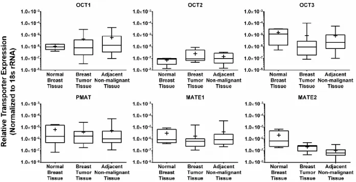
Gene Expression of Cation-selective Transporters in Human Breast Tissues. The expression of OCT1, OCT2, OCT3, PMAT, MATE1 and MATE2 genes was assessed by RT-PCR in human breast tumor tissues, their corresponding adjacent non-malignant tissues, and in normal breast tissues obtained from mammoplasty surgeries. OCT3 and PMAT were the
predominant transporter genes expressed in all three tissue types (Figure 2.2), with lower expression of OCT1 and MATE1 genes, and negligible expression of the OCT2 gene. MATE2 gene expression was negligible in breast tumor tissues, and was low in normal breast tissues and in tissues adjacent to breast tumors. The expression of OCT1, OCT3, PMAT and MATE1 genes was down regulated in all 15 pairs of breast tumor tissues analyzed compared to the corresponding adjacent non-malignant tissues, although this decrease was not statistically significant. No comparison was made between OCT2 and MATE2 gene expression in breast tumor tissues and their corresponding adjacent non-malignant tissues as these transporters were below detectable levels in several tissues examined.
Metformin Uptake in Human Breast Cancer Cell Lines. [14C]Metformin (50 µM) uptake was first assessed in the low transporter-expressing MCF-7 cell line, a widely used in vitro model for breast cancer. Uptake was inefficient, and was not inhibited by the pan cation-selective