THE ROLE OF BRAIN INTERLEUKIN-1 IN STRESS-ENHANCED FEAR LEARNING
Meghan E. Jones
A thesis submitted to the faculty of the University of North Carolina at Chapel Hill in partial fulfillment of the requirements for the degree of Master of Arts in the Department of
Psychology (Behavioral Neuroscience).
Chapel Hill 2014
Approved by:
iii ABSTRACT
MEGHAN E JONES: The role of brain interleukin-1 in stress-enhanced fear learning (Under the direction of Donald T. Lysle)
Post-Traumatic Stress Disorder (PTSD) has been shown to be associated with pro-inflammatory markers, including elevated plasma levels of interleukin-1β (IL-1β). However, the precise role of neuroinflammation and central immune signaling on the development of this debilitating psychological disorder is not known. Here, we employed stress-enhanced fear learning, an animal model of the disorder, to examine the role of central IL-1β in PTSD. The results show that the severe stressor in SEFL induces a time-dependent increase in IL-1β immunoreactivity and mRNA expression within the dentate gyrus of the dorsal hippocampus. There was no increase in IL-1β in the basolateral amygdala or the perirhinal cortex.
TABLE OF CONTENTS
LIST OF FIGURES………...………....v
Chapter I INTRODUCTION……….….…...1
II METHODS………..….…...…...4
Animals……….…….….………..…...…..4
Experiment 1………..……….………...…4
. Experiment 2………..………...………..…5
Experiment 3………....……….…...……...….6
Statistical Analysis………...….…...………..……...…...16
III RESULTS……….……….…………...…....……...…10
Experiment 1………..………..………...…………...…..10
Experiment 2………...……….……….……...…..11
Experiment 3……...……….………...…….11
IV DISCUSSION………..……...….13
LIST OF FIGURES
Figure
CHAPTER I INTRODUCTION
Psychopathology and disease states involving depression and anxiety have been associated with pro-inflammatory markers in both human populations and preclinical rodent models (Spivak, Shohat et al. 1997; Gill, Saligan et al. 2009; Stepanichev, Dygalo et al. 2014). However, the precise role of neuroinflammation and immune signaling in this context remains unclear. Cytokines, traditionally known for their role in the periphery in defense against infection and disease, have recently been revealed as important signaling molecules in neuro-glial communication pathways. For example, transgenic mouse lines deficient in central signaling involving pro-inflammtaory cytokines, such as interleukin-1β (IL-1β) or tumor necrosis factor-α (TNF-α), consistently exhibit altered anxiety behavior in the elevated plus maze or open field test. (Silverman, Macdougall et al. 2007; Koo and Duman 2009). Further, an infusion of IL-1β into the lateral ventricle is sufficient to induce anxiety like behavior in the elevated plus maze (Koo and Duman 2009). Thus, the immune system has the capability to modulate central signaling and play an important role in behavior and the
development of psychopathology.
and insufficient amounts of IL-1β impair memory formation (Goshen, Kreisel et al. 2007). Further, converging evidence suggests that many of the neurobiological parallels between a disease state and a state of stress, both of which are often characterized by
sickness behavior-like phenotypes, may be explained by IL-1β signaling (Maier 2003). There is evidence that IL-1β is up regulated throughout the brain after stress exposure (Nguyen, Deak et al. 1998; O'Connor, Johnson et al. 2003) and is important in stress-induced Hypothalamic-Pituitary-Adrenal (HPA) axis regulatory responses (Goshen, Kreisel et al. 2008).
The overall goal of the present study was to examine the role of brain IL-1β in the development of stress-enhanced fear learning (SEFL), an animal model of Post-Traumatic Stress Disorder (PTSD) developed by Fanselow and colleagues (Rau, DeCola et al. 2005). In PTSD, a severe traumatic event leads to debilitating psychological and physiological
consequences characterized by chronic or exaggerated fear and anxiety (Gill, Saligan et al. 2009). PTSD affects 7.7 million Americans (NIMH), including up to 17% of combat veterans (Richardson, Frueh et al. 2010). However, there is no consistently effective treatment. While several animal models of PTSD have been developed (van der Kolk 1987; Yamamoto, Morinobu et al. 2009; Kaouane, Porte et al. 2012), the stress-enhanced fear learning
3
The present studies test the hypothesis that the severe stressor in SEFL is capable of inducing alterations in IL-1β expression in the brain and that this expression is functionally relevant to the development of enhanced fear learning. In Experiment 1, we examined the time course of IL-1β expression following the severe stressor in SEFL. Our analysis focused on the dorsal hippocampus (DH, including the dentate gyrus (DG), CA1, and CA3),
basolateral amygdala (BLA), and perirhinal cortex (PrhC) - three regions that have been shown to be critical to fear conditioning (Anagnostaras, Gale et al. 2001; Kim, Loucks et al. 2011; Acheson, Gresack et al. 2012; Kent and Brown 2012). In Experiment 2, we tested whether blocking central IL-1β signaling with an infusion of interleukin-1 receptor
antagonist (IL-1ra) in the 48 hours following the severe stressor prevented the development of SEFL. In Experiment 3 we tested whether morphine administration attenuated the induction of IL-1β following the severe stressor in SEFL. This third experiment is based upon prior studies in our laboratory using this model showing that morphine administered after the severe stressor is effective in preventing the development of SEFL (Szczytkowski-Thomson, Lebonville et al. 2013). Moreover, clinical studies also show that morphine administration in the hours after a combat injury or single event trauma requiring
hospitalization was associated with a reduced risk of PTSD (Bryant, Creamer et al. 2009; Stoddard, Sorrentino et al. 2009; Holbrook, Galarneau et al. 2010; Nixon, Nehmy et al. 2010; Melcer, Walker et al. 2014). Thus, in this third experiment, we hypothesize that IL-1β
CHAPTER II
METHODS AND MATERIALS METHODS
Animals
Male Sprague-Dawley rats (250-400g, Charles River Laboratories, Raleigh, NC) were housed individually under a reversed light-dark (12-hour) cycle with ad libitum access to food and water. Animals were handled regularly during a one week acclimation period prior to experimentation. All procedures were conducted in accordance with and approval by the UNC Institutional Animal Care and Use Committee.
Experiment 1 - Analysis of IL-1β immunoreactivity and mRNA expression following the severe stressor in SEFL
from mere exposure to a novel context would be evident after 24 hours (Goshen and Yirmiya 2009). All other animals were exposed to Context A and sacrificed via transcardial perfusion at the appropriate times.
In Experiment 1b brain tissue was processed for quantitative polymerase chain reaction (qPCR) analysis of mRNA. These animals (N = 32, n = 8) were assigned to either a
no foot shock group (NS Control) or a foot shock group and one of three time points, 0, 24, or 48 hours after the stressor. Again, animals in the NS control group were sacrificed 24 hours after Context A exposure and all other animals were sacrificed via cervical dislocation at the appropriate times.
Experiment 2 - Effect of a central infusion of IL-1ra on the development of SEFL Surgical Procedures
Animals were surgically implanted with intracerebroventricular (i.c.v.) cannulae. Animals were anesthetized with a 1.0 mg/kg intraperitoneal injection of 9:1 (vol:vol)
ketamine hydrochloride (100 mg/ml) mixed with xylazine (100 mg/ml). Guide cannulae (26 Gauge, Plastics One, Roanoke, VA) were directed at the right lateral ventricle (AP -0.9 mm, ML -1.5 mm, DV -3.4 mm, relative to bregma). Animals were given three weeks for post-operative recovery. Correct placements were verified and animals with incorrect placement were dropped from the analysis.
Procedures
Animals (N = 36, n = 9) were assigned to a Context A treatment (foot shock or no foot shock) and a drug treatment (IL-1ra or vehicle) and exposed to the SEFL paradigm. The first day of the SEFL paradigm is the foot shock treatment in Context A, as described in
6
and auditory characteristics, as described previously (Szczytkowski-Thomson, Lebonville et al. 2013). Context B was set-up to record the animals’ behavior using a video recording system (Sony Video Camera Model HDR-CX150). Seven days after Context A exposure, animals were placed into Context B for 30 minutes. On Day 8, animals were placed into Context B for a single scrambled foot shock (1mA, 1s) at 3 minutes, 12 seconds. On days 9, 10, 15 and 23 (Test Days 1, 2, 7 and 14), animals were placed in Context B for 8 minutes, 32 seconds and behavior was recorded to measure freezing behavior, a measure of learned fear, defined as a lack of all movement except that required for breathing. No animals in any group demonstrated freezing behavior to Context B during habituation/prior to the single foot shock, suggesting that there was no generalization of fear between contexts. Thus, any
differences observed between treatment groups would reflect altered learning to the single foot shock in Context B. Videos were analyzed by raters blind to treatment condition.
IL-1ra (GenScript, Piscataway, NJ) was reconstituted in sterile saline (2.5 µg/µl). Twenty-four and 48 hours after removal from Context A, animals were microinfused with 4 μl of either IL-1ra (10 µg) or sterile saline vehicle at a rate of 2 µl/min. Injectors were left in place for 4 minutes to allow for diffusion. These time points were based on our earlier finding that morphine administration prevents the development of SEFL when administered 48 hours after, but not immediately after, Context A (Szczytkowski-Thomson, Lebonville et al. 2013).
Experiment 3 - Analysis of IL-1β immunoreactivity following the severe stressor of SEFL in combination with morphine treatment
7
obtained from the National Institute on Drug Abuse (NIDA) and dissolved in sterile saline (7.5 mg/ml). Immediately, 24, and 48 hours after removal from Context A, animals were administered either 7.5 mg/kg morphine or saline vehicle subcutaneously. One hour after the final injection, animals were sacrificed via transcardial perfusion.
Immunohistochemical Analysis
In Experiments 1a and 3, animals were deeply anesthetized with 9:1 (vol:vol) ketamine hydrochloride (100 mg/ml) mixed with xylazine (100 mg/ml) and transcardially perfused with cold phosphate buffer (PB; pH = 7.4) followed by 4% paraformaldehyde in 0.1 M PB. Brains were placed in 30% sucrose for cryoprotection and sliced into 40 µm sections. Tissue sections were washed for 15 minutes with 0.1 M PB, and incubated for 30 minutes at 80°C in sodium citrate (pH = 8.5) for antigen retrieval. Subsequently, sections were
incubated for 30 minutes with streptavidin and biotin blocks (Vector Laboratories,
Burlingame, CA, USA) and pre-incubated for 60 minutes with 3% normal goat serum and 0.3% Triton X-100 in 0.1 M PB. Sections were incubated overnight with rabbit anti-IL-1β (1:1000; Abcam, Cambridge, Ma) in 0.1 M PB with 3% normal goat serum and 0.3%
Tween20. Similar to Johnson & Kan (Johnson and Kan 2010), sections were then incubated for 60 minutes with biotinylated goat anti-rabbit IgG (1:1000, Vector Laboratories,
Burlingame, CA) in 1% normal goat serum and a streptavidin- Alexafluor488-conjugated tertiary antibody (1:1000, Life Technologies, Grand Island, NY) was used for visualization. Tissue sections labeled with only secondary and tertiary antibodies were used as secondary controls to ensure specificity of our primary antibody. Sections were mounted onto
8
Color images were captured through a digital camera (Roper Scientific), mounted on an optical microscope (BX-51, Olympus), and positive fluorescence was quantified using ImageJ (NIH). Images captured were between -2.76 mm and -4.2 mm relative to bregma (Paxinos and Watson, 1998). Three to six sections were analyzed per animal for each brain region and values were averaged and expressed as positive stain per 5 µm2. Images were normalized to background fluorescence using a manual thresholding procedure such that the binary overlay completely covered all positive stain (similar to (Sugama, Takenouchi et al. 2011)). In addition, the number of IL-1β-positive cells in the all images taken from the dentate gyrus of the DH was counted manually. Images with high background that resulted from poor perfusion were dropped from the analysis. This decision was made at the time of thresholding and was made blind to the treatment condition. All thresholding and counting was conducted blind to the treatment condition. Publication images were compiled with Adobe Photoshop CS software. Color levels and background were reduced for optimal
representation with levels and curves tools. Images from all experimental groups were treated equally.
Quantitative Polymerase Chain Reaction (qPCR) analysis
9
nucleic acid workstation (PE Biosystems) according to the manufacturer’s protocol. Real-time RT-PCR reactions were performed in the ABI Prism 7700 sequence detector (PE Biosystems) in a total volume of 30 µl (10 µl RNA, 20 µl reaction mixture). Each qPCR amplification was performed in duplicate: 30 min at 48°C for the RT reaction and 10 min at 94°C followed by 40 temperature cycles (15 sec at 94°C and 1 min at 60°C). Signal intensity was normalized to β-actin as an endogenous control. The nucleotide sequences of the PCR primers and fluorogenic probes used for the IL-1β and β-actin genes were as follows: IL-1β forward: 5’-GCC TCA AGG GGA AGA ATC TA-3’, reverse: 5’-ATC CAC ACT CTC CAG CTG C-3’, probe: 5’-FCT GTG TAA TGA AAG ACG GCA CAC CCA CQ-3’; β-actin forward: 5’-TGC CTG ACG GTC AGG TCA-3’, reverse: 5’-CAG GAA GGA AGG CTG GAA G-3’, probe: 5’-FCA CTA TCG GCA ATG AGC GGT TCC GQ-3’.
Statistical Analysis
For Experiments 1a and 1b, positive fluorescence and cell counts were analyzed using a one-way ANOVA with treatment group as the between subjects factor. For Experiment 2, a 2 x 2 ANOVA with Context A treatment and drug treatment as between subjects factors was used to analyze baseline freezing behavior. A 2 x 2 x 4 ANOVA with Context A treatment and drug treatment as between subjects factors and test day as a within subjects factor was used to analyze freezing behavior across test days. Lastly, for Experiment 3, positive
CHAPTER III RESULTS RESULTS
Experiment 1: Severe stressor enhances IL-1β immunoreactivity and mRNA expression in the dorsal hippocampus but not basolateral amygdala or perirhinal cortex
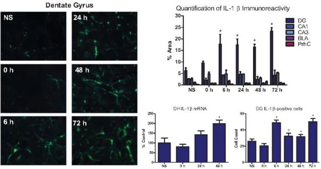
In Experiment 1a, IL-1β immunoreactivity was significantly enhanced by exposure to the severe stressor in a time-sensitive manner such that an increase was observed beginning 6 hours after the severe stressor and persisted through 72 hours (Figure 1). Severe stress
significantly enhanced IL-1β immunoreactivity in the dentate gyrus (DG) both in terms of the percent area of positive fluorescence, F (5, 25) = 6.472, p < .01, and in terms of
IL-1β-positive cell counts, F (5, 16) = 13.544, p < .01 . LSD post hoc tests revealed that compared to the no foot shock group, IL-1β expression was significantly enhanced at 6, 24, 48, and 72 hours (p < .01). In contrast, IL-1β expression in CA1, CA3, the BLA and the PrhC was not altered by the severe stressor at any of the time points (Figure 1).
In Experiment 1b, exposure to the severe stressor significantly enhanced IL-1β mRNA expression in the dorsal hippocampus in a time-dependent manner, F (3, 26) = 6.927,
the DH since the CA1/3 did not achieve significance. Still, Experiment 1b further confirms an increase in IL-1β on a delayed timeline in the DH following foot shock. Experiment 2: IL-1ra prevents the development of stress-enhanced fear learning
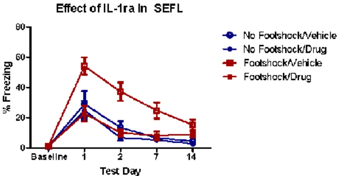
There was no effect of Context A treatment or drug treatment on baseline freezing in Context B, indicating no generalization of fear between contexts, F (3, 38) = 1.91, p > .05. A 2 x 2 x 4 ANOVA revealed a significant main effect of Context A treatment, F (1, 36) = 11.08, p < .01, and a significant main effect of drug treatment, F (1, 36) = 12.99, p < .01. There was also a significant effect of test day, indicating that conditioned fear diminished over time, F (3, 34) = 21.76, p < .01. Most interestingly, there was a significant Context A treatment by drug treatment interaction, F (1, 36) = 6.497, p < .05. LSD post hoc
comparisons showed that the animals that received foot shock followed by vehicle exhibited significantly more freezing behavior compared to animals that received no foot shock in Context A followed by vehicle (p < .01), confirming a significant stress-enhanced fear
learning effect. The animals that received foot shock in Context A followed by IL-1ra and the animals that received no foot shock in Context A followed by IL-1ra did not differ in
freezing behavior exhibited in Context B, p > .05. Thus, these results show that stress-enhanced fear learning was prevented by an i.c.v. administration of IL-1ra (Figure 2). Experiment 3: Morphine administration attenuates severe stressor-induced IL-1β immunoreactivity in the dorsal hippocampus
12
Context A treatment by morphine treatment interaction, F (1, 19) = 31.050, p < .01. LSD post hoc comparisons revealed that animals that received foot shock in Context A followed by vehicle treatment exhibited significantly enhanced IL-1β expression compared to animals that received no foot shock in Context A followed by vehicle treatment (p < .001), replicating the effect observed in Experiment 1a. Animals that received no foot shock in Context A followed by morphine treatment were not significantly different from animals that received no foot shock in Context A followed by vehicle. However, animals that received foot shock in Context A followed by the morphine treatment exhibited significantly less IL-1β
expression compared to the group that received foot shock in Context A followed by vehicle,
CHAPTER IV DISCUSSION
The current studies demonstrate for the first time that exposure to a severe stressor that is capable of producing a PTSD-like phenotype in rats induces a time-dependent increase in IL-1β in the DH. Furthermore, central administration of IL-1ra prevents subsequent effects of that stressor, namely, the development of SEFL. Previously our laboratory established that morphine following the severe stressor also blocks the development of SEFL (Szczytkowski-Thomson, Lebonville et al. 2013). Here, we show that the same systemic morphine treatment also attenuates stress-induced IL-1β expression in the DH. These studies suggest that
morphine may act through an IL-1β-dependent mechanism to alter SEFL. Collectively, the experiments provide new evidence that the expression of IL-1β in the brain following a severe stressor is a critical component of the development of SEFL, a model of PTSD.
In the present study, IL-1β was detected in three brain regions known to be important in PTSD and fear acquisition and expression—the hippocampus, amygdala, and perirhinal cortex (Kim, Loucks et al. 2011; Acheson, Gresack et al. 2012). These structures are
hippocampus is thought to be important in the emotional and affective component of memory (Fanselow and Dong 2010) but it is not known what role IL-1 might have in the ventral hippocampus.
Several cell types are capable of producing IL-1β, and both neurons and glia express IL-1 receptors (Yabuuchi, Minami et al. 1994; Zhang, Sun et al. 2010). In astrocytes, IL-1β initiates the formation of IL-1β signaling intermediates, such as IL-1 receptor-associated kinase (IRAK; (Ringwood and Li 2008; Flannery and Bowie 2010)) and the subsequent activation of p38, extracellular signal-regulated kinase (ERK) and nuclear factor kappa-light-chain-enhancer of activated B cells (NFkB; (Guasch, Blanco et al. 2007; Huang, Smith et al. 2011)). Conversely, in hippocampal neurons, IL-1β activates p38 and ERK, but not NFkB (Huang, Smith et al. 2011). It should also be acknowledged that IL-1ra blocks multiple forms of IL-1 including IL-1α as well as IL-1β. The relative contributions of these different forms of IL-1 should be considered when determining the cellular sources of IL-1.
15
number of studies indicating the cytokines are involved in both short- and long-term changes in synaptic plasticity.
Physiological evidence that cytokines influence synaptic plasticity is also supported by behavioral evidence that central cytokine signaling influences learning in several
paradigms, including fear conditioning (Yirmiya and Goshen 2011; Szczytkowski, Lebonville et al. 2013). For example, acute administration of either TNF-α or IL-1β and chronic administration of interleukin-6 (IL-6) enhanced memory performance in a passive avoidance paradigm (Matsuda, Wen et al. 1996; Yirmiya, Winocur et al. 2002; Brennan, Beck et al. 2004). Furthermore, there is evidence for an effect of IL-1ra in step-down passive avoidance learning, but some report memory improvement (Depino, Alonso et al. 2004) while others have reported memory impairment(Yirmiya, Winocur et al. 2002). Early studies have also shown that morphine administration, which we show here to reduce hippocampal IL-1β, reduces memory for step down avoidance(Izquierdo 1979). Thus, it is clear that IL-1β can alter learning and memory, but the direction of the effects may depend upon such factors as the timing of the administration, the severity of stressor, and the type of learning being examined. The present findings are unique in that they begin to address the role of IL-1β in the neural plastic changes required to alter future learning, i.e., SEFL.
The present findings suggest that key neurobiological changes that render the animal hypersensitive to future fear learning occur on a delayed timeline, in concert with the
16
immediately, after severe stress significantly reduced the prevalence of the extreme
behavioral response in the elevated plus maze and the acoustic startle response test, which is thought to reflect PTSD-like behavior (Kozlovsky, Zohar et al. 2012). This is consistent with prior data from our laboratory showing that the 48 hour time point is key (Szczytkowski-Thomson, Lebonville et al. 2013). Interestingly, the effect of morphine on the prognosis of PTSD in combat veterans was also time-dependent such that the relationship between morphine administration and a reduced likelihood of PTSD was strongest when it was administered in level 2 care, which occurs not immediately on the battlefield but within 72 hours of the initial trauma (Melcer, Walker et al. 2014). Frankland and Bontempi suggested a model of memory consolidation in which components are integrated in the hippocampus into a single memory trace which, over time, becomes consolidated to cortical structures and integrated with other experiences (termed systems consolidation) (Frankland and Bontempi 2005). One hypothesis is that SEFL (and PTSD) is (are) driven by an alteration in the later phases of memory consolidation following the trauma that results in the loss of contextual detail and renders the animal hypersensitive to future fear learning.
The present studies do not test whether morphine or IL-1ra impair contextual fear learning to Context A. We cannot assume that manipulations that attenuate SEFL, such as IL-1ra or morphine, also impair memory for Context A. Fanselow and colleagues (2005)
showed that contextual fear learning to the context where the initial severe shock occurs is not critical to the expression of SEFL. They found that eliminating fear learning to Context A and extinction of Context A did not affect the expression of SEFL in Context B (Rau,
17
influences fear learning to Context A and further whether IL-1β expression is directly involved in the learning/enhanced learning to the subsequent foot shock in Context B.
18
19
Figure 2. IL-1ra prevents the development of SEFL. IL-1ra infusion (i.c.v., 10 µg at 24 and
20
Figure 3. Morphine attenuates stress-induced IL-1β in the dorsal hippocampus.
Representative images (20x) of IL-1β (green) in the dentate gyrus of the dorsal hippocampus from animals in each of the four groups are shown in (a). ImageJ analysis of positive
fluorescence stain (b) revealed that in vehicle treated animals (NS/Veh and Shock/Veh), foot shock significantly enhanced IL-1β; however, morphine administration suppressed the induction of IL-1β in animals that received foot shock followed by morphine treatment (Shock/M). IL-1β- positive cells were also counted in the dentate gyrus of the dorsal hippocampus (c) and the same pattern was confirmed. (* greater than NS/Veh, p < .05; †
21
WORKS CITED
1. Acheson, D. T., J. E. Gresack, et al. (2012). "Hippocampal dysfunction effects on context memory: possible etiology for posttraumatic stress disorder."
Neuropharmacology 62(2): 674-685.
2. Anagnostaras, S. G., G. D. Gale, et al. (2001). "Hippocampus and contextual fear conditioning: recent controversies and advances." Hippocampus 11(1): 8-17.
3. Avital, A., I. Goshen, et al. (2003). "Impaired interleukin-1 signaling is associated with deficits in hippocampal memory processes and neural plasticity." Hippocampus
13(7): 826-834.
4. Bai, Y. M., T. P. Su, et al. (2014). "Comparison of inflammatory cytokine levels among type I/type II and manic/hypomanic/euthymic/depressive states of bipolar disorder." J Affect Disord 166: 187-192.
5. Brennan, F. X., K. D. Beck, et al. (2004). "Proinflammatory cytokines differentially affect leverpress avoidance acquisition in rats." Behav Brain Res 153(2): 351-355.
6. Bryant, R. A., M. Creamer, et al. (2009). "A study of the protective function of acute morphine administration on subsequent posttraumatic stress disorder." Biol
Psychiatry 65(5): 438-440.
7. Dantzer, R. (2001). "Cytokine-induced sickness behavior: where do we stand?" Brain Behav Immun 15(1): 7-24.
8. Depino, A. M., M. Alonso, et al. (2004). "Learning modulation by endogenous hippocampal IL-1: blockade of endogenous IL-1 facilitates memory formation."
Hippocampus 14(4): 526-535.
9. Fanselow, M. S. and H. W. Dong (2010). "Are the dorsal and ventral hippocampus functionally distinct structures?" Neuron 65(1): 7-19.
10. Flannery, S. and A. G. Bowie (2010). "The interleukin-1 receptor-associated kinases: Critical regulators of innate immune signalling." Biochemical Pharmacology 80(12): 1981-1991.
22
12. Gill, J. M., L. Saligan, et al. (2009). "PTSD is associated with an excess of inflammatory immune activities." Perspect Psychiatr Care 45(4): 262-277.
13. Goshen, I., T. Kreisel, et al. (2008). "Brain interleukin-1 mediates chronic stress-induced depression in mice via adrenocortical activation and hippocampal neurogenesis suppression." Mol Psychiatry 13(7): 717-728.
14. Goshen, I., T. Kreisel, et al. (2007). "A dual role for interleukin-1 in hippocampal-dependent memory processes." Psychoneuroendocrinology 32(8-10): 1106-1115.
15. Goshen, I. and R. Yirmiya (2009). "Interleukin-1 (IL-1): a central regulator of stress responses." Front Neuroendocrinol 30(1): 30-45.
16. Guasch, R. M., A. M. Blanco, et al. (2007). "RhoE participates in the stimulation of the inflammatory response induced by ethanol in astrocytes." Exp Cell Res 313(17): 3779-3788.
17. Holbrook, T. L., M. R. Galarneau, et al. (2010). "Morphine use after combat injury in Iraq and post-traumatic stress disorder." N Engl J Med 362(2): 110-117.
18. Huang, Y., D. E. Smith, et al. (2011). "Neuron-specific effects of interleukin-1beta are mediated by a novel isoform of the IL-1 receptor accessory protein." J Neurosci
31(49): 18048-18059.
19. Izquierdo, I. (1979). "Effect of naloxone and morphine on various forms of memory in the rat: possible role of engogenous opiate mechanisms in memory consolidation."
Psychopharmacology (Berl) 66(2): 199-203.
20. Johnson, E. A. and R. K. Kan (2010). "The acute phase response and soman-induced status epilepticus: temporal, regional and cellular changes in rat brain cytokine concentrations." J Neuroinflammation 7: 40.
21. Kaouane, N., Y. Porte, et al. (2012). "Glucocorticoids can induce PTSD-like memory impairments in mice." Science 335(6075): 1510-1513.
23
23. Kim, H. S., G. Lee, et al. (2002). "Molecular phenotyping for analyzing subtle genetic effects in mice: application to an angiotensinogen gene titration." Proc Natl Acad Sci U S A 99(7): 4602-4607.
24. Kim, M. J., R. A. Loucks, et al. (2011). "The structural and functional connectivity of the amygdala: from normal emotion to pathological anxiety." Behav Brain Res
223(2): 403-410.
25. Koo, J. W. and R. S. Duman (2009). "Interleukin-1 receptor null mutant mice show decreased anxiety-like behavior and enhanced fear memory." Neurosci Lett 456(1): 39-43.
26. Kozlovsky, N., J. Zohar, et al. (2012). "Microinfusion of a corticotrophin-releasing hormone receptor 1 antisense oligodeoxynucleotide into the dorsal hippocampus attenuates stress responses at specific times after stress exposure." J Neuroendocrinol
24(3): 489-503.
27. Maier, S. F. (2003). "Bi-directional immune-brain communication: Implications for understanding stress, pain, and cognition." Brain Behav Immun 17(2): 69-85.
28. Matsuda, S., T. C. Wen, et al. (1996). "Interleukin-6 prevents ischemia-induced learning disability and neuronal and synaptic loss in gerbils." Neurosci Lett 204(1-2): 109-112.
29. Melcer, T., J. Walker, et al. (2014). "Glasgow Coma Scores, early opioids, and posttraumatic stress disorder among combat amputees." J Trauma Stress 27(2): 152-159.
30. Nguyen, K. T., T. Deak, et al. (1998). "Exposure to acute stress induces brain interleukin-1beta protein in the rat." J Neurosci 18(6): 2239-2246.
31. Nixon, R. D., T. J. Nehmy, et al. (2010). "Predictors of posttraumatic stress in children following injury: The influence of appraisals, heart rate, and morphine use."
Behav Res Ther 48(8): 810-815.
32. O'Connor, K. A., J. D. Johnson, et al. (2003). "Peripheral and central
24
33. Rau, V., J. P. DeCola, et al. (2005). "Stress-induced enhancement of fear learning: an animal model of posttraumatic stress disorder." Neurosci Biobehav Rev 29(8): 1207-1223.
34. Richardson, L. K., B. C. Frueh, et al. (2010). "Prevalence estimates of combat-related post-traumatic stress disorder: critical review." Aust N Z J Psychiatry 44(1): 4-19.
35. Ringwood, L. and L. Li (2008). "The involvement of the interleukin-1 receptor-associated kinases (IRAKs) in cellular signaling networks controlling inflammation."
Cytokine 42(1): 1-7.
36. Schneider, H., F. Pitossi, et al. (1998). "A neuromodulatory role of interleukin-1beta in the hippocampus." Proc Natl Acad Sci U S A 95(13): 7778-7783.
37. Silverman, M. N., M. G. Macdougall, et al. (2007). "Endogenous glucocorticoids protect against TNF-alpha-induced increases in anxiety-like behavior in virally infected mice." Mol Psychiatry 12(4): 408-417.
38. Spivak, B., B. Shohat, et al. (1997). "Elevated levels of serum interleukin-1 beta in combat-related posttraumatic stress disorder." Biol Psychiatry 42(5): 345-348.
39. Stepanichev, M., N. N. Dygalo, et al. (2014). "Rodent Models of Depression: Neurotrophic and Neuroinflammatory Biomarkers." Biomed Res Int 2014: 932757.
40. Stoddard, F. J., Jr., E. A. Sorrentino, et al. (2009). "Preliminary evidence for the effects of morphine on posttraumatic stress disorder symptoms in one- to four-year-olds with burns." J Burn Care Res 30(5): 836-843.
41. Sugama, S., T. Takenouchi, et al. (2011). "Immunological responses of astroglia in the rat brain under acute stress: interleukin 1 beta co-localized in astroglia."
Neuroscience 192: 429-437.
42. Szczytkowski-Thomson, J. L., C. L. Lebonville, et al. (2013). "Morphine prevents the development of stress-enhanced fear learning." Pharmacol Biochem Behav 103(3): 672-677.
43. Szczytkowski, J. L., C. Lebonville, et al. (2013). "Heroin-induced conditioned immunomodulation requires expression of IL-1beta in the dorsal hippocampus."
25
44. Tancredi, V., G. D'Arcangelo, et al. (1992). "Tumor necrosis factor alters synaptic transmission in rat hippocampal slices." Neurosci Lett 146(2): 176-178.
45. van der Kolk, B. A. (1987). "The drug treatment of post-traumatic stress disorder." J Affect Disord 13(2): 203-213.
46. Yabuuchi, K., M. Minami, et al. (1994). "Localization of Type-I Interleukin-1 Receptor Messenger-Rna in the Rat-Brain." Molecular Brain Research 27(1): 27-36.
47. Yamamoto, S., S. Morinobu, et al. (2009). "Single Prolonged Stress: Toward an Animal Model of Posttraumatic Stress Disorder." Depression and Anxiety 26(12): 1110-1117.
48. Yirmiya, R. and I. Goshen (2011). "Immune modulation of learning, memory, neural plasticity and neurogenesis." Brain Behav Immun 25(2): 181-213.
49. Yirmiya, R., G. Winocur, et al. (2002). "Brain interleukin-1 is involved in spatial memory and passive avoidance conditioning." Neurobiol Learn Mem 78(2): 379-389.
50. Zhang, R., L. Sun, et al. (2010). "Acute p38-mediated inhibition of NMDA-induced outward currents in hippocampal CA1 neurons by interleukin-1beta." Neurobiol Dis