Received: August 12, 2015.
Accepted: January 22, 2016.
Pre-published: January 27, 2016.
©2016 Ferrata Storti Foundation
Check the online version for the most updated information on this article, online supplements, and information on authorship & disclosures: www.haematologica.org/content/101/5/607
Material published in Haematologica is cov-ered by copyright. All rights reserved to the Ferrata Storti Foundation. Copies of articles are allowed for personal or internal use. Permission in writing from the publisher is required for any other use.
Correspondence:
[email protected]
Association of acute myeloid leukemia’s
most immature phenotype with risk groups
and outcomes
Jonathan M. Gerber,1Joshua F. Zeidner,2Sarah Morse,3Amanda L. Blackford,3
Brandy Perkins, Breann Yanagisawa,3Hao Zhang,3Laura Morsberger,3Judith
Karp,3Yi Ning,3Christopher D. Gocke,3Gary L. Rosner,3B. Douglas Smith,3and
Richard J. Jones3
1Levine Cancer Institute, Charlotte, NC; 2Lineberger Comprehensive Cancer Center,
University of North Carolina, Chapel Hill, NC; and 3The Sidney Kimmel Comprehensive
Cancer Center at Johns Hopkins, Johns Hopkins University, Baltimore, MD, USA
*JMG and JFZ contrbuted equally to this work.
Ferrata Storti Foundation
EUROPEAN HEMATOLOGY ASSOCIATION
Haematologica
2016
Volume 101(5):607-616
doi:10.3324/haematol.2015.135194
T
he precise phenotype and biology of acute myeloid leukemia stem
cells remain controversial, in part because the “gold standard”
immunodeficient mouse engraftment assay fails in a significant
frac-tion of patients and identifies multiple cell-types in others. We sought to
analyze the clinical utility of a novel assay for putative leukemia stem cells
in a large prospective cohort. The leukemic clone’s most primitive
hematopoietic cellular phenotype was prospectively identified in 109
newly-diagnosed acute myeloid leukemia patients, and analyzed against
clinical risk groups and outcomes. Most (80/109) patients harbored
CD34
+CD38
–leukemia cells. The CD34
+CD38
–leukemia cells in 47 of the
80 patients displayed intermediate aldehyde dehydrogenase expression,
while normal CD34
+CD38
–hematopoietic stem cells expressed high levels
of aldehyde dehydrogenase. In the other 33/80 patients, the CD34
+CD38
–leukemia cells exhibited high aldehyde dehydrogenase activity, and most
(28/33, 85%) harbored poor-risk cytogenetics or FMS-like tyrosine kinase 3
internal tandem translocations. No CD34
+leukemia cells could be detected
in 28/109 patients, including 14/21 patients with nucleophosmin-1
muta-tions and 6/7 acute promyelocytic leukemia patients. The patients with
CD34
+CD38
–leukemia cells with high aldehyde dehydrogenase activity
manifested a significantly lower complete remission rate, as well as poorer
event-free and overall survivals. The leukemic clone’s most immature
phe-notype was heterogeneous with respect to CD34, CD38, and ALDH
expression, but correlated with acute myeloid leukemia risk groups and
outcomes. The strong clinical correlations suggest that the most immature
phenotype detectable in the leukemia might serve as a biomarker for
“clin-ically-relevant” leukemia stem cells. ClinicalTrials.gov:
NCT01349972
.
ABSTRACT
Introduction
More than two decades ago, Lapidot et al. reported that acute myeloid leukemia
(AML) cells capable of engrafting immunodeficient mice expressed a CD34+CD38–
nor-mal hematopoietic stem cell (HSC) phenotype.1These so-called leukemia stem cells
(LSCs) gave rise to partially differentiated progeny that constituted the bulk of the
leukemia, but possessed only limited proliferative potential.2More recently, leukemic
cells of varying surface phenotypes, even within the same patient, have been shown to be capable of engrafting immunodeficient mice, the generally accepted “gold standard”
for LSC activity.3,4 However, this traditional approach for LSC identification has proven
in a significant fraction of AML patients no leukemia cell
sub-set will engraft5-10 even using the newer mouse models.11 This
inability to confirm the identity of LSCs in many patients is at least in part the reason that the clinical relevance of LSCs
remains uncertain.12
Regardless of their tumorigenic potential in immunodefi-cient mice, leukemic cells that persist after therapy [i.e. min-imal residual disease (MRD)] are arguably the most clinically
important.13,14 We recently showed that MRD during
com-plete remission (CR) was enriched for CD34+CD38–leukemic
cells, and their presence after therapy was highly associated
with subsequent clinical relapse.13 Others found that
CD34+CD38–leukemia cell frequency correlated with
prog-nosis.7,15Thus, accumulating evidence now suggests that
ini-tial clinical responses likely reflect the behavior of the bulk leukemia, while long-term survival/cure requires the
eradica-tion of LSCs.7,13-15 We also showed that the leukemic
CD34+CD38– cells from most patients, particularly those
with core-binding factor (CBF) AMLs, could be separated from normal HSCs by their expression of aldehyde dehydro-genase 1 (ALDH). Normal HSCs exhibited high ALDH
expression (CD34+CD38–ALDHhigh), while the putative LSCs
expressed intermediate levels (CD34+CD38–ALDHint).13These
findings have recently been confirmed.16,17
Clinical outcomes in AML are highly diverse with some patients curable with standard therapies, others initially refractory to all known therapies, and the majority eventually relapsing and succumbing to the disease after initially achiev-ing CRs. While patient factors such as age and performance status may influence the heterogeneous outcomes, the underlying biology - currently best reflected by cytogenetic and molecular markers - is the major determinant. AML’s highly diverse biology suggests that the LSCs are also hetero-geneous. Accordingly, our previous report identified two patients, both primary refractory to induction, whose puta-tive LSCs demonstrated high ALDH expression
indistin-guishable from normal HSCs.13 We could not detect any
CD34+leukemia cells in two other patients.13 Other groups
have also described heterogeneous CD34 and ALDH
expres-sion in AMLs.8,16-20
Since no leukemia subset from many patients will engraft
immunodeficient mice,5-11 and no leukemic CD34+CD38–
cells can be identified in some patients,4,13,15,16,21other means
for LSC identification are needed to allow for their study
clin-ically.14 Based on our smaller study of mostly CBF AML
patients,13 we hypothesized that the most primitive
hematopoietic cell phenotype that could be found in leukemia cells might have important clinical relevance. Thus, we prospectively assessed the leukemia’s most immature phenotype in a multi-institutional randomized clinical trial comparing two induction therapies in patients lacking favor-able-risk cytogenetics: standard cytarabine-based “7+3"
ther-apy22 and a novel regimen called FLAM (flavopiridol,
cytara-bine, mitoxantrone).23,24 To fully assess heterogeneity of the
leukemic clone’s most immature phenotype, we also studied patients who initially agreed to the trial but were ultimately ineligible because they were found to have favorable-risk cytogenetics. Here we find that the most primitive hematopoietic cellular phenotype present in leukemia cells is not only heterogeneous for CD34, CD38, and ALDH expres-sion, but also that this phenotypic heterogeneity correlates with both AML risk groups and outcomes. Moreover, the robust clinical correlations suggest that the most immature phenotype detectable in the leukemia might serve as a bio-marker for “clinically-relevant” LSCs.
Methods
Patients
Patients aged 18-70 with newly-diagnosed AML, excluding CBF AMLs and APL, were eligible for this multicenter clinical study ( clin-icaltrials.gov NCT01349972).24 Patients were randomized 2:1 to
FLAM or the standard “7+3” regimens, respectively.24 Patients who
achieved complete or partial responses to the first cycle were eligible to receive a second cycle of FLAM or high-dose cytarabine (HiDAC), and/or could undergo allogeneic bone marrow transplantation (alloBMT) as per physician discretion. Johns Hopkins patients who were study ineligible because their cytogenetics proved favorable were also included in this analysis. Informed consent for participa-tion in NCT01349972, as well as for the bone marrow donaparticipa-tions by the patients not treated on trial, was obtained in accordance with the Declaration of Helsinki as approved by the Johns Hopkins Institutional Review Board.
Isolation of cells
Specimens were collected between April 2011 and April 2013. Marrow mononuclear (MMNC) and CD34+cell subsets were
iden-tified and isolated as previously described.13,25At least 500,000 cells
from each AML specimen were then stained with Aldefluor (Aldagen, Durham, NC, USA) to assess ALDH activity according to the manufacturer’s instructions utilizing diethylaminobenzaldehyde (DEAB) controls. Next, cells were labeled with monoclonal phyco-erythrin-conjugated anti-CD34 and allophycocyanin (APC)-conjugated anti-CD38 (BD Biosciences, San Jose, CA, USA) and analyzed with a MoFlo cell sorter (Beckman Coulter, Brea, CA, USA). Gating for CD34 and CD38 populations was based on clearly distinguishable populations, or in the absence of such, the negative antibody control.25 A representative example of gating is shown in
Online Supplementary Figure S1.
Fluorescence
in situ
hybridization (FISH) and molecular
analyses
For patients with cytogenetic abnormalities detectable by FISH, 250-1000 cell aliquots were sorted directly onto slides and fixed with 3:1 methanol-glacial acetic acid (Sigma-Aldrich, St. Louis, MO, USA). FISH was performed and analyzed by the Johns Hopkins Kimmel Cancer Center Cytogenetics Core, using probes specific for the patients’ known cytogenetic abnormalities per manufacturer’s guidelines (Abbot Molecular, Des Plaines, IL, USA) as we previously described.13Real-time polymerase chain reaction for FLT3 internal
tandem duplication (ITD) (qPCR) and NPM1 mutations (reverse transcriptase-qPCR) was performed by Johns Hopkins Molecular Hematopathology Laboratory.
Data analysis
The AML’s most immature phenotype was scored in a blinded fashion by RJJ, BP, and SM as we previously described.13 Any
differ-ences in scoring were to be decided by a simple majority, but there was complete concordance on all observations. The samples were then de-identified by the Johns Hopkins Kimmel Cancer Center Specimen Accessioning Core for statistical analysis. Clinical out-comes were determined by the NCT01349972 clinical study team24
blinded to the AML phenotypic data. Event-free survival (EFS) was defined as the date of treatment to the occurrence of persistent AML, relapse, or death. Poor-risk cytogenetics [> 3 clonal abnormal-ities, -5, 5q-, -7, -7q, t(3;3), inv 3, non-t(9;11) 11q23 excluding t(6;9), t(9;22)] and molecular abnormalities (FLT3-ITD mutation) were clas-sified according to the European LeukemiaNet reporting system.26
Statistical analysis
Fisher’s exact tests or T-tests, and for differences in outcome, strati-fied by treatment arm (FLAM vs.7+3), by Mantel-Haenszel tests. Overall survival (OS) and EFS were estimated using the Kaplan-Meier method. Differences in OS and EFS according to the leukemic clone’s most primitive hematopoietic cellular phenotypes were ana-lyzed with hazard ratios (HR) from Cox proportional hazards mod-els that adjust for treatment arm, and tested for significance using likelihood ratio tests. Analyses were completed using R version 3.1.1.27
Results
Patient characteristics
The leukemia clone’s most primitive hematopoietic cellu-lar phenotype was assessed in all patients entered in
NCT0134997224with adequate bone marrow specimens for
analyses. Of the 147 patients entered in the clinical trial, bone marrow samples from 98 patients were analyzed. The main reason for patients not being analyzed was the absence of a research sample because not enough cells could be obtained with the diagnostic marrow (43 patients). The specimen arrived in the laboratory but was not adequate for analysis in 4 patients, and consent for the laboratory study was with-drawn in 2 patients. Over the same time frame, seven patients with CBF AML and 14 with APL were newly diag-nosed and treated at Johns Hopkins. Bone marrow samples from 4 of the CBF patients and 7 of the APL patients were available for analysis. The clinical characteristics of the 98 patients on trial and the 11 favorable-risk patients not eligible for the trial are shown in Table 1.
The leukemia’s most immature phenotype was
heterogeneous
We defined the most immature phenotype present in the AML based on CD34, CD38, and ALDH expression, as we
previously described.25,28,29 As CD34+CD38–ALDHhigh
HSCs16,28,30 differentiate into more committed progenitors,
both CD34 and ALDH expression decrease while CD38
expression increases.29,31-33 Thus, CD34+CD38-ALDHint,
CD34+CD38+, and CD34-cells were considered increasingly
more differentiated phenotypes. The leukemic versusnormal
origin of the hematopoietic phenotypes was determined by cytogenetic (FISH) or molecular (FLT3-ITD or NPM1) mark-ers when present.
CD34+cells comprised a median of 12% (range 0.07 - 81%)
of total MMNCs from the 98 patients in NCT01349972. In
22/98 of the patients, the AML phenotype was clinically
determined to be CD34-by standard flow cytometry
crite-ria:7,16,34 i.e., CD34+ cells represented
< 1% of the MMNCs (Table 1, Online Supplementary Figure
S2A). In all 22 patients with < 1% CD34+ cells in the
MMNCs, the small fraction (mean + SEM - 0.52+0.08) of
CD34+cells was completely CD38-ALDHhigh(Table 2, Figure
1A), and displayed low forward (FSC) and side (SSC) scatter
on flow cytometry (data not shown). Only a small percentage
(2.2+1.6%) of the CD34+CD38-ALDHhighcells contained the
leukemia-specific marker present in the five CD34–
leukemias with cytogenetics detectable by FISH (Table 2).
Likewise, when an AML with < 1% CD34+ cells was
FLT3-ITD or NPM1-mutated (14/22 patients), the CD34+cells did
not harbor the mutation (Figure 1B).
CD34+cells comprised a mean of 25.3+3.1% of MMNCs
in the 76 patients from NCT01349972 who harbored CD34+
leukemia cells; the CD34+CD38–cells comprised 44.8+3.4%
of the CD34+cells in these patients. In 43 of these 76 patients,
the majority (65.1+3.4%) of CD34+CD38–cells were ALDHint
(Table 2, Online Supplementary Figure S2B), while the ALDHhigh
population represented 1.7+0.5% of the CD34+CD38–cells
(Figure 2A, Table 2). In the 11/43 cases with leukemia-specific cytogenetics scorable by FISH, we confirmed that the
CD34+CD38–ALDHint cells were predominantly leukemic
(Table 2). In contrast, the small number of
CD34+CD38–ALDHhighcells predominantly lacked the FISH
marker that characterized the leukemia (Table 2). Likewise,
when AMLs with prominent CD34+CD38–ALDHint
popula-tions exhibited FLT3-ITD mutapopula-tions (3 patients) or were
NPM1-mutated (4 cases), the CD34+CD38–ALDHint cells
exhibited the mutation while the CD34+CD38–ALDHhighcells
did not (Figure 2B). The CD34+CD38–ALDHhighHSCs
exhib-ited much lower FSC (data not shown) and SSC than the
CD34+CD38–ALDHintAML cells (Figure 2A). These data are
consistent with the CD34+CD38–ALDHhighcells representing
normal HSCs as we previously demonstrated.13 The 4 CBF
patients also displayed prominent leukemic CD34+CD38–
ALDHint populations harboring, and small CD34+CD38–
ALDHhighfractions lacking, the FISH abnormality (Tables 1, 2).
Only ALDHhighCD34+CD38–cells were present in 26 of the
76 patients in NCT01349972 with > 1% CD34+cells (Tables
1, 2 and Figure 3A). In the 14 patients with leukemia-specific
mutations scorable by FISH, the CD34+CD38–ALDHhigh
pop-ulation contained mostly (78+6.7%) leukemic cells (Table 2). Similarly, this population was mostly leukemic in those
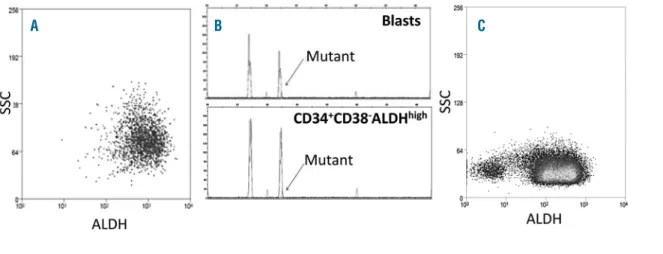
Figure 1. Assessment of CD34+cells from an NPM1 and FLT3-ITD mutat-ed AML patient with < 1% CD34+ cells.(A) Representative flow cyto-metric staining pattern of ALDH activity by CD34 is displayed on
MMNCs from patient. All the CD34+
cells are CD34+CD38–ALDHhigh. The
CD34+ALDHhigh cells are shown in
rectangle. (B) FLT3-ITD status of
iso-lated cell fractions. The CD34–blasts
harbored the FLT3-ITD mutation,
while the CD34+cells exclusively
dis-played the 330bp wild-type gene.
patients with AML-specific mutations (6 patients with FLT3-ITD and 2 with NPM1 mutations) (Figure 3B). The
CD34+CD38–ALDHhigh leukemia cell population contained
many more cells, and also exhibited much higher FSC (data
not shown) and SSC on flow cytometry, than
CD34+CD38–ALDHhighHSC populations (Figure 2A) as others
have also found.7In 7 of the 76 patients with CD34+AML
cells, two nearly equal-sized (or dual) CD34+CD38–ALDHint
(34.4+3.3 of CD34+CD38-cells) and CD34+CD38–ALDHhigh
(40.2+5.2% of CD34+CD38– cells) populations were seen
(Figure 3C, Tables 1, 2). Adequate numbers of cells were sort-ed for FISH in 4 of these 7 patients, and both the
CD34+CD38–ALDHintandCD34+CD38–ALDHhighpopulations
were leukemic (Table 2).
Table 1.Clinical characteristics of patients studied. Clinical Trial NCT01349972 Patients.
Patient Total (%) CD34– CD34+CD38–ALDHint CD34+CD38–ALDHhigh Dual ALDHhigh& ALDHint
characteristics (n=98) (n=22) (n=43) (n=26) (n=7)
Median Age 60 (Range: 29-70) 58 (Range: 31-70) 62 (Range: 30-70) 60 (Range: 32-70) 58 (Range: 29-65)
Male 50 (51%) 9 (41%) 25 (58%) 14 (54%) 2 (29%)
WBC>50,000/mm3 9 (9%) 2 (9%) 4 (9%) 2 (8%) 1 (14%)
Adverse Cytogenetics 41 (42%) 1 (5%) 14 (33%) 17 (65%) 7 (100%) Complex Karyotype 29 (30%) 1 (5%) 9 (21%) 14 (54%) 4 (57%) Monosomal Karyotype 23 (23%) 0 5 (12%) 14 (54%) 3 (43%)
FLT3-ITD mutation 9 (9%) 3 (14%) 3 (7%) 6 (23%) 0
NPM1 mutation 22 (22%) 14 (64%) 6 (14%) 2 (8%) 0
Secondary AML (prior MDS or MPN) 39 (40%) 5 (23%) 16 (37%) 15 (58%) 3 (43%)
Treatment-related 10 (10%) 2 (9%) 4 (9%) 4 (15%) 0
Favorable-risk 12 (12%) 10 (45%) 2 (5%) 0 0
Intermediate-1 risk 30 (31%) 7 (32%) 15 (35%) 8 (31%) 0 Intermediate-2 risk 18 (18%) 3 (14%) 14 (33%) 1 (4%) 0 Adverse-risk 38 (39%) 1 (5%) 14 (33%) 17 (65%) 7 (100%)
FLAM 69 (70%) 16 (73%) 29 (67%) 19 (73%) 5 (71%)
7+3 29 (30%) 6 (27%) 14 (33%) 7 (27%) 2 (29%)
Complete remissions 63 (64%) 19 (86%) 29 (67%) 13 (50%) 2 (29%)
Concomitant CBF and APL patients
Patient characteristics Total (%) CD34– CD34+CD38–ALDHint CD34+CD38–ALDHhigh Dual ALDHhigh& ALDHint
(n=11) (n=6) (n=4) (n=0) (n=0)
Median Age 55 (Range: 21-70) 60 (Range: 21-70) 40 (Range: 31-65) NA NA
Male 2 (18%) 2 (33%) 0 (0%) NA NA
WBC>50,000/mm3 0 (%) NA NA NA NA
t(8;21)(q22;q22) 2 0 2 0 0
inv (16) 2 0 2 0 0
APL* 7 6 0 0 0
Complete Remissions 11 (100%) 6 (100%) 4 (100%) NA NA
ALDH: aldehyde dehydrogenase; NA: not applicable; WBC: white blood count; AML: acute myeloid leukemia; MDS: myelodysplastic syndrome; MPN: myeloid proliferative neoplasm; FLAM: flavopiridol, cytarabine, mitoxantrone; CBF: core binding factor; APL: acute promyelocytic leukemia; *the most primitive leukemic phenotype detectable in one APL patient was CD34+CD38+ALDHint.
Table 2.Characterization of the most immature phenotype present in the leukemia by CD34, CD38, and ALDH.
AML Subtype # % CD34+ *% CD34+ CD34+CD38–ALDHint CD34+CD38–ALDHhigh
CD38– ^% %FISH+ ^% %FISH+
CD34– 22 0.52+0.08 100 0 NA 100 2.2±1.6
APL 7 0.15±0.04# 100 0 NA 100 0
CD34+CD38–ALDHint 43 26.1±4.1 45.3±4.5 65.1±3.4 69.4±13.6 1.7±0.5 2.9±1.8
CBF 4 13.8±7.7 22.4±9.1 78.1±5.6 98±0.3 3.8±3.5 0
CD34+CD38–ALDHhigh 26 25.2±4.6 43.1±5.9 0 NA 65.6±2.5 78±6.7 CD34+CD38–dual 7 21.2±16.9 40±8.7 34.4±3.3 94±4 40.2±5.2 92±5 ALDHint/ALDHhigh
Absence of detectable CD34
+AML cells is associated with
NPM1 mutations or
APL
Of the 22 patients in NCT01349972 with < 1% CD34+ cells
in the MMNCs, 14 harbored NPM1 mutations compared to
8 of the 76 patients with CD34+ AML cells (Table 1; Online
Supplementary Figure S2A, S2B, P<0.001). Of the 12 patients
with NPM1 mutations as the sole abnormality, no CD34+
leukemia cells could be detected in 11 (Online Supplementary
Figure S2A) and one harbored CD34+ CD38–ALDHintleukemia
cells (Online Supplementary Figure S2B) (P<0.002). The only
two patients in the series with t(9;11) were among the other
8 non-NPM1-mutated patients in this CD34– group
(P<0.001), as were 4 patients with normal cytogenetics
(Online Supplementary Figure S2A). Only one CD34–patient
harbored poor-risk cytogenetics, and 3 of the CD34–
NPM1-mutated patients also manifested FLT3-ITD mutations (Online Supplementary Figure S2A). Of the 8 CD34+ NPM1-mutated patients, 6 had a predominant population of
CD34+CD38–ALDHint(5 had additional detectable mutations)
and 2 (both with complex cytogenetics) had
CD34+CD38–ALDHhigh leukemia cells (Online Supplementary
Figure S2B).
Of the 7 APL patient specimens available for study, 6 also
had < 1% (0.15+0.04) CD34+ cells (Tables 1, 2). These 6
patients showed exactly the same pattern as the other AMLs
with <1% CD34+: i.e., the CD34+ cells were exclusively
CD38–ALDHhigh and lacked the t(15;17) by FISH (Table 2).
CD34+cells comprised 27.3% of the MMNCs in the other
APL patient (Table 2); very few (0.9%) of the CD34+ cells
from this patient were CD38–, and they all lacked t(15;17) by
FISH. In contrast, the CD34+CD38+ cells did harbor the
translocation.
CD34
+CD38
–ALDH
highleukemia cells are associated with
poor-risk AML
Of the 26 patients in NCT01349972 displaying a
predom-inant CD34+CD38–ALDHhigh leukemic population, 17 had
poor-risk cytogenetics and an additional 4 patients had FLT3-ITD mutations (Table 1). All 7 of the patients with dual
CD34+CD38–ALDHint and CD34+CD38–ALDHhigh leukemia
populations also harbored poor-risk cytogenetics: 4 had high-ly complex cytogenetic changes, two del 7q, and one del 5q
(Table 1). Thus, 28/33 (85%) patients with
CD34+CD38–ALDHhighAML cells harbored poor-risk genetic
markers, while only 4 of the 22 (18%) patients with <1%
CD34+cells and 16 out of 43 (37%) patients with
predomi-nant CD34+CD38–ALDHint populations harbored poor-risk
cytogenetics or FLT3-ITD mutations (P<0.0001). The patients
with CD34+CD38–ALDHhighAML cells were also more likely
to have AML arising out of myelodysplastic syndrome or myeloproliferative disease (18/33, 55%) than the
CD34+CD38–ALDHint and CD34– groups (21/65, 32%,
P=0.04).
The leukemias’ most primitive hematopoietic cell
phenotype correlates with outcomes
Not surprisingly, given the strong association with
poor-risk genetics, patients harboring CD34+CD38–ALDHhigh
leukemic populations displayed relative drug resistance. There was a significantly lower CR rate for patients
harbor-ing CD34+CD38–ALDHhigh leukemic populations when
com-pared to patients with CD34+CD38–ALDHint or no CD34+
cells AML cells (Table 3, P=0.007). The CR rates for the
patients with CD34+CD38–ALDHhigh leukemic populations
were similar with FLAM (11/24, 46%) and 7+3 (4/9, 44%). However, there was a trend for more CRs on the FLAM arm (36/45, 80%) than on the 7+3 arm (12/20, 60%) in the other
65 patients (P=0.1).
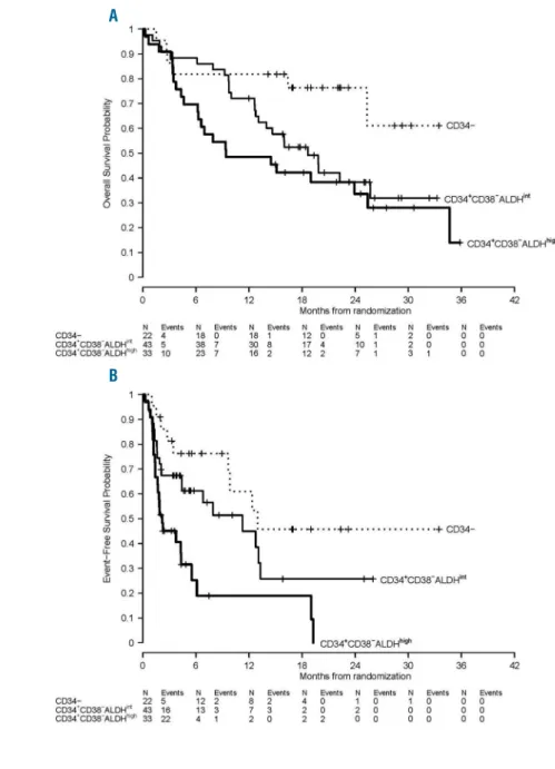
We next studied if the most immature phenotype present in the leukemia also showed a correlation with survival. OS was significantly different according to most immature
leukemia phenotype present in the leukemia (P=0.02, Table
3, Figure 4A), with patients harboring CD34+CD38–ALDHhigh
AML cells demonstrating the worst OS. There was also a
sig-Figure 2. Prominent ALDHintpopulation of CD34+CD38–cells from a patient with FLT3-ITD AML.(A) Representative flow cytometric staining pattern of ALDH activity
by side scatter (SSC) is displayed for CD34+CD38–cells isolated from patient. (B) FLT3-ITD status of isolated cell fractions. The CD34+CD38–ALDHintpopulation (oval)
harbored the FLT3-ITD mutation, while the CD34+CD38–ALDHhigh cells (square) exclusively displayed the 330bp wild-type gene.
nificant difference in EFS according to the most primitive
leukemia phenotype (P<0.001, Table 3, Figure 4B). The EFS
probability at 1-year was 61% (95% CI, 41-90%), 45% (95% CI 29-69%), and 19% (95% CI 8-47%) for patients without
detectable CD34+ leukemia cells and those with
CD34+CD38–ALDHint and CD34+CD38–ALDHhigh leukemia
cells, respectively (P<0.001).
As others have found a strong correlation between just
leukemic CD34+CD38–cell numbers (without using ALDH
expression) at diagnosis and outcome,7,15 we analyzed the
prognostic impact of total CD34+CD38–numbers. There was
a trend for total CD34+CD38–cell numbers at diagnosis to
correlate with outcome. For patients with detectable CD34+
AML cells in trial NCT01349972, CD34+CD38–cells
repre-sented 5.6+1.5% of the MMNCs for those who entered a CR
compared to 11+3% in those who did not (P=0.08, t-test). Of
those patients with < 5% CD34+CD38–cells, 23% remained
event-free compared to 9.5% with > 5% CD34+CD38–cells
(P=0.2, Fisher's exact test). The mean frequency of
CD34+CD38–cells was the same in both the ALDHint and
ALDHhighgroups at 7.6+1.8% and 7.5+2.5%, respectively.
The type of postremission therapy was not specifically mandated on this trial, and many of the patients went onto alloBMT (Table 3). AlloBMT was very effective in all patients in NCT01349972, regardless of their most primitive
leukemia phenotype. Of the patients with ALDHhighleukemia
cells, 8/15 who achieved a CR underwent alloBMT in CR1 and 5 remain alive and disease-free (Table 3). Similarly, 16 of
the CD34+CD38–ALDHint and 6 of the CD34–patients
under-went alloBMT in CR1, and 7 and 5 patients remain alive and
disease-free, respectively (Table 3, P=0.2). In contrast, the
outcomes of the patients who did not receive alloBMT in CR1, with most receiving cytarabine-based consolidation therapy, differed significantly by the most primitive pheno-type present in leukemia cells. Seven patients with
CD34+CD38–ALDHhigh leukemia cells did not undergo
alloBMT in CR1, and all relapsed including 3 with normal cytogenetics and wild-type FLT3/NPM1 (Table 3). In con-trast, 4/13 (3/9 with normal cytogenetics and wild-type
FLT3/NPM1) patients with CD34+CD38–ALDHint leukemia
cells and 7/13 (1 of 2 with normal cytogenetics and wild-type
FLT3/NPM1) CD34–patients who did not undergo alloBMT
remain alive and disease-free in CR1 (Table 3, P=0.06).
Discussion
The failure of CRs to reliably translate into cures in AML35,36
can be explained by the LSC paradigm. However, the true clinical relevance of LSCs remains the focus of considerable
debate.3-20,37 Several groups have shown that CD34+CD38–
leukemia cell numbers present at diagnosis have strong prog-nostic significance, providing support for a clinical relevance
for LSCs.7,15Patients with increased numbers of CD34+CD38–
at diagnosis in clinical trial NCT01349972 showed a trend toward worse outcomes. Our inability to show a stronger
clinical correlation between CD34+CD38–leukemia cell
num-bers at diagnosis and outcome may relate to the exclusion of favorable-risk cytogenetic-risk groups from the study. We also did not use the same methodology as others who showed a stronger correlation; we analyzed only total
CD34+CD38– numbers, while others further refined the
CD34+CD38–subset to include the expression of leukemic
stem cell associated markers7or CD123.15 We did find that
the most immature hematopoietic cellular phenotype pres-ent in leukemia cells was heterogeneous, ranging from
CD34–to that of primitive HSCs (i.e., CD34+CD38–ALDHhigh),
but was relatively consistent across AML risk groups. Perhaps most importantly, the strong association between the leukemic clone’s most immature phenotype and out-come in this prospective patient cohort supports further test-ing of this clinical biomarker in future studies.
The vast majority of AML patients (80/109) in our series
harbored CD34+CD38–leukemia cells, as initially reported
by Lapidot et al.1Moreover, we confirmed our prior data13
that the majority of non poor-risk AMLs, including all of the
CBF patients, harbored CD34+CD38–leukemia cells that
could be separated from normal HSCs by their lower ALDH activity. However, 33 out of 98 (34%) of patients
from NCT01349972 harbored CD34+CD38–ALDHhigh
leukemia cells. This group of patients was more likely to harbor poor-risk cytogenetics or FLT3-ITD mutations, and had a statistically lower chance of achieving CRs than the other AML patients. Importantly, the presence of
CD34+CD38–ALDHhigh leukemia cells was associated with a
significantly lower EFS and OS, even when no unfavorable genetic or cytogenetic abnormalities could be identified.
Even though patients with CD34+CD38–ALDHhigh LSCs did
Table 3.Clinical outcomes of patients in NCT01349972 by leukemia’s most primitive hematopoietic cellular phenotype.
CD34+CD38–ALDHhigh CD34+CD38–ALDHint CD34– P*
(including dual) (n=43) (n=22)
(n=33)
Complete remission 15/33 (45%) 29/43 (67%) 19/22 (86%) 0.007 Median OS (months) 9.4 (95% CI: 6-36) 18.7 (95% CI: 13-36) Not reached 0.02
HR – 1.3 (95% CI: 0.7-1.4) HR – 1 HR - 0.4 (95% CI: 0.2–1)
Median EFS (months) 2.2 (95% CI: 2-6) 11.3 (95% CI, 4-36) 13 (95% CI: 4-36) 0.001 HR = 2.2 (95% CI: 1.2-3.9) HR - 1 HR - 0.6 (95% CI: 0.3-1.3)
AlloBMT in CR1 8/15 (53%) 16/29 (55%) 6/20 (30%)
Continuously EF 5/8 (63%) 7/16 (44%) 5/6 (83%) 0.2
Median EFS (months) Not reached Not reached 23.1 (95% CI: 23 – 35) No BMT in CR1 7/15 (47%) 13/29 (45%) 13/20 (65%)
Continuously EF 0/7 4/13 (31%) 7/13 (54%) 0.06
Median EFS (months) 4.6 (95% CI: 3 – 32) 11.9 (95% CI: 5 – 32) Not reached
poorly overall, 5/8 of those patients who got to alloBMT remain alive and disease-free. Several groups have also
described ALDHhighLSCs in a fraction of AML patients who
appeared to have a worse prognosis.8,17-20
No CD34+ leukemia cells could be detected in 22/98
patients from NCT01349972. As others have also
described,7,16,34 the CD34+ cells in these patients represented
<1% of the cells at diagnosis, were exclusively
CD38–ALDHhigh, and lacked the leukemic mutation. Thus, the
CD34+cells in such patients likely represented residual
nor-mal HSCs. NPM1 mutations were detected in 14 (64%) of
the 22 AML patients who lacked detectable CD34+cells, and
11/12 AML patients with NPM1 mutation as a sole
abnor-mality were in this group. No CD34+cells were detected in
6/7 APL patients, as others have also found.38The one APL
patient in this series with CD34+ AML cells only had the
t(15;17) detected in the CD34+CD38+ cells. Other groups
have reported that CD34 expression is a bad prognostic
fac-tor for both NPM1-mutated AMLs39and APLs;40-42the small
numbers of these patients in our cohort may have hindered demonstrating similar statistical significance.
The phenotype of the LSCs in NPM1-mutated AML has been somewhat controversial. Two other groups also found
that most NPM1-mutated AMLs were CD34–, with the
CD34+cells lacking leukemia mutations.4,16However, Martelli
et al. found that the NPM1 mutation was present in
CD34+CD38–cells, and these cells generated AML in
immun-odeficient mice.43 Interestingly, CD34+ cells represented
>1.5% of the MMNCs in all the NPM1-mutated AMLs
trans-planted into mice in that report.43We also found the mutation
in the CD34+CD38–cells from all 8 NPM1-mutated AML
patients with >1% CD34+ MMNCs. Of note, 7 of these
patients had cytogenetic or FLT3-ITD mutations in addition to NPM1. Thus, it appears that the most immature leukemic
cell in NPM1-mutated AMLs can be either CD34+or CD34–;
it is possible that the differences can be explained by the fact
that Martelli et al. did not perform immunodeficient mouse
transplants with any of the 18 patients in their series
harbor-ing < 1% CD34+cells.43
Despite our inability to detect AML cells by PCR in the
small population of CD34+ cells present in the diagnostic
marrows of the CD34–AMLs, a very small number (2.2%) of
CD34+CD38–ALDHhighcells had the FISH marker that
charac-terized the AML (Table 2). Zeijlemaker et al. recently
suggest-ed that although the vast majority of AML patients with <
1% CD34+cells in their diagnostic marrow lacked CD34+
AML cells, a small number did harbor neoplastic CD34+
cells.21It is similarly possible that CD34+CD38–ALDHhigh AML
cells may be present at very low levels in the patients whose leukemias’ most immature phenotype appeared to be
CD34+CD38–ALDHint; however, based on the low FSC/SSC
of these CD34+CD38–ALDHhighcells , we believe that these
small leukemic populations by FISH represent flow sorting contamination. We also previously found that the
CD34+CD38–ALDHhighcells present in AMLs harboring large
CD34+CD38–ALDHint populations only produced normal
hematopoiesis when transplanted into
immunocompro-mised mice.13Importantly, the phenotype of the most
primi-tive hematopoietic cells found to harbor predominately leukemia-specific mutations correlated with AML risk groups and outcomes.
Our data raise the possibility that the most immature phe-notype present in leukemia may be a function of the stage of hematopoietic differentiation at which the leukemogenic mutation develops. Those AMLs harboring leukemia cells sharing a phenotype with primitive normal HSCs
(CD34+CD38–ALDHhigh) had the worst prognosis, while CBF
and intermediate-risk AMLs’ most primitive phenotype was that of more differentiated hematopoietic progenitors
(CD34+CD38–ALDHint). The most immature hematopoietic
phenotype found in the most favorable-risk AMLs, APLs and those with NPM1-mutations as sole abnormalities, expressed
even more differentiated phenotypes: CD34+CD38+ and
CD34–CD38+.
These findings suggest that the leukemia clones’ most primitive hematopoietic cellular phenotype might serve as a biomarker for risk-stratifying patients at diagnosis. About 30-40% of AML patients lack any cytogenetic or usual genetic
prognostic factors,44and even when present such prognostic
factors may not be available for days or weeks. The most immature phenotype present in leukemia cells can be readily determined in essentially all patients by flow cytometry within hours of diagnosis. Rapid risk-stratification may be
particularly useful for patients harboring
CD34+CD38–ALDHhighleukemia cells, which appear to
iden-tify high-risk patients often refractory to induction
Figure 3. Prominent ALDHhighpopulations of CD34+CD38–cells.(A) Representative flow cytometric staining pattern of ALDH activity by side scatter (SSC) is displayed
for CD34+CD38–cells isolated from patient. The CD34+CD38-ALDHhighcells represented essentially all of the total CD34+CD38- cells. (B) FLT3-ITD status of isolated
cell fractions. The CD34+CD38-ALDHhighpopulation harbored the FLT3-ITD mutation. (C) Representative flow cytometric staining pattern of ALDH activity by side scatter
(SSC) is displayed for CD34+CD38–cells isolated from a patient with dual CD34+CD38–ALDHint and CD34+CD38–ALDHhighpopulations.
chemotherapy. Although the phenotype of
CD34+CD38–ALDHhighleukemia cells is the same as normal
HSCs, the flow cytometric pattern of the
CD34+CD38–ALDHhighpopulation at AML diagnosis allows
the primitive leukemic phenotype to be clearly distinguished from HSCs even in the absence of cytogenetic or genetic
markers. The ALDHhighcells represent the vast majority of the
CD34+CD38– cells and had higher FSC/SSC at diagnosis
when leukemic (Figures 3A, Table 2), while the low FSC/SSC
ALDHhighHSCs represented only a very small percentage (on
average 1-2%) of the total CD34+CD38– cells (Figure 2A,
Table 2). Others have published similar findings.7 Should
studies confirm the adverse prognosis of a
CD34+CD38–ALDHhighleukemia phenotype, rapid
identifica-tion of such patients could allow them early access to clinical trials studying novel induction approaches. Moreover, a
CD34+CD38–ALDHhighleukemic phenotype could be used to
guide patients toward alloBMT when no prognostic cytoge-netic or gecytoge-netic abnormalities are present.
Acknowledgments
The authors thank the patients who contributed research samples,
investigators who enrolled patients on this clinical trial and graciously shared patient samples (Matthew C. Foster: University of North Carolina, Mark R. Litzow: Mayo Clinic-Rochester, MN, Lawrence E. Morris: The Blood and Marrow Transplant Group at Northside Hospital, Stephen Strickland: Vanderbilt University Medical Center, Jeffrey E. Lancet: H. Lee Moffitt Cancer and Research Institute, Prithviraj Bose: Virginia Commonwealth University, M. Yair Levy: Texas Oncology, Baylor Charles A. Simmons Cancer Center, and Raoul Tibes: Mayo Clinic-Scottsdale, AZ, USA), the Cancer Therapy Evaluation Program (L. Austin Doyle, John J. Wright, Richard F. Little) at the NCI for sponsoring and supporting the clinical study, and the research staff at the Johns Hopkins Kimmel Cancer Center who assisted in specimen procurement. J.F.Z. is a recipient of a 2013 Young Investigator Award, in memory Dr. John R. Durant, and a 2014-2017 LLS Special Fellow in Clinical Research Award.
Funding
This work was supported by the Leukemia & Lymphoma Society (LLS) (TRP R6459-13, R.J.J. and J.M.G.), and the National Institutes of Health [grants P01 CA015396 (R.J.J.), U01 A70095 (J.E.K.), 5T32 HL007525 (J.M.G.), and P30 CA006973].
Figure 4. (A) OS and (B) EFS in clinical trial NCT01349972 by the most immature phenotype detectable in leukemia cells. With a median
follow-up of 22 (range 12-36) months, OS (P=0.02) and
EFS (P<0.001) were significantly different
accord-ing to the leukemias’ most primitive hematopoietic phenotype.
A
References
1. Lapidot T, Sirard C, Vormoor J, et al. A cell initiating human acute myeloid leukaemia after transplantation into SCID mice. Nature. 1994;367(6464):645-648.
2. Bonnet D, Dick JE. Human acute myeloid leukemia is organized as a hierarchy that originates from a primitive hematopoietic cell. Nat Med. 1997;3(7):730-737.
3. Sarry JE, Murphy K, Perry R, et al. Human acute myelogenous leukemia stem cells are rare and heterogeneous when assayed in NOD/SCID/IL2Rgammac-deficient mice. J Clin Invest. 2011;121(1):384-395.
4. Taussig DC, Vargaftig J, Miraki-Moud F, et al. Leukemia-initiating cells from some acute myeloid leukemia patients with mutated nucleophosmin reside in the CD34(-) frac-tion. Blood. 2010;115(10):1976-1984. 5. Pearce DJ, Taussig D, Zibara K, et al. AML
engraftment in the NOD/SCID assay reflects the outcome of AML: implications for our understanding of the heterogeneity of AML. Blood. 2006;107(3):1166-1173.
6. Cheung AM, Chow HC, Kwong YL, Liang R, Leung AY. FLT3/internal tandem duplication subclones in acute myeloid leukemia differ in their engraftment potential in NOD/SCID mice. Leuk Res. 2010;34(1):119-122. 7. Terwijn M, Zeijlemaker W, Kelder A, et al.
Leukemic stem cell frequency: a strong bio-marker for clinical outcome in acute myeloid leukemia. PLoS One. 2014;9(9):e107587. 8. Cheung AM, Wan TS, Leung JC, et al.
Aldehyde dehydrogenase activity in leukemic blasts defines a subgroup of acute myeloid leukemia with adverse prognosis and superior NOD/SCID engrafting poten-tial. Leukemia. 2007;21(7):1423-1430. 9. Monaco G, Konopleva M, Munsell M, et al.
Engraftment of acute myeloid leukemia in NOD/SCID mice is independent of CXCR4 and predicts poor patient survival. Stem Cells. 2004;22(2):188-201.
10. Ailles LE, Gerhard B, Kawagoe H, Hogge DE. Growth characteristics of acute myelogenous leukemia progenitors that initiate malignant hematopoiesis in nonobese diabetic/severe combined immunodeficient mice. Blood. 1999;94(5):1761-1772.
11. Feuring-Buske M, Gerhard B, Cashman J, Humphries RK, Eaves CJ, Hogge DE. Improved engraftment of human acute myeloid leukemia progenitor cells in beta 2-microglobulin-deficient NOD/SCID mice and in NOD/SCID mice transgenic for human growth factors. Leukemia. 2003;17(4):760-763.
12. Rombouts WJ, Martens AC, Ploemacher RE. Identification of variables determining the engraftment potential of human acute myeloid leukemia in the immunodeficient
NOD/SCID human chimera model.
Leukemia. 2000;14(5):889-897.
13. Gerber JM, Smith BD, Ngwang B, et al. A clinically relevant population of leukemic CD34+CD38 - cells in acute myeloid leukemia. Blood. 2012;119(15):3571-3577. 14. Ghiaur G, Gerber J, Jones RJ. Concise review:
Cancer stem cells and minimal residual dis-ease. Stem Cells. 2012;30(1):89-93. 15. Vergez F, Green AS, Tamburini J, et al. High
levels of CD34+CD38low/-CD123+ blasts are predictive of an adverse outcome in acute myeloid leukemia: a Groupe Ouest-Est des Leucemies Aigues et Maladies du Sang
(GOELAMS) study. Haematologica.
2011;96(12):1792-1798.
16. Schuurhuis GJ, Meel MH, Wouters F, et al. Normal hematopoietic stem cells within the AML bone marrow have a distinct and higher ALDH activity level than co-existing leukemic stem cells. PLoS One. 2013; 8(11):e78897.
17. Hoang VT, Buss EC, Wang W, et al. The rar-ity of ALDH(+) cells is the key to separation of normal versus leukemia stem cells by ALDH activity in AML patients. Int J Cancer. 2015;137(3):525-536.
18. Pearce DJ, Taussig D, Simpson C, et al. Characterization of cells with a high alde-hyde dehydrogenase activity from cord blood and acute myeloid leukemia samples. Stem Cells. 2005;23(6):752-760.
19. Ran D, Schubert M, Pietsch L, et al. Aldehyde dehydrogenase activity among pri-mary leukemia cells is associated with stem cell features and correlates with adverse
clin-ical outcomes. Exp Hematol.
2009;37(12):1423-1434.
20. Ran D, Schubert M, Taubert I, et al. Heterogeneity of leukemia stem cell candi-dates at diagnosis of acute myeloid leukemia and their clinical significance. Exp Hematol. 2012;40(2):155-165.
21. Zeijlemaker W, Kelder A, Wouters R, et al. Absence of leukaemic CD34 cells in acute myeloid leukaemia is of high prognostic value: a longstanding controversy deci-phered. Br J Haematol. 2015;171(2):227-238. 22. Estey EH. How to manage high-risk acute
myeloid leukemia. Leukemia.
2012;26(5):861-869.
23. Karp JE, Blackford A, Smith BD, et al. Clinical activity of sequential flavopiridol, cytosine arabinoside, and mitoxantrone for adults with newly diagnosed, poor-risk acute myel-ogenous leukemia. Leuk Res. 2010;34(7):877-882.
24. Zeidner JF, Foster MC, Blackford AL et al. Randomized multicenter phase 2 study of flavopiridol (alvocidib), cytarabine, and
mitoxantrone (FLAM) versus
cytarabine/daunorubicin (7+3) in newly
diag-nosed acute myeloid leukemia.
Haematologica. 2015;100(9):1172-9. 25. Gerber JM, Qin L, Kowalski J et al.
Characterization of chronic myeloid leukemia stem cells. Am J Hematol. 2011;8631-37.
26. Dohner H, Estey EH, Amadori S et al. Diagnosis and management of acute myeloid leukemia in adults: recommendations from an international expert panel, on behalf of the
European LeukemiaNet. Blood.
2010;115(3):453-474.
27. R: R Core Team. R: A language and environ-ment for statistical computing. R Foundation for Statistical Computing, Viena, Austria. 2014.
28. Ghiaur G, Yegnasubramanian S, Perkins B, Gucwa JL, Gerber JM, Jones RJ. Regulation of human hematopoietic stem cell self-renewal by the microenvironment's control of retinoic acid signaling. Proc Natl Acad Sci USA.
2013;110(40):16121-16126.
29. Gerber JM, Gucwa JL, Esopi D et al. Genome-wide comparison of the transcrip-tomes of highly enriched normal and chronic myeloid leukemia stem and progenitor cell populations. Oncotarget. 2013;4(5):715-728. 30. Storms RW, Trujillo AP, Springer JB et al.
Isolation of primitive human hematopoietic progenitors on the basis of aldehyde dehy-drogenase activity. Proc Natl Acad Sci U S A. 1999;96(16):9118-9123.
31. Krause DS, Fackler MJ, Civin CI, May WS. CD34: structure, biology, and clinical utility. Blood. 1996;87(1):1-13.
32. Jones RJ, Barber JP, Vala MS, et al. Assessment of aldehyde dehydrogenase in viable cells. Blood. 1995;85(10):2742-2746. 33. Jones RJ, Collector MI, Barber JP, et al.
Characterization of mouse lymphohe-matopoietic stem cells lacking colony-form-ing activity. Blood. 1996;88(2):487-491. 34. van der Pol MA, Feller N, Roseboom M, et al.
Assessment of the normal or leukemic nature of CD34+ cells in acute myeloid leukemia with low percentages of CD34 cells. Haematologica. 2003;88(9):983-993. 35. Appelbaum FR, Gundacker H, Head DR, et
al. Age and acute myeloid leukemia. Blood. 2006;107(9):3481-3485.
36. Cheson BD, Bennett JM, Kopecky KJ, et al.
Revised recommendations of the
International Working Group for Diagnosis, Standardization of Response Criteria, Treatment Outcomes, and Reporting Standards for Therapeutic Trials in Acute Myeloid Leukemia. J Clin Oncol. 2003;21(24):4642-4649.
37. Becker MW, Jordan CT. Leukemia stem cells in 2010: current understanding and future directions. Blood Rev. 2011;25(2):75-81. 38. Turhan AG, Lemoine FM, Debert C, et al.
Highly purified primitive hematopoietic stem cells are PML-RARA negative and generate nonclonal progenitors in acute promyelocytic leukemia. Blood. 1995; 85(8):2154-2161. 39. Dang H, Chen Y, Kamel-Reid S, Brandwein J,
Chang H. CD34 expression predicts an adverse outcome in patients with NPM1-pos-itive acute myeloid leukemia. Hum Pathol. 2013;44(10):2038-2046.
40. Lee JJ, Cho D, Chung IJ, et al. CD34 expres-sion is associated with poor clinical outcome in patients with acute promyelocytic leukemia. Am J Hematol. 2003;73(3):149-153. 41. Breccia M, De Propris MS, Stefanizzi C, et al. Negative prognostic value of CD34 antigen also if expressed on a small population of acute promyelocitic leukemia cells. Ann Hematol. 2014;93(11):1819-1823.
42. Ahmad EI, Akl HK, Hashem ME, Elgohary TA. The biological characteristics of adult CD34+ acute promyelocytic leukemia. Med Oncol. 2012;29(2):1119-1126.
43. Martelli MP, Pettirossi V, Thiede C, et al. CD34+ cells from AML with mutated NPM1 harbor cytoplasmic mutated nucleophosmin and generate leukemia in immunocompro-mised mice. Blood. 2010;116(19):3907-3922. 44. Walker AR ,Marcucci G. Management of