RESEARCH ARTICLE
A COMPARITIVE CLINICAL STUDY OF SUBJECTIVE AND OBJECTIVE EVALUATION OF
DENTURES FABRICATED USING DIFFERENT IMPRESSION TECHNIQUES
*Swathi Selvaraj, Roseline Meshramkar, Lekha K.P. and Ramesh Nadiger
Department of Prosthodontics, SDM College of Dental Sciences, Dharwad, India
ARTICLE INFO ABSTRACT
Background: Impression techniques for complete denture fabrication have evolved considerably over
decades. However, it needs to be assessed whether the retention provided by different techniques is adequate enough to establish them as an alternative to conventional techniques. The purpose of this study was to evaluate the retention of complete dentures fabricated using two different impression techniques like selective pressure and functional impression techniques both subjectively and
objectively. Materials and methods: Maxillary dentures were fabricated for 10 edentulous patients
using two different impression techniques. Each patient had two maxillary dentures and its retention was checked both subjectively and objectively. For subjective evaluation, a questionnaire was given and satisfaction level between the two dentures were compared. For objective evaluation, retention was measured using an instrument which uses the principle of class I pulley and a spring balance apparatus. During insertion, a metallic framework was attached at the centre of the denture using autopolymerising acrylic resin keeping it perpendicular to the occlusal plane of edentulous ridge. A small hook was engaged to the metal framework and weights were added on the spring balance. The weight at which both maxillary dentures dislodged was noted. The values were tabulated and
subjected to statistical analysis. Results: The statistical analysis shows that the difference between
modified functional impression and selective pressure is statistically significant (P< 0.05) in objective
evaluation. But there was no significant difference in subjective evaluation of dentures. Conclusion:
The two impression techniques yeilded adequately retentive dentures. However, retention of dentures fabricated from modified functional impression technique was better compared to selective pressure technique objectively.
Copyright©2019, Swathi Selvaraj et al. This is an open access article distributed under the Creative Commons Attribution License, which permits unrestricted
use, distribution, and reproduction in any medium, provided the original work is properly cited.
INTRODUCTION
Complete denture is a prosthesis that replaces the entire dentition and associated structures of maxilla and mandible. The fabrication of complete denture requires a number of steps, the first being impression making. An impression is a record, a facsimile of mouth tissues taken at an unstrained rest position or in various positions of displacement (Academy of prosthodontics, 2005). In the case of an edentulous arch, this requires a unique combination of managing movable soft tissue commensurate with integrating different materials and a technique for accurate reproduction (DeVan, 1952). The history of complete denture impression procedures has been influenced largely by the development of impression materials from which new ideas and techniques arose. A complete denture impression is a negative registration of the entire denture bearing, stabilizing and border seal areas present in edentulous mouth. The objectives of an impression provides stability, retention, comfort and support to denture. An impression also acts as an foundation for the improved appearance of the patient and at the same time maintains health
of oral tissues. The impression techniques are numerous but may be classified according to jaw position and the degree of pressure used when making the impression, that is open or closed mouth, pressure, non pressure or negative pressure or selective pressure. The selective pressure technique was proposed by Boucher in 1950. It combines the principles of both pressure and minimal pressure techniques. It confines the forces acting on denture to stress bearing areas. These tissues were recorded under selective pressure while others are relieved under minimal pressure. Functional impression materials are those which when applied to the tissue surface of a denture base or impression tray, recorded the topography and the position of basal seat and border tissues as they existed in a functional state. Tissue conditioning had been found useful as functional impression materials. The functional Impression technique utilized the property of tissue conditioner material to allow time for the tissues to reposition themselves as they had an ability to compress under pressure but rebound when pressure was released. The introduction of newer impression materials and techniques has made it necessary to evaluate whether these are efficient and accurate enough to substitute the materials and techniques that have been used since
ISSN: 0975-833X
International Journal of Current Research Vol. 11, Issue, 10, pp.7590-7594, October, 2019
DOI: https://doi.org/10.24941/ijcr.36940.10.2019
INTERNATIONAL JOURNAL OF CURRENT RESEARCH
Article History:
Received 04th July, 2019
Received in revised form 19th August, 2019
Accepted 15th September, 2019
Published online 30th October, 2019
Citation: Swathi Selvaraj, Roseline Meshramkar, Lekha K.P. and Ramesh Nadiger, 2019. “Evaluation of various marketed formulations of boswellia by
RP-HPLC and HPTLC”, International Journal of Current Research, 11, (10), 7590-7594.
Key Words:
Complete denture, Retention,
Impression Techniques.
decades. The retention achieved in a denture is an important criteria to check the accuracy and efficacy of an impression material or technique. Hence this study was planned to evaluate the selective pressure and modified open mouth functional impression techniques and correlate the retention achieved for the complete dentures fabricated using these techniques.
MATERIALS AND METHODS
Ten completely edentulous patients who reported to the department of prosthodontics, SDM college of dental sciences
Inclusion criteria: Healthy patients with firm resilient oral
mucosa.
Exclusion criteria:
1. With systemic illness
2. Poor neuromuscular control
3. With undercuts or flabby ridge
The selected patient’s history, examination both intra oral and extra oral were carried out to assess patient’s tissues. An appropriate edentulous stock metal tray with 5mm clearance between the tray and maxillary ridge was selected and primary impression was made with impression compound. The impression was poured in type II dental plaster and primary cast was obtained. The primary cast was duplicated using putty (Aquasil) to obtain two primary casts for each patient. Then full wax spacer was adapted in both the casts with 4 tissue stops (2 stops in canine region and 2 in the molar region).The custom tray was fabricated for both the primary casts using autopolymerizing acrylic resin by sprinkle on technique.
Selective pressure technique (Group I): The custom tray for
this technique was trimmed 2mm short of sulcus. The border molding was done with low fusing compound by sectional method. The wax spacer was removed and definitive final impression was made using zinc oxide eugenol impression paste.
A) Primary impression made with impression compound
B) Wax spacer adapted on custom tray for modified functional and selective pressure
impression technique
C) Custom trays fabricated using autopolymerising acrylic resin
D) Group I- Selective Pressure Impression technique made using ZnOE Eugenol Impression paste.
E) D-Soft Tissue Conditioner F) Group II-Border moulding and final
impression made using Tissue conditioner
E) A metallic hook attached to the anterior part of the maxillary denture using sticky wax keeping it perpendicular to the occlusal plane
of edentulous ridge
F) Retention measuring apparatus
G) Objective evaluation of Retention of dentures fabricated using selective pressure
Modified open mouth functional impression technique
(Group II): The custom tray for this technique extended upto
the sulcus and no handle was made so that the patients could make functional movements easily. The tray extension was checked in patient’s mouth and overextensions were trimmed. The powder and liquid of tissue conditioner was mixed in correct ratio and was applied to the borders and posterior palatal seal area. Patient was asked to do functional movements like smile, yawn, whistle, speak “ooo” and “eeee” in regular fashion and then patient was asked to pucker the mouth for 5 seconds for border seal. To record the posterior palatal seal, patient was asked to say ‘ah’ in short vigorous bursts, swallow and cough. Patient was asked to repeat all these movements for 8 to 10 minutes. Then wax spacer was removed and tissue conditioner was applied to make definitive final impression. The two definitive impressions were poured with dental stone. The tissue conditioner impression was coated with dental stone using paint brush so as not to disturb tissue conditioner material. After this layer of plaster was set, dental stone was poured. Then the procedure was continued conventionally till polishing of complete denture.
Objective evaluation
Retention of maxillary dentures fabricated using two different impression techniques was measured using an instrument created in our department of prosthodontics. It uses the
principle of class I pulley and as pring balance apparatus.
During insertion, a metallic hook was attached at the anterior part of the maxillary denture using sticky wax keeping it perpendicular to the occlusal plane of edentulous ridge. A
string was adapted over the class I pulley in such a way that
one end which is away from the patient was attached to spring
balance and the other end which has a small hook was engaged to the metal framework and hence the maxillary denture. The weights were added on the spring balance and more weight was added till the denture dislodges at a certain weight. The weight at which the denture dislodged was noted. The dislodging forces were calculated for both dentures fabricated using selective pressure and modified functional impression techniques. The values were tabulated and subjected to statistical analysis.
Subjective evaluation
To compare the patient satisfaction level between two dentures, a questionnaire was given to them after denture insertion. Different answers to each question were assigned scores and the total score was calculated for evaluation and analysis.
RESULTS
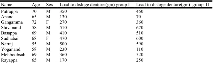
[image:3.595.120.480.481.588.2]Table 1. Shows the values for load in gms that were required to dislodge the dentures using two different impression techniques. For Objective evaluation, (Table.3) the statistical analysis shows that the difference between the mean loads to dislodge the complete dentures fabricated by modified functional impression technique and selective pressure impression technique is statistically significant (P=0.031). For subjective evaluation, Patients were asked to firstly grade their dentures in general, and then they were asked to provide separate grades on the retention, aesthetics, ability to speak and masticate with their dentures and comfort of wearing dentures. The difference in satisfaction level between both the dentures were not statistically significant.
Table 1. Load required to dislodge the complete dentures
Name Age Sex Load to disloge denture (gm) group I Load to disloge denture(gm) group II
Putrappa 70 M 350 460
Anand 65 M 130 70
Gangamma 72 F 270 360
Shivanand 58 M 510 670
Basappa 69 M 410 510
Sudhabai 68 F 470 600
Natraj 55 M 500 590
Yoganand 58 M 230 110
Mehboobsab 69 M 360 520
Rayappa 65 M 170 250
Table 2. Results for Paired Samples Statistics
Mean N Std. Deviation Std. Error Mean
Pair 1 Group1 340.0000 10 ######## 43.05036
Group2 414.0000 10 ######## 66.18493
Table 3. Results for Paired Samples Correlations
N Correlation Sig.
Pair 1 Group1 & Group2 10 .946 .000
Paired Differences
t df Sig.
(2-tailed)
Mean Std. Deviation Std. Error
Mean
95% Confidence Interval of the Difference
Lower Upper
Pair 1 Group1 -
H) Subjective evaluation of retention of dentures using Questionnare
DISCUSSION
Construction of a retentive complete denture for various edentulous patients is one of the goals of a prosthodontist. As irretentive denture disturbs all other goals as speech, mastication and in turn affects patient’s psychology. Patients with well-formed alveolar ridges were selected to improve the retention and stability of the denture bases which is an important variable during the research. On the other hand, patients with atrophied or thin mucoperiosteum were excluded since there might be a source of soreness that may affect the retentive quality. The result of the study emphasizes that both the impression techniques yielded adequately retentive dentures. However the retention of complete dentures fabricated using modified functional impression technique was better than the selective pressure technique. This is in accordance with Abdul hakim et al who has recommended that maintenance of viscoelastic property is a key to clinical success to be used as functional impression. It is a material that flows for a period of time and gives the exact border morphology of tissues. A functional impression should flow readily under functional stress, with minimal elastic recovery ensuring continual adaptation to underlying soft tissues as they
are altered under stress (Abdel-Hakim et al., 1994). Similarly,
Drago et al. also concluded that denture bases fabricated using
functional impression technique showed higher retention (Drago, 2003). For some authors, however, zinc oxide paste is still the final impression material of choice in most instances
(Zar et al. ; Weng and Khlevnoy) as it records the accurate
surface detail. It is a mixed study involving the use of a retention measuring apparatus to measure the load needed to dislodge the dentures, and the administration of a subjective patient questionnaire to assess chewing, comfort and confidence. Most of the patient scored all the examined variables to best score category for both the dentures. The variety of impression materials and the range of working characteristics of these materials made it possible to develop impression procedures best suited for specific conditions in
each area in a given mouth. Whatever method is used form a king impression, it should be based on the basic principles of maximum tissue area coverage and intimate contact so as to achieve the objectives of retention, support, stability, esthetics, and preservation of ridge.
Conclusion
Thus within the limitations of study, it was concluded that
i) The dentures fabricated using modified functional
impression showed highest retentive values compared to selective pressure technique.
ii) Both the techniques, modified functional impression
technique and selective pressure technique yielded adequately retentive dentures.
iii)For subjective evaluation, the difference in satisfaction
level between both the dentures were not statistically significant.
REFERENCES
Abdel-Hakim AM, Al-Dalgan SA, Al-Bishre GM. 1994. Displacement of border tissues during final impression
procedures. J Prosthet Dent., 71:133–8.
Academy of prosthodontics, 2005. Glossary of Prosthodontic
terms. J Prosthet Dent., 94(1):10-92.
Appelbaum EM, Mehra RV. 1984. Clinical evaluation of
polyvinylsiloxane for complete denture impressions. J
Prosthet Dent., 52(4):537-539.
Boucher CO. 1951. A Critical analysis of mid century
impression techniques for full dentures. J Prosthet Dent.,
1(4):472-491.
Chaffe NR, Cooper LF, Felton DA. 1999. A technique for border molding edentulous impressions using vinyl
polysiloxane impression material. J Prosthodont.,
8(2):129-134.
Chase WW. 1961. Tissue conditioning utilizing dynamic
adaptive stress. J Prosthet Dent., 11(5):804-815.
Chee DT, Donovan T. 1992. Polyvinyl siloxane impression
materials. A review of properties and techniques. J
Prosthet Dent., 68(5):728-732.
Chee DT, Donovan T. 1992. Polyvinylsiloxane Impression
materials:a review of properties and techniques. J Prosthet
Dent., 68(5):728-732.
DeVan MM. 1952. Basic principle of impression making. J
Prosthet Dent., 2(1):26-35.
Drago, C. J. 2003. “A retrospective comparison of two definitive impression techniques and their associated post insertion adjustments in complete denture prosthodontics,”
Journal of Prosthodontics, vol.12, no.3, pp.192–197.
Dwivedi A, Vyas R. 2013. Theories of impression making and
their rationale in complete denture prosthodontics. J Orofac
Res., 3(1):34-37.
Edwards LF, Boucher CO. 1942. Anatomy of mouth in
relation to complete dentures. J Am Dent Assoc.,
29:331-339.
Hayawaka I, Wantanbe I. 2003. Impressions for complete dentures using new silicone impression materials. Quintessence Int., 34(3):177-180.
Hickey JC, Zarb GA, Bolender CL.1985. Boucher’s
Prosthodontic treatment for edentulous patients.9th ed.St
Louis, MO: CV Mos by Company, p.119-230.
Edentulous impressions. Compend cont Educ Dent.,
28(8):452-460.
Massad JJ. 2008. Complete denture Prosthodontics: modern
approaches to old concern. Inside Dent., 4(8):2-6.
Rao S, Chowdhary R, Mahoorkar S. 2010. A Systematic review for impression technique for conventional complete
denture. J Ind Prosthodon Soc., 10(2):105-111.
Smith, D. E.et al. 1979. “One-step border molding of complete denture impressions using a polyether impression
material,” J., Prosthetic Dent., mar; 41 (3): 347-51.
Solomon EG. 1973. Open mouth functional impression technique for complete dentures with silicone elastomer.
Ind Dent Assoc., 45(1):29-35.
Starcke EN, Marcroft KR. 1972. Physical properties of tissue
conditioning materials as used in functional impressions. J
Prosthet Dent., 27(2):111-119.
Tan H. K., P. M. Hooper, and C. G. Baergen, 1996. “Variability in the shape of maxillary vestibular impressions recorded with modeling plastic and a polyether
impression material,” Int., J Prosthodontics. May-Jun; 9
(3); 282-9