J. exp. Biol: (1981), 91, 165-177 165 With 9 figures
Great Britain
AN ELECTROMYOGRAPHIC ANALYSIS OF THE
ELEVATOR/DEPRESSOR MUSCLE MOTOR PROGRAMME
IN THE FREELY-WALKING SCORPION,
PARUROCTONUS MESAENSIS
BY ROBERT F. BOWERMAN
Department of Zoology and Physiology, University of Wyoming, Laramie, WY 82071
{Received 6 May 1980)
SUMMARY
Electromyograms from the elevator and depressor muscles, together with tarsal claw receptor activity, were recorded from the fourth legs of freely walking scorpions. The slope of the depressor burst duration versus step cycle time was less for short cycle times, below about 600 ms, than it was for longer cycles. The opposite was true for the elevator burst duration versus step cycle relationship, and the slope for longer cycle times was not significantly different from zero. The switching of motor activity between antagonists at the stance to swing phase transition was different from that of the swing to stance phase. The depressor burst invariably terminated before the elevator burst, while the elevator burst frequently did not terminate until after the depressor burst had begun. A similar asymmetry of the elevator/depressor motor programme has been described for insect and crustacean preparations. The termination of the depressor muscle burst represents the initial peripheral indicator that the decision to step has been made centrally. The latency between the central decision and the time when the leg is lifted, as determined by tarsal claw receptor burst termination, can be as much as 125 ms. This observation is of importance when considering both intrasegmental and intersegmental neural control mechanisms of scorpion locomotion.
INTRODUCTION
Scorpions possess the most primitive arachnid nervous system, both in terms of the organization of the central neuropile and in the low-level of ganglionic fusion (Bullock & Horridge, 1965). The segmental local control centres for all eight scorpion walking legs are located in the single suboesophageal ganglion (Babu, 1965). This consolidation may be advantageous for the generation of quick, well co-ordinated movements when hunting (Bub & Bowerman, 1979; Polis, 1979).
must contract at particular times, for certain durations, with sufficient intensity t | achieve the co-ordinated movement of the entire leg (Root & Bowerman, 1978; Bowerman & Root, 1978). Obviously, different leg movements are controlled by different motor programmes (Sherman, Novotny & Camhi, 1977; Ayers & Davis, 1977; Reingold & Camhi, 1977). Locomotor motor programmes have been described in a number of insects and crustaceans, by recording electroneurograms (ENG's) or electromyograms (EMG's) in freely walking (Ewing & Manning, 1966; Macmillan, 1975; Delcomyn & Usherwood, 1973; Pearson, 1972; Clarac & Ayers, 1977; Ayers & Davis, 1977; Burns & Usherwood, 1979) or tethered preparations (Evoy & Fourtner, 1973; Delcomyn, 1973; Barnes, 1977; Spirito, Evoy & Barnes, 1972; Clarac, 1978). No comparable studies on arachnid walking motor programmes have been reported. In this paper, activity of leg muscles is related to stepping movements in freely walking scorpions, Paruroctonus mesaensis. Electromyograms were recorded from the elevator and depressor muscles in the trochanter-femur joint, in the fourth leg, since movements of this joint account for the major elevator-depressor movements in the closely related species Hadrurus arizonensis (Root & Bowerman, 1978). Leg contact with the substrate was monitored by recording from tarsal mechanoreceptors. A similar technique has been employed for insects (Runion & Usherwood, 1968; Burns, 1973) and scorpions (Brownell & Farley, 1979 a, b).
METHODS
Specimens, Paruroctonus mesaensis, were collected near Yuma, Arizona, and main-tained in individual gallon jars partially filled with sand. Each was fed a cricket or a cockroach every several weeks.
The tarsal receptor axons and cell bodies were located by cobalt backfills (see Gwilliam & Cole, 1979). The cut end of the nerve was immersed in a pool of distilled water for about 2 min, then 2 % CoCl2 overnight at 4-6 °C. The leg was washed
in saline for about an hour to remove any cobalt that had leaked down the leg via the blood vessel that is associated with the leg nerve. In order to expedite fluid exchange, the cuticle was punctured several times in each segment with a minuten pin. Cobalt was precipitated by immersing the preparation for an hour in a solution of (NH4)2S. The cobalt-filled nerves were made visible by dehydrating and clearing
the preparation in methyl salicylate. Drawings were made at 25 and 100 power with a dissecting microscope and camera lucida.
Analysis of motor activity in the scorpion
B
167
100 jam
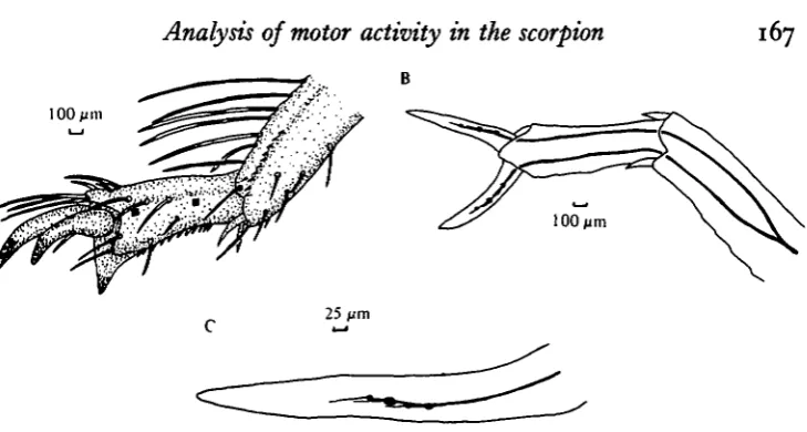
Fig. 1. (A) Camera lucida drawing of the posterior aspect of the tarsus of the left fourth walking leg. The approximate sites where the electrodes were inserted in the tarsal 2 seg-ment are denoted by the solid squares. (B) Dorsal view of the tarsus of the left fourth leg. The sensory nerve was cut near the tibia-tarsal 1 joint for backfilling with cobalt. The nerve bifurcates in the tarsal 1 segment yielding two separate distal sensory nerves which innervate the anterior and posterior faces of the tarsal segments. Within the lumen of each tarsal claw, three to four bipolar sense cells are present. (C) Drawing of the tarsal claw to show cobalt-filled, bipolar sensory neurones. The neurones are distributed in a linear fashion along the lumen of the tarsal claw. The dendrites can be seen to extend distally from the cell body for 50-75 /wn. It is not known if this represents the true limit of their distal projection.
an additional wax anchoring before coursing to the Grass P-15 preamplifiers situated overhead. The scorpion could walk freely in a rectangular plexiglas chamber on a grounded steel plate without pulling the electrode leads taut. Occasionally, the preparation was mechanically perturbed to evoke higher rates of stepping.
Electrical recordings were filmed from an oscilloscope, sometimes after recording a TEAC four-channel tape recorder. Most measurements from the film were on
accurate to within ± 5 ms. Data for scoprions walking in a straight line were punched on to cards for subsequent computer analysis. Regression lines were assessed as being significantly different from one another or from zero slope at the 95 % confidence level using Student's t test.
RESULTS
Anatomy and physiology of tarsal receptors
Sensory receptors of the tarsal claws and the numerous tactile hairs located on the tarsal 2 segment were stained by cobalt backfilling (Fig. 1 A). Axons from these receptors join to form two sensory leg nerves which innervate the anterior and posterior aspects of the tarsal 2 segment (Fig. 1B). The tactile hairs are each innervated by a single bipolar neurone. Clusters of 5-10 smaller cells of unknown function were sometimes noticed close to a tarsal hair cell body. Each tarsal claw has 3-4 bipolar neurones located within the lumen of the claw. One cell was consistently larger than the others (Fig. 1C).
[image:3.451.52.417.48.243.2]i segment, the ventral aspect of the tarsal 2 segment, and the tips of the tarsal clavM are generally in contact with the substrate. Activity recorded over this period by
electrodes implanted in the tarsal 2 segment, was exclusively sensory (no moto-neurones or muscles are located this far distally) and closely delineated the time period over which the leg was on the substrate. Stimulation of the tarsal claw with a mechanical probe showed that very little activity was recorded unless the tip of the claw was touched, and that this activity was recorded within 4 ms of stimulation (Fig. 2 A). There was very little activity in response to imposed or natural movements of the tarsal claw relative to the tarsal 2 segment. Although the receptors are phasic, they discharge over the entire stance phase provided the animal is moving (Fig. 2B).
EMGs
Electromyograms of the elevator muscle are characterized by several features which distinguish them from those of the depressor muscle (Fig. 2B). For the elevator, the timing of burst onset is clearcut, whereas burst termination is frequently indistinct, particularly at slower stepping rates. Such indistinct bursting has been observed in lobster (Macmillan, 1975) and crayfish (Barnes, 1977). Even though the T - F joint for leg 4 exhibits only one cycle of elevation-depression movement during each step cycle (Root & Bowerman, 1978), the elevator muscle is often activated by a burst phased with the swing phase initiation and a second burst that occurs phased with the depressor burst during the stance phase. The significance of this second burst, which occurs more often at higher rates of stepping, is unknown. A similar absence of strict reciprocity between antagonist walking leg muscles has been described in other arthropods, particularly for more distal joints (Macmillan, 1975; Barnes, 1977; Clarac & Ayers, 1977; Clarac, 1978; Burns & Usherwood, 1979). As noted in Fig. 3, elevator burst durations tend to increase in length at progressively slower rates of stepping. However, since the scattergram levels off at the longer step cycle times, the data were subdivided and treated as two separate populations.
The slopes of regression lines for data points to the left and data points to the right of a particular cycle time were compared for six different dividing values between 500 and 750 ms. The 600 ms value appeared the best choice for an inflection point for all relationships. The relationship of increasing elevator burst duration for increasing cycle times still holds below 600 ms (Fig. 3). Above 600 ms, elevator burst duration is constant on the average, i.e. statistically independent of cycle time.
Analysts of motor activity in the scorpion
169
1 1 t i t I
100 ms
Fig. 2. (A) The activity of a tarsal claw receptor as recorded by implanted leads in response to electromechanical stimulation of the single tarsal claw. An insect pin coupled to a bi-morph crystal driven by a square wave of variable duration and voltage was used to precisely stimulate the preparation. The timing of the applied stimulating pulse is presented over the nerve record. Since the electromechanical stimulator was close to the recording leads, there are on and off stimulus artifacts on the nerve recorded in addition to the sensory responses. (1) Stimulation of the tip of the claw for 50 ms produced eight spikes with an average frequency of about 125 spikes/s. The receptor adapts to this type of stimulation. The last interspike interval in the burst is about three times as long as the first interval. (2) In order to measure latency between stimulus onset and the first spike, a short 10 ms pulse was applied to the mechanical stimulator. The stimulator was positioned so as to stimulate just the tip of the claw. The amplitude of the square wave was adjusted so as to evoke just one spike over this time period. The maximal latency between stimulus onset and first spike was about 4 ms. With stronger stimuli, as during walking, the latency would be less than this value. (3) Stimulation of the base of the claw produced no comparable discharge. The entire claw was moved relative to the tarsal 2 segment, just as when the claw tip was stimulated. With this stimulation, some activity of low amplitude was recorded, which may be from the smaller cells in the tarsal claw lumen. Again, five sweeps were superimposed. (B) Electrical records of depressor muscle EMG (Dep.), elevator muscle EMG (Elev.), and tarsal claw receptor activity (T.C.R.) all from the right fourth leg. On the left, where the records start, the animal is standing still. As was typical, the first peripheral sign of walking was the onset of the depressor burst. Tarsal claw receptors were activated a few ms later. Between the termination of the depressor burst (•) and the onset of the elevator burst (•)» there is little electrical activity in either muscle. Shortly after the onset of the elevator EMG (•)» the leg is lifted ( f ) as indicated by the termination of the tarsal claw burst. These three events which occur during the stance to swing transition, depressor burst termination (•), elevator burst onset (•), and then leg lift ( f ), invariably occur in this sequence. Near the end of the swing phase, the elevator burst is terminated, the depressor muscle is activated, and the leg touches down ( | ) . While the onset of the tarsal claw burst is clearly delineated, the depressor burst onset, and particularly the elevator burst termi-nation are frequently indistinct. At the end of this stepping sequence, the preparation stops walking. Instead of abruptly terminating the fourth depressor EMG burst and acti-vating the elevator muscle for another step, the depressor activity merely fades away. The tarsal claw burst is terminated when the preparation stops walking.
Swing-stance phase transitions
1000
250 500 750 Elevator cycle time (ms)
1000 1250 1500
Fig. 3. Composite scattergram-regression lines for elevator burst duration versus step cycle time divided at the 600 ms cycle time. Data points for the regression ( ) above 600 ms are indicated by A ; those for the regression below 600ms ( ) indicated by O. The slope of the over 600 ms data is not significantly different from zero. This indicates that elevator burst duration is constant, on the average, for stepping frequencies slower than about 1 -66 c/s. The 196 data points for this figure were obtained from 4 different preparations.
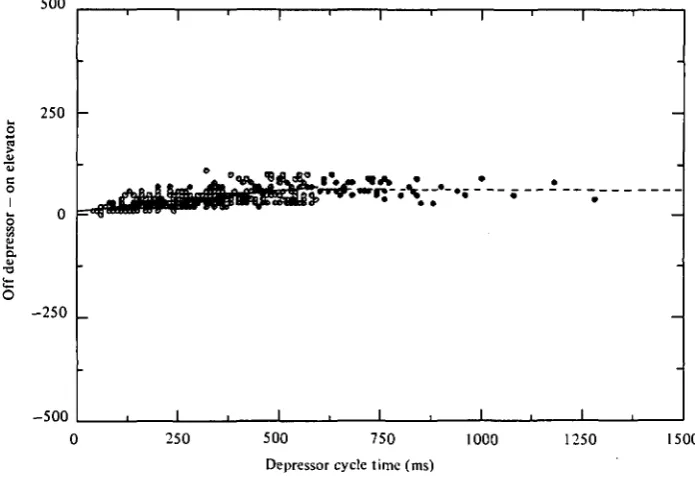
depressor and elevator activity. The depressor burst terminates abruptly; then about 40 ms later elevator onset occurs. Secondly, there is a statistically significant relation-ship such that switching latencies are increasingly greater at longer cycle times, up to 600 ms, where latency values level off.
For the swing to stance phase transition, the latency between time of elevator burst termination and depressor burst onset versus cycle time is illustrated in Fig. 6. Even though the average latency is about 12 ms, there are many instances where depressor onset precedes elevator burst termination.
Clearly, the motor programme is asymmetrical in that the stance to swing phase transition is more tightly controlled with no periods of antagonist co-activation, whereas the swing to stance phase transition is loosely coupled with brief but frequent
periods of co-activation.
Timing of EMG's relative to swing and stance phase movements
Analysis of motor activity in the scorpion
1711000
1250 1500
[image:7.451.50.396.38.301.2]500 750 1000 Depressor cycle time (ms)
Fig. 4. Composite scattergram-regression lines for depressor burst duration versus step cycle time for data divided at 6oo ms and displayed as for Fig. 3. The slope for the over 600 ms data is not significantly different from unity. This indicates that, on the average, all variation in cycle time for step frequencies slower than about i-66 c/s can be accounted for by variation in depressor burst variation. The 480 data points for this figure were obtained from 6 different preparations.
500
250
0
- 2 5 0
500 1
1 ' 1
-e
-1 . -1
• 1 1 1 . 1 .
-1 , 1 , 1 .
250 500 750 1000 1250 1500
Depressor cycle time (ms)
[image:7.451.39.387.369.607.2]o
250
--500
500 750 1000 1250 1500
Elevator cycle time (ms)
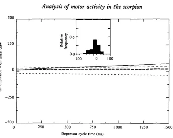
Fig. 6. A composite scattergram-regression line illustrating the relationship for the latency between elevator burst termination and depressor burst onset versus step cycle time. The 249 data points were obtained from six different preparations and are displayed as for Fig. 3. The negative data points indicate occurrences of elevator-depressor co-activation.
500
1«»
I o
250
--500
1 1 '
iv
e
J3
a:
-nc
y
<D
3
O*
a
10
0-5
0 0
-100
1
•
1
m•
•
•
0
1
100
1 1 1
1
-1
0 250 500 750 1000 1250 1500
Elevator cycle time (ms)
Analysis of motor activity in the scorpion
J73
500
* 250
o
--•a O
250
--500
1 1 '
>
3
u
-c
3 rr
f
| 0-5
0 0
--100
• M "H»
•
0
' tm — —
-1
100
, 1
1 1 '
-1 -1 -1
250 500 750 Depressor cycle time (ms)
[image:9.451.45.389.45.319.2]1000 1250 1500
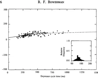
Fig. 8. The latency between the onset of the depressor burst and the onset of the tarsal claw receptor burst plotted against cycle time, for four different preparations. There are significant differences between different preparations for this relationship. The 201 data points from the four preparations are consolidated in the inset, which displays the relative frequency histogram. The mean latency is 9-3 ms, with a standard deviation of 27-8 ms.
this relationship, the specific timing of elevator muscle burst onset represents an accurate and reliable indicator of the time of swing phase initiation.
During the stance phase, both the depressor muscle and tarsal claw receptors are active. Although the depressor burst generally begins before the tarsal claw burst, there is considerable scatter in the relationship (Fig. 8). The depressor muscle may be activated from 60 ms before to 60 ms after re-establishment of leg contact with the substrate. This variation is due to variability both within and between preparations. In those instances where depressor muscle activity lags leg contact, the swing to stance phase switching must be due to the action of muscles controlling other joints and/or the influence of gravity on the leg after the elevator muscles have been turned off.
DISCUSSION
Elevator burst duration/depressor burst duration versus step cycle time
While the slope of the depressor relationship is steeper than that for the elevate relationship, neither relationship is truly linear, provided the breadth of cycle times sampled is wide enough. For example, consider the published figures for depressor burst direction versus step cycle time for the terrestrial preparations (Pearson, 1972; Fig. 6; Delcomyn & Usherwood, 1973, Fig. 6; Reingold & Camhi, 1977, Fig. 4; current paper, Fig. 4). In each case, the slope is steeper over the slower step cycles than the faster cycles. Even though there is no clear specific transition point in the scattergram, the slope transition appears to be near 300-400 ms for the cockroach and 500-600 ms for the scorpion. The basic relationship for the aquatic crustacean preparations is basically the same. The slope of the depressor burst duration versus step cycle time is steeper for the longer cycle times studied by Ayers & Davis (1977, Fig. 10) than for the shorter cycle times (Macmillan, 1975, Fig. 2A). The slope transition occurs between cycle times of 1-5 to 2-0 s. At the slower stepping rates, the slope of the elevator burst duration versus step cycle time decreases and may reach zero (Pearson, 1972, Fig. 5; Reingold & Camhi, 1977, Fig. 4; current paper, Fig. 3). Macmillan (1975) did not provide a figure, but stated that elevator burst durations were shorter cycle times. Delcomyn & Usherwood (1973) stated that there was no relationship between elevator burst and cycle time for the cockroach, but did not provide a figure. It would be interesting to see if such a relationship could be demonstrated over the shorter cycle times, since this was the case for the other two studies on cockroach P. americana preparations (Pearson, 1972, Reingold & Camhi, 1977). Clarac (1978, Fig. 3) and Ayers & Davis (1977, Fig. 10) found no relationship for elevator burst versus cycle time in their crustacean preparations. Perhaps this is because the step cycles analysed were longer than the 1 "5-2-0 s transition time noted for these slow walking aquatic crustaceans.
The elevator and depressor burst duration versus cycle time relationships are not linear. Protraction time/retraction time ratios (p/r) versus cycle times (Burns, 1973, Fig. 5; Macmillan, 1975, Table 8) and elevator/depressor phase scattergrams (Reingold & Camhi, 1977, Fig. 5) should be considered in this light. For example, the phase of the elevator burst within the depressor cycle time for the scorpion (current paper) is consistent at about 0-70 for cycle times below 600 ms, but becomes progressively larger at longer step cycle times. This is because the elevator burst duration remains at a constant value, so that the increasing depressor burst length occupies a successively greater portion of the total step cycle.
Analysts of motor activity in the scorpion 175
stepping (Pearson, 1972; Burrows & Hoyle, 1973; Delcomyn & Usherwood, 1973; Ayers & Davis, 1977).
Swing-stance phase transitions
There are also striking similarities between the various arthropods studied in the transition of motoneurone activity from one muscle to that of its antagonist. In all cases, the stance to swing phase shift is accomplished by a nearly synchronous depressor burst termination and elevator burst onset. This is noted in arachnids (Fig. 5), insects (Pearson & lies, 1970, Fig. 10; Delcomyn & Usherwood, 1973, no figure; Reingold & Camhi, 1977, Fig. 6), and crustaceans (Macmillan, 1975, Fig. 30; Ayers & Davis, 1977, Fig. 11). The relationships are relatively tight, i.e. low variation about mean values, with either no periods or only very short periods where both antagonists are active. The different preparations are also similar to one another in the swing to stance phase transition. The timing of the elevator burst termination is generally not as tightly related to the timing of depressor burst onset as for the reciprocal relationship described above (same figures except in the current paper where Fig. 6 instead of Fig. 5). There are also frequent periods of simultaneous co-contraction, with the elevator discharge continuing well into the stance phase in certain instances.
Therefore, for almost all arthropods studied, the output of the segmental oscillator is asymmetrical. There is little or no antagonist co-activation at the stance to swing phase transition and considerably more co-activation at the swing to stance phase transition. It may be that this asymmetry can be appreciated by considering the different functions of the muscles involved. The depressor muscles are active during the stance phase when the propulsive forces for locomotion are generated. They are generally larger and presumably capable of generating greater tensions than the smaller elevator muscles, which just lift the legs. If the tension in the depressor muscles during the stance phase is relatively large, there may be a premium on turning them off before or shortly after the elevator muscles are activated. This may help the elevator muscles to lift the leg as commanded. On the other hand, the tension in the elevator muscles may be relatively small and easily overcome at the end of the swing phase by contraction of the depressor muscles. Also, movements due to the elevator muscle must work against the forces of gravity, while those of the depressor muscles are aided by gravitational forces. Both factors would reduce the need for the elevators to be turned off before the depressor muscles could carry out their responsibilities.
s fa ? T3 O 500 250 _ 0
2 5 0
--500
1 '
_
1 1
•I •* •
1 * * lativ e <u & u 3 C U 1 ' 10 0-5 0 0 00 i 1
L
100 1 -— -— 200250 500 750 1000 1250 1500
[image:12.451.47.386.50.319.2]Depressor cycle time (ms)
Fig. 9. A composite scattergram-regression line illustrating the relationship for the latency between depressor burst termination and tarsal claw termination versus step cycle time. Step cycles were measured as the time between successive depressor burst terminations. This latency value reflects the time between the decision to step and the actual step itself. The 143 data points from the three preparations are consolidated in the relative frequency histo-gram shown in the inset.
that the decision to step had been made centrally (Fig. 2 B). The actual lifting of the leg may lag the central decision to step by over 100 ms at the slower step cycles (Fig. 9). If the segmental oscillator is viewed as a relaxation oscillator (Graham, 1977), then the most important point in the cycle is the time at which the oscillator is reset. This is the time at which the decision to step is made. The leg then completes the swing phase and is maintained in the stance phase until the next step decision is made. Therefore, depressor burst termination represents the best reference point for considering the temporal features of EMG activity for other muscles within the leg. The same is true for consideration of intersegmental stepping phase relationships, where the questions of interest concern the coupling of segmental oscillators.
REFERENCES
ALEXANDER, C. G. (1972). Locomotion in the isopod crustacean Ligia oceanica (Linn.). Comp. Biochem.
Physiol. 42A, 1039-1047.
AYERS, J. L. & DAVIS, W. J. (1977). Neuronal control of locomotion in the lobster. I. Motor programs for forward and backward walking. J. comp. Physiol. H5> I — 2
7-BABU, K. S. (1965). Anatomy of the central nervous system of arachnids. Zool. Jb. Anat. Bd. 8a,
I-I54-BARNES, W. J. P. (1975). Leg coordination during walking in the crab, Uca pugnax. J. comp. Physiol. 96, 237-256.
BARNES, W. J. P. (1977). Proprioceptive influences on motor output during walking in the crayfish.
543~s63-Analysis of motor activity in the scorpion 177
WERMAN, R. F. (197s). The control of walking in the scorpion. I. Leg movements during normal ing. J. comp. Physiol. ioo, 183-196.
BOWERMAN, R. F. & ROOT, T. M. (1978). External anatomy and muscle morphology of the walking legs of the scorpion Hadrurus arizonensis. Comp. Biochem. Physiol. 59A, 49-56.
BROWNELL, P. & FARLEY, R. D. (1979 a). Detection of vibrations in sand by tarsal sense organs of the nocturnal scorpion Paruroctonus mesaensis. J. comp. Physiol. 131, 23-30.
BROWNELL, P. & FARLEY, R. D. (19796). Orientation to vibrations in sand by the nocturnal scorpion,
Paruroctonus mesaensis: Mechanism of target location. J. comp. Physiol. 131, 31-38.
BUB, K. & BOWERMAN, R. F. (1979). Prey capture by the scorpion Hardurus arizonensis Ewing (Scor-piones: Vaejovidae). J. Arachnol. 7, 243—253.
BULLOCK, T. H. & HORRIDGE, G. A. (1965). Structure and function in the nervous system of inverte-brates. San Francisco: W. H. Freeman.
BURNS, M. D. (1973). The control of walking in orthoptera. I. Leg movements in normal walking.
J. exp. Biol. 58, 45-58.
BURNS, M. D. & USHERWOOD, P. N. R. (1979). The control of walking in orthoptera. II. Motor neuron activity in normal free walking animals. J. exp. Biol. 79, 69-98.
BURROWS, M. & HOYLE, G. (1973). The mechanism of rapid running in the ghost crab, Ocypope
ceratophthalma. J. exp. Biol. 58, 327-349.
CLARAC, F. (1978). Locomotory programs in basal leg muscles after limb autonomy in the Crustacea.
Brain Res. 145, 401-405.
CLARAC, F. & AYERS, J. (1977). La marche chez les Crustacea: activite motrice programmee et regulation peripherique. J. Physiol., Paris 73, 523-544.
DELCOMYN, F. (1973). Motor activity during walking in the cockroach Periplaneta americana. II. Tethered walking. J. exp. Biol. 59, 643-654.
DELCOMYN, F. & USHERWOOD, P. N. R. (1973). Motor activity during walking in the cockroach
Periplaneta americana. I. Free walking. J. exp. Biol. 59, 629-642.
EVOY, W. H. & FOURTNER, C. R. (1973). Nervous control of walking in the crab, Cardisoma guanhumi. III. Proprioceptive influences on intra- and intersegmental coordination. J. comp. Physiol. 83, 303-318.
EWING, A. W. & MANNING, A. (1966). Some aspects of the efferent control of walking in three cock-roach species. J. Insect Physiol. 12, 1115-1118.
GRAHAM, D. (1972). A behavioral analysis of the temporal organization of walking movements in the first instar and adult stick insect {Carausius morosus). J. comp. Physiol. 81, 23-52.
GRAHAM, D. (1977). Simulation of a model for the coordination of leg movements in free-walking . insects. Biol. Cybernetics 26, 187-198.
GRAHAM, D. (1978). Unusual step patterns in the free-walking grasshopper Neoconocephalus robustus I. General features of the step pattern. J. exp. Biol. 73, 147-157.
GWILLIAM, G. F. & COLE, E. S. (1979). The morphology of the central nervous system of the barnacle,
Semibalanus cariosus (Pallas), jf. Morph. 159, 297-310.
MACMILLAN, D. L. (1975). A physiological analysis of walking in the American lobster (Homarus
americanus). Phil. Trans. R. Soc. B 270, 1-59.
PEARSON, K. G. (1972). Central programming and reflex control of walking in the cockroach. J. exp.
Biol. 56,
173-193-POLIS, G. (1979). Prey and feeding phenology of the desert sand scorpion Paruroctonus mesaensis (Scorpionidae: Vaejovidae). J. Zool., Lond. 188, 333-346.
REINCOLD, S. C. & CAMHI, J. M. (1977). A quantitative analysis of leg movements during three different behaviors in the cockroach, Periplaneta americana. J. Insect Physiology 23, 1407—1420. ROOT, T. M. & BOWERMAN, R. F. (1978). Intra-appendage movements during walking in the scorpion
Hadrurus arizonensis. Comp. Biochem. Physiol. 59A, 57-63.
RUNION, H. I. & USHERWOOD, P. N. R. (1968). Tarsal receptors and leg reflexes in the locust and grasshopper. J. exp. Biol. 49, 421-436.
SHERMAN, E., NOVOTNY, M. & CAMHI, J. M. (1977). A modified walking rhythm employed during righting behavior in the cockroach Gromphadorhina portentosa. J. comp. Physiol. 113, 303-316. SPIRITO, C. P., EVOY, W. H. & BARNES, W. J. P. (1972). Nervous control of walking in the crab,