ISSN Online: 2165-7467 ISSN Print: 2165-7459
DOI: 10.4236/ojepi.2018.82007 May 25, 2018 76 Open Journal of Epidemiology
Characterization of Clinical and Environmental
Isolates of
Cryptococcus
neoformans/Cryptococcus gattii
Complex
Maintained in Yeast Culture Collection in São
Paulo, Brazil
Pedro Henrique Magalhães Cardoso
1, Bosco Christiano Maciel da Silva
2, Jairo Ivo dos Santos
3,
Rennan Luiz Oliveira dos Santos
4, Diniz Pereira Leite Júnior
5,6, Marcos Ereno Auler
7,
Luciana da Silva Ruiz
8, Eriques Gonçalves da Silva
4, Débora Moreira
4, Carina Domaneschi
4,
Francisco de Assis Baroni
9, Marcia de Souza Carvalho Melhem
10, Marilena dos Anjos Martins
10,
Claudete Rodrigues Paula
4*1Faculty of Veterinary Medicine and Zootechny-FMVZ-USP, University of São Paulo (USP), São Paulo, Brazil 2Faculty of Medicine/FMUSP, University of São Paulo (USP), São Paulo, Brazil
3Federal University of Santa Catarina (UFSC), Florianópolis, Brazil 4Faculty of Dentistry, University of São Paulo (FOUSP), São Paulo, Brazil 5Faculty of Medicine, Federal University of Mato Grosso (UFMT), Cuiabá, Brazil 6University Center of Várzea Grande (UNIVAG), Várzea Grande, Brazil
7Faculty of Pharmacy, University of the WesternCenter of Paraná (UNICENTRO), Guarapuava, Brazil 8Institute Adolfo Lutz (IAL), Bauru, Brazil
9Federal Rural University of Rio de Janeiro (UFRRJ), Seropédica, Brazil 10Institute Adolfo Lutz (IAL), São Paulo, Brazil
Abstract
Objective: As isolates of Cryptococcus are frequently kept collection stocks in institutions, sometimes without proper characterization, we sought to deter-mine the genotype profiles, protease and phospholipase activities “in vitro”
and the susceptibility testing for azoles and amphotericin B. Methodology: 84 isolates from several regions of Brazil (40 samples from clinical origin and 44 isolates from environmental origin) were maintained at the microorganism’s bank of the Biomedical Science Institute (ICB-USP) of the São Paulo Univer-sity, in São Paulo, Brazil. This isolates was submitted fungal strains determi-nation, DNA extraction and purification, determination of genotype by URA5 gene RFLP of CGB-positive isolates, protease and phospholipase activity and susceptibility to antifungals. Results: Of six CGB positive isolates tested by RFLP-PCR, only four presented a genomic profile consistent C. gattii species How to cite this paper: Cardoso, P.H.M.,
Da Silva, B.C.M., Dos Santos, J.I., Dos Santos, R.L.O., Leire-Jr, D.P., Auler, M.E., Ruiz, L.S., Da Silva, E.G., Moreira, D., Domaneschi, C., Baroni, F.A., Melhem, M.S.C., Martins, M.A. and Paula, C.R. (2018) Characterization of Clinical and Environmental Isolates of Cryptococcus neoformans/Cryptococcus gattii Complex Maintained in Yeast Culture Collection in São Paulo, Brazil. Open Journal of Epide-miology, 8, 76-92.
https://doi.org/10.4236/ojepi.2018.82007
Received: April 8, 2018 Accepted: May 22, 2018 Published: May 25, 2018
DOI: 10.4236/ojepi.2018.82007 77 Open Journal of Epidemiology (VGII), while two other were C. neoformans (VNI and VNIII), indicating the existence of canavanine-resistante C. neoformans isolates in the culture col-lections. The clinical isolates secreted higher levels of phospholipase and en-vironmental isolates but no differences were observed for the protease levels. Almost all isolates were sensible to azoles and amphotericin B. Conclusion: We point out in this research the existence of C. neoformans strains resistant to canavanine and intrinsic characteristic of C. gatti. These results demon-strate the importance to perform a detailed characterization of isolates kept in culture collections.
Keywords
Cryptococcus Complex, Genotyping, Protease, Phospholipase, Etest, Culture Collection
1. Introduction
Basidiomycetous yeasts of the genus Cryptococcus comprise seventy species, some of which are associated with invasive infections in humans and animals, such as Cryptococcus neoformans and Cryptococcus gattii which belong to the
Cryptococcus neoformans/Cryptococcus gattii complex [1] [2] [3]. C. neofor-mans comprises two varieties named variety grubii (serotype A) and variety
neoformans (serotype D), while C. gattii has two serotypes (B and C). They have distinct, though occasionally overlapping, ecological niches, with C. neoformans
often been isolated from pigeon (Columba livia) nests and droppings, and C. gattii often been isolated from decaying trees and soil [4][5][6][7].
C. neoformans and C. gattii can be phenotypically differentiated by L-canavanine glycine bromothymol blue (CGB) agar medium [8][9] while its genotype-based differentiation can be performed by PCR fingerprinting [10], restriction frag-ment length polymorphism (RFLP-PCR) typing [11][12], intergenic spacer se-quencing [13], amplified fragment length polymorphism analysis (AFLP) [14] and multilocus sequence typing (MLST) [15][16].
Phenotypic characteristics that are associated with the virulence of Crypto-coccus have also been studied since they play important roles in the fungal pa-thogenesis [17]. These include the capsule thickness [18] [19] [20], melanin production [21], urease, protease and phospholipase activities [22][23][24] and susceptibility profile to different antifungals [12][25][26][27].
Considering that the biological characteristics of many C. neoformans com-plex stocks in Brazil are not generally tested in detail, and that some isolates are recorded with outdated taxonomic nomenclatures or even misidentified, they need to be constantly reevaluated.
2. Objective
We sought to determine the genotype profiles, in vitro protease and phospholi-Copyright © 2017 by authors and
Scientific Research Publishing Inc. This work is licensed under the Creative Commons Attribution International License (CC BY 4.0).
http://creativecommons.org/licenses/by/4.0/
DOI: 10.4236/ojepi.2018.82007 78 Open Journal of Epidemiology pase activities and the susceptibility testing to azoles and amphotericin B, in clinical and environmental isolates of the C. neoformans/C. gattii complex, which are kept stored at the microorganism’s bank of the Instituto de Ciências Biomédicas (ICB-USP) of the Universidade de São Paulo (USP), in São Paulo, Brazil.
3. Material and Methods
3.1. Fungal Strains
This study was done on 84 isolates from several regions of Brazil. Of them, 40 samples were isolated from either cerebrospinal fluid or blood samples of human and animal cryptococcosis cases and 44 samples were isolated from environment bird droppings or the leaves of Eucalyptus. They were formerly identified as C. neoformans and were maintained in chloramphenicol-supplemented Sabouraud dextrose agar slants plus mineral oil, at the microorganism’s bank at the Labor-atory of Pathogenic Yeasts of the ICB-USP. Standard strains ICB 110—C. neo-formans var. grubii, ICB134—C. neoformans var. neoformans, ICB 162—C. gat-tii and ICB 12A—Candida albicans were used in all tests. The isolates were iden-tified by their urease activity, growth in L-canavanine glycine bromothymol blue (CGB) medium and assimilation of nitrate [3]. The institutional review board of ICB-USP committee approved this study (Protocol 367/2010).
3.2. DNA Extraction and Purification
DNA genotyping was done on CGB positive isolates. The DNA extraction was done according Martins et al. (2007) [28]. Briefly, the isolates were dissolved in a lysis buffer containing 10 mM Tris-HCl pH 8.0, 10 mM ethilenediaminetetraa-cetic acid (EDTA) pH 8.0, 0.5% sodium dodecyl sulfate (SDS), 0.01% N-laurilsarcozyl and 100 μg/mL proteinase K.
The mixtures were briefly vortexed and incubated at 56˚C for 2 hours or until the cell lysis was completed. DNA was extracted by phenol/chloroform/isoamyl alcohol (25:24:1) method and precipitated with isopropanol [29]. The DNA pel-let was washed with 70% ethanol, centrifuged for 10 min at 10,000 g and air-dried. The DNA was suspended in 200 μL of sterile ultra-pure water taining RNase 20 μg/mL, kept at 4˚C overnight and stored at −20˚C. DNA con-centrations were determined in O.D. at 260 nm, while DNA purity was deter-mined by the ratio of O.D. at 260 and 280 nm. For PCR amplification one μL of each DNA sample (85 - 90 ng) was used as DNA template [29][30].
3.3. Determination of Genotype by
URA
5 Gene RFLP of
CGB-Positive Isolates
DOI: 10.4236/ojepi.2018.82007 79 Open Journal of Epidemiology (5'-ATG/TCC/TCC/CAA/GCC/CTC/GAC/TCC/G-3') and SJ01
(5'-TTA/AGA/CCT/CTG/AAC/ACC/GTA/CTC-3') (Invitrogen, Carlsbad, CA, USA), 1.0 μL of DNA and 10.1 μL of nuclease-free water. The amplification of the fragments was performed in a Veriti® 96-Well Fast Thermal Cycler (Applied Biosystems TM, Thermo Fisher Scientific, Waltham, MA, USA). The cycling condition was 95˚C for 4 min initial denaturation, followed by 34 cycles of 94˚C for 45 sec of denaturation, 57˚C for 1 min of annealing, 72˚C for 1 min of exten-sion, followed by a final cycle of 72˚C for 10 minutes of final extension. Subse-quently, amplification products were mixed with one fifth volume of loading buffer (15% ficoll 400, 0.25% orange G, MiliQ water; 2.0 μL), 15.0 μL of ampli-fied PCR products double digested using Cfr13I (isoschizomer of Sau96I) (10 U μL; 1.0 μL) and HhaI (20 U μL, 1.0 μL), and 1.0 μL of sterile MiliQ water in a fi-nal volume of 20.0 μL (Thermo Fis her Scientific, Waltham, MA, USA), and then incubated in dry at 37˚C for 3 h or overnight. The restriction fragments were then separated by 2% agarose gel electrophoresis at 100 V for 40 min. RFLP pat-terns were assigned visually by comparison with the patpat-terns obtained from the standard reference isolates from eight molecular types: VNI-VNIV for C. neo-formans,and VGI-VGIV for C gattii [30].
3.4. Protease and Phospholipase Activity
Protease activity was performed in triplicate according to Rüchel et al. (1982) [31]. Briefly, a culture medium dish containing sterile bovine albumin fraction V (Sigma-Aldrich, St. Louis, MO, USA) was distributed into Petri dishes. Each iso-late was inocuiso-lated on the central surface of the piso-late and they were incubated at 32˚C in 5% CO2 for 15 days and examined every two days until the 15th day. A
translucent halo formed around the yeast colony indicated protease activity. Proteolytic activity (Pz value) was calculated as the ratio of the diameter of the colony (Dc) and to the diameter of the colony plus that of the degradation zone diameter (Dzd) (Pz = Dc/Dc + Dzd). Therefore, Pz = 1.00 indicated no activity (negative); Pz < 1.00 and > 0.64 indicated median activity (positive) and Pz < 0.64 indicated a strong activity (strongly positive) for protease activity.
Phospholipase activity was performed in triplicate by the egg yolk agar test [32]. Briefly, the isolates were previously cultured during 48 hours Sabouraud Dextrose Agar Supplemented with chloramphenicol (Difco Laboratories Inc., Detroit, MI, USA) and then inoculated on the central surface of the phospholi-pase test plate and were incubated at 32˚C in 5% CO2 for 15 days. An opaque
DOI: 10.4236/ojepi.2018.82007 80 Open Journal of Epidemiology
3.5. Susceptibility to Antifungals
Etest strips for fluconazole, itraconazole, voriconazole, posaconazole and am-photericin B were provided by AB Biodisk (Solna, Sweden) and the assay was done according the manufacturer’s instructions (http://www.abbiodisk.com/). Sterile fungal cell suspensions were adjusted to the turbidity of a 0.5 McFarland standard. Sterile and non-toxic swabs were dipped in these cell suspensions and inoculated on the surface of Petri dishes containing RPMI 1640 (Gibco-BRL, Grand Island, NY, USA) supplemented with 1.5% agar and 2% glucose (J.T.Baker® Chemicals, Center Valley, PA, USA), buffered with 3-(N-morpholino) propanesulfonic acid (MOPS) (Sigma-Aldrich, St. Louis, MO, USA) [33][34].
Etest strips were applied to each Petri dish, and the plates were incubated at 35˚C in 5% CO2. The strip MICs were read at 48 and 72 hours of incubation.
The azole minimum inhibitory concentration (MIC) was read at the lowest con-centration at which the border of the elliptical inhibition zone intercepted the scale on the strip. Any growth, such as microcolonies, throughout a discernible inhibition ellipse was ignored [34]. The amphotericin B MICs were read at the point at which the zone of almost complete inhibition intersected the strip. All susceptibility tests were repeated twice.
The interpretative criteria used for susceptibility to fluconazole, itraconazole, voriconazole, posaconazole and amphotericin B antifungals was done according to Clinical and Laboratory Standards Institute (CLSI) [35]: for fluconazole, sus-ceptible (S) ≤ 8 μg/mL; sussus-ceptible dose dependent (S-DD) 16 - 32 μg/mL; resis-tant (R) ≥ 64 μg/mL; for itraconazole, S ≤ 0.125 μg/mL; S-DD 0.25 - 0.5 μg/mL; R ≥ 1 μg/mL; for voriconazole S ≤ 1 μg/mL; S-DD 2 μg/mL; R ≥ 4 μg/mL; for posaconazole S ≤ 1 μg/mL; S-DD 2 μg/mL; R ≥ 4 μg/mL. Since no breakpoints have been published for amphotericin B, breakpoints chosen for it were (S ≤ 1 μg/ml, S; intermediate (I) 2 μg/mL; R ≥ 4 μg/mL) [36].
4. Results
The isolates of clinical and environmental origin were re-identified and were consistent with the colonial and cellular morphology and physiological characte-ristics of C. neoformans complex, as proposed by Kurtzman et al. (2011) [3]. The isolates were also cultured on CGB medium, for differentiation of C. neoformans
and C. gattii. Of the 40 isolates of clinical origin, 36 (90.0%) were identified as C. neoformans and four (10.0%) as C. gattii. On the other hand, of the 44 isolates of environment origin, 42 (95.5%) were identified as C. neoformans and two iso-lates (4.5%) as C. gattii.
4.1. RFLP of
URA
5 Gene of CGB-Positive Isolates
DOI: 10.4236/ojepi.2018.82007 81 Open Journal of Epidemiology
Figure 1. RFLP profiles of the URA5 genes from Cryptococcus gattii
obtained by double-digestion with HhaI and Cfr13I. Lanes 1 - 5: iso-lates 161, 182, 183, 184 e 189 respectively. Lanes 6 - 9 standards ge-notypes VGI, VGII, VGIII and VGIV respectively. M-100bp DNA ladder (Invitrogen).
[image:6.595.270.478.348.653.2]DOI: 10.4236/ojepi.2018.82007 82 Open Journal of Epidemiology
4.2. Protease and Phospholipase Activity
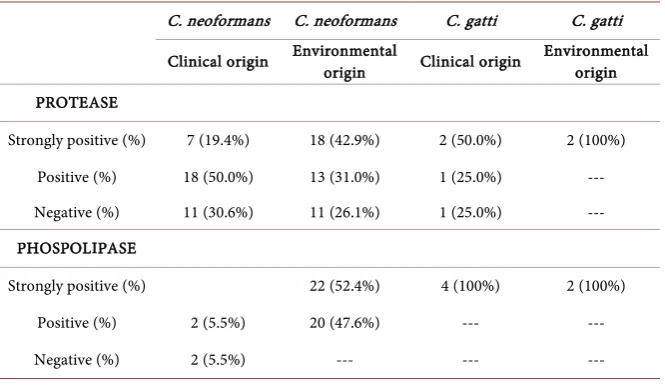
As shown in Table 1, of the 40 isolates of clinical origin nine (22.5%) were strongly positive for protease, 19 (47.5%) were positive and 12 (30.0%) were negative for protease. On the other hand, of the 44 isolates of environmental ori-gin 20 (45.5%) were strongly positive for protease, 13 (29.5%) were positive for protease and 11 (25.0%) were negative for protease. No statistically differences were observed in the groups for protease.
Of the 40 isolates of clinical origin 37 (92.5%) were strongly positive, one (2.5%) was positive and two (5.0%) were negative for phospholipase (Table 1). Of the 44 isolates of environmental origin 24 (54.5%) were strongly positive and 20 (45.5%) were positive for phospholipase. The difference between the groups was statistically significant for phospholipase (P = 0.0003).
4.3. Susceptibility to Antifungals
Fluconazole—In isolates of clinical origin, the MIC values ranged from 0.0940 to 4.000 μg/mL, with a mean value of 0.8091 μg/mL. In isolates of envi-ronment origin, the values ranged from 0.0320 to 8.000 μg/mL, with a mean value of 0.6200 μg/mL.
Itraconazole—In isolates of clinical origin, the MIC values ranged from 0.0080 to 0.2500 μg/mL, with a mean value of 0.0661 μg/mL. In isolates of envi-ronment origin, the values ranged from 0.0080 to 0.5000 μg/mL, with a mean value of 0.0388 μg/mL.
Voriconazole—In isolates of clinical origin, the MIC values ranged from 0.0020 to 0.0320 μg/mL, with a mean value of 0.0065 μg/mL. In isolates of envi-ronment origin, the values ranged from 0.0020 to 0.0640 μg/mL, with a mean value of 0.0057 μg/mL.
[image:7.595.208.541.533.728.2]Posaconazole—In isolates of clinical origin, the MIC values ranged from 0.0020 to 0.0940 μg/mL, with a mean value of 0.0214 μg/mL. In isolates of
Table 1. Production of protease and phospholipase by the isolates.
C. neoformans C. neoformans C. gatti C. gatti
Clinical origin Environmental origin Clinical origin Environmental origin
PROTEASE
Strongly positive (%) 7 (19.4%) 18 (42.9%) 2 (50.0%) 2 (100%) Positive (%) 18 (50.0%) 13 (31.0%) 1 (25.0%) --- Negative (%) 11 (30.6%) 11 (26.1%) 1 (25.0%) --- PHOSPOLIPASE
Strongly positive (%) 22 (52.4%) 4 (100%) 2 (100%)
Positive (%) 2 (5.5%) 20 (47.6%) --- ---
DOI: 10.4236/ojepi.2018.82007 83 Open Journal of Epidemiology environment origin, the values ranged from 0.0020 to 0.0640 μg/mL, with a mean value of 0.0178 μg/mL.
Amphotericin B—In isolates of clinical origin, the MIC values ranged from 0.0120 to 0.1250 μg/mL, with a mean value of 0.0490 μg/mL. Isolates of envi-ronmental origin presented MIC values ranging from 0.0020 to 0.1900 μg/mL, with a mean value of 0.0348 μg/mL.
5. Discussion
In this study all isolates of C. neoformans were retested in relation to biochemi-cal tests and the six isolates, four from clinibiochemi-cal origin and two from environ-mental origin, which were positive in CGB-positive, were submitted to molecu-lar typing by RFLP-PCR. Only four presented a genomic profile consistent with the C. gattii species (VGII), while two other were, in fact, C. neoformans (VNI and VNIII).
Traditionally, C. neoformans has been grouped in three varieties: C. neofor-mans var. gattii (serotype B and C), C. neoformans var. grubii (serotype A) and
C. neoformans var. neoformans (serotype D and AD) [37]. However, the gattii
variety has been renamed as C. gattii [3][38]. According this nomenclature, all our strains of C. neoformans were retested from the biochemical tests and the six strains that were positive in the CGB medium were then presumptively rec-lassified as C. gattii [3].
Recently, a new classification of C. neoformans complex has been proposed [39], adding new species, and the former species C. neoformans and C. gattii are now considered to be a complex of seven species: C. neoformans (former C. neoformans var. grubii genotypes VNI and VNII), Cryptococcus deneoformans
(former C. neoformans var. neoformans, genotype VNIV), C. gattii (former C. gattii genotype VGI), Cryptococcus bacillisporus (former C. gattii genotype VGIII), Cryptococcus deuterogattii (former C. gattii genotype VGII), Crypto-coccus tetragattii (former C. gattii VGIV), Cryptococcus decagattii (former C. gattii VGIV/VGIIIc), plus at least four hybrid of these species [39]. Considering this new classification, four strains of C. gattii of our stock could be named as
Cryptococcus deuterogattii and one as C. neoformans.
In relation to the methods used in the phenotypic characterization, Klein et al.
(2009) [8] analyzed 102 strains of yeast to verify urease activity, melanin produc-tion and glycine assimilaproduc-tion in the CGB medium as well as sequencing. The 17
C. gattii strains tested were positive in the CGB medium, the 54 C. neoformans
strains were negative in the CGB medium, one of which had weak staining activ-ity in the medium, six of 20 other Cryptococcus species were also positive in the CGB medium, but negative for melanin production. This demonstrates that there is a limitation in the exclusive use of CGB culture for identification of
DOI: 10.4236/ojepi.2018.82007 84 Open Journal of Epidemiology Indeed, in our study we observed that two strains of C. neoformans displayed urease activity which is not a common phenomenon. However, there are been some reports on urease activity by well characterized C. neoformans isolates [40] [41] [42]. The possible explanation is that these strains suffered some kind of mutations and became canavanine-resistant [40][41][42].
There are several published works that describe the importance of different extracellular enzymes, such as urease, protease, phospholipase and phenolox-idase, among others, that allow the yeast to alter pH in the phagolysosome, de-grades proteins of importance for the immune response of the host, favoring in-vasion [23][43]. In our study, most of the Cryptococcus strains, both of clinical origin and environmental origin, presented high protease values, although some of them, in the two groups, were not observed to produce significant amounts of this enzyme. This demonstrates the difficulty of correlating proteolytic activity observed by in vitro experiments with possible in vivo effects, for example, de-termining their actual contribution to disease development.
When the production of phospholipase was evaluated, it was observed that most of the isolates of clinical origin showed high production of the enzyme (92.5% were strongly positive and 2.5% positive), at a higher frequency than that observed in isolates of environmental origin (54.5% were strongly positive and 45.5% positive). Vidotto et al. (1996) [44] tested 23 isolates of C. neoformans for phospholipase production and found positivity in 95.7% (22) isolates, with a Pz ranged between 0.271 and 0.949.
According to the authors, there was a correlation between the phospholipase production and the size of the capsule in the isolated from AIDS patients. How-ever, in this study, it was not possible to establish a relationship between capsu-lar thickness and phospholipase production. In addition, genetic disruption stu-dies have revealed the importance of the phospholipase B gene in yeast virulence in animal models, observing a decrease in the activity of this enzyme when the gene is altered [45] [46]. Thus, phospholipase activity would be a more useful phenotypic marker to determine and evaluate the potential degree of virulence among isolates from clinical and environmental isolates of C. neoformans com-plex [45][46][47].
Extracellular phospholipases are a heterogeneous group of enzymes produced by several bacteria and fungi and have been implicated in their pathogenicity because they can cause damage to host cell membranes that result in the destabi-lization of membranes, cell lysis and release of lipid second messengers [48][49] [50]. Studies of Candida albicans demonstrated that phospholipase activity was correlated with mucosal invasion [51] and increased mortality in mice [52]. Al-so, production of phospholipases A, B, C and D by Aspergillus fumigatus has been reported [52][53].
condi-DOI: 10.4236/ojepi.2018.82007 85 Open Journal of Epidemiology tion as AIDS patients and presents the pigeon droppings and soil of as the main reservoir. In our study, the majority of isolates of environmental origin used came from pigeon droppings and other birds, where the most frequent species is
C. neoformans [6][59][60][61][62]. Although C. neoformans and C. gattii oc-cupy similar ecological niches, C. gattii is mainly associated with deteriorated wood, plant remains and soil. In particular, eucalyptus trees represent the most common substrate for C. gattii [62]-[67], and we could confirm this, as our sam-ples isolated from Eucalyptus leaves belong to the C. gattii species.
Cryptococcal infections in humans usually are treated with amphotericin B, associated or not with 5-flucytosine, almost followed by maintenance therapy with some azole antifungal, mainly fluconazole. High rates of cryptococcosis re-cidiva suggest a potential emergence of antifungal resistance, clinical or other-wise, among C. neoformans isolates [68][69][70]. In view of this situation, it is necessary to routinely determine the sensitivity profile of C. neoformans isolates to different antifungal agents and, in addition, to allow an evaluation of the effi-cacy of the treatments used and the evolution of the disease.
In this present study, we used disk diffusion tests, the commercial Etest kit that has been described as a good method for routine laboratory use, proving to have a good alternative for in vitro sensitivity tests for azoles [71][72][73]. This method allows the determination of MICs, and has shown excellent benefits, such as easy execution and quick results [74]. It also demonstrates good correla-tion with the standard method by CLSI [35] which is more labor-intensive and a long reading time. Although CLSI methods are not considered ideal for testing
C. neoformans[75] [76], broth microdilution and agar diffusion method have been applied worldwide to study the antifungal sensitivities of C. neoformans
isolates.
Both clinical and environmental isolates were highly sensible to the antifun-gals amphotericin B, posaconazole and voriconazole. Several studies have re-ported that amphotericin B as well as new triazoles (voriconazole and posaco-nazole) has potent activity against C. neoformans, where more than 99% of the isolates show MICs ≤ 1.0000 μg/mL, regardless of geographic origin of isolates [77] [78] [79], suggering that this new triazoles may bealternative treatements when similar isolates are finded in clinical practice [79][80].
DOI: 10.4236/ojepi.2018.82007 86 Open Journal of Epidemiology one can be better assess their characteristics as new laboratory tools become available.
6. Conclusion
We point out in this research the existence of C. neoformans strains resistant to canavanine and intrinsic characteristic of C. gatti. The other important results obtained were the higher levels of phospholipase by clinical strains than envi-ronmental strains but no differences were observed for the protease levels. Se-lected phenotypic characteristics of C. neoformans and C. gatti complex must be compared to molecular studies to perform a correct diagnosis to the species.
Acknowledgements
The authors would like to thank for the funds provided by Conselho Nacional de Desenvolvimento Científico e Tecnológico (CNPq), Coordenação de Aper-feiçoamento de Pessoal de Nível Superior (CAPES) and by Fundação de Amparo à Pesquisa do Estado de São Paulo (FAPESP)Grant 11/06950-5.
Conflict of Interest
All authors declared that there is no conflict of interest.
References
[1] Brandt, M.E., Hutwagner, L.C., Klug, L.A., Baughman, W.S., Rimland, D., Gravis, E.A., Hamill, R.J., Thomas, C., Pappas, P.G., Reingold, A.L. and Pinner, R.W.(1996) Molecular Subtype Distribution of Cryptococcus neoformans in Four Areas of the United States. Cryptococcal Disease Active Surveillance Group. Journal of Clinical Microbiology, 34, 912-917.
[2] Iqbal, N., DeBess, E.E., Wohrle, R., Sun, B., Nett, R.J., Ahlquist, A.M., Chiller, T., Lockhart, S.R., et al. (2010) Correlation of Genotype and In Vitro Susceptibilities of
Cryptococcus gattii Strains from the Pacific Northwest of the United States. Journal of Clinical Microbiology, 48, 539-544. https://doi.org/10.1128/JCM.01505-09
[3] Kurtzman, C.P., Fell, J.W. and Boekhout, T. (2011) The Yeasts: A Taxonomic Study. 5th Edition, ACM Press, NY.
[4] Emmons, C.W. (1951) Isolation of Cryptococcus neoformans from Soil. Journal of Bacteriology, 62, 685-690.
[5] Emmons, C.W. (1955) Saprophytic Sources of Cryptococcus neoformans Associated with the Pigeon (Columba livia). American Journal of Hygiene, 62, 227-232.
https://doi.org/10.1093/oxfordjournals.aje.a119775
[6] Baroni, F.A., Paula, C.R., Silva, E.G., Viani, F.C., Rivera, I.N., Oliveira, M.T.B. and Gabale, W.(2006) Cryptococcus neoformans Strains Isolated from Church Towers in Rio de Janeiro City, RJ, Brazil. The Revista do Instituto de Medicina Tropical de São Paulo, 48, 71-75. https://doi.org/10.1590/S0036-46652006000200003
[7] Lin, X. and Heitman, J. (2006) The Biology of the Cryptococcus neoformans Species Complex. Annual Review of Microbiology, 60, 69-105.
https://doi.org/10.1146/annurev.micro.60.080805.142102
DOI: 10.4236/ojepi.2018.82007 87 Open Journal of Epidemiology
(2009) Identification of Cryptococcus gattii by Use of L-Canavanine Glycine Bro-mothymol Blue Medium and DNA Sequencing. Journal of Clinical Microbiology, 47, 3669-3672. https://doi.org/10.1128/JCM.01072-09
[9] Kwon-Chung, K.J., Polacheck, I. and Bennett, J.E. (1982) Improved Diagnostic Me-dium for Separation of Cryptococcus neoformans var. neoformans (Serotypes A and D) and Cryptococcus neoformans var. gattii (Serotypes B and C). Journal of Clinical Microbiology, 15, 535-537.
[10] Ellis, D., Marriott, D., Hajjeh, R.A., Warnock, D., Meyer, W. and Barton, R.(2000) Epidemiology: Surveillance of Fungal Infections. Medical Mycology, 38, 173-182.
https://doi.org/10.1080/mmy.38.s1.173.182
[11] Meyer, W., Castañeda, A., Jackson, S., Huynh, M., Castañeda, E., et al. (2003) Mo-lecular Typing of IberoAmerican Cryptococcus neoformans Isolates. Emerging In-fectious Diseases, 9, 189-195. https://doi.org/10.3201/eid0902.020246
[12] Silva, D.C., Martins, M.A., Szeszs, M.W., Bonfietti, L.X., Matos, D. and Melhem, M.S.(2012)Susceptibility to Antifungal Agents and Genotypes of Brazilian Clinical and Environmental Cryptococcus gattii Strains. Diagnostic Microbiology and Infec-tious Disease, 72, 332-339. https://doi.org/10.1016/j.diagmicrobio.2011.11.016
[13] Diaz, M.R., Boekhout, T., Kiesling, T. and Fell, J.W. (2005) Comparative Analysis of the Intergenic Spacer Regions and Population Structure of the Species Complex of the Pathogenic Yeast Cryptococcus neoformans. FEMS Yeast Research, 5, 1129-1140.
https://doi.org/10.1016/j.femsyr.2005.05.005
[14] Boekhout, T., Theelen, B., Diaz, M., Fell, J.W., Hop, W.C., Abeln, E.C., Dromer, F. and Meyer, W. (2001) Hybrid Genotypes in the Pathogenic Yeast Cryptococcus neoformans. Microbiology, 147, 891-907.
https://doi.org/10.1099/00221287-147-4-891
[15] Kidd, S.E., Guo, H., Bartlett, K.H., Xu, J. and Kronstad, J.W. (2005) Comparative Gene Genealogies Indicate that Two Clonal Lineages of Cryptococcus gattii in Brit-ish Columbia Resemble Strains from Other Geographical Areas. Eukaryotic Cell, 4, 1629-1638. https://doi.org/10.1128/EC.4.10.1629-1638.2005
[16] Fraser, J.A., Giles, S.S., Wenink, E.C., Geunes-Boyer, S.G., Wright, J.R., Diezmann, S., Allen, A., Stajich, J.E., Dietrich, F.S., Perfect, J.R. and Heitman, J. (2005) Same-Sex Mating and the Origin of the Vancouver Island Cryptococcus gattii Out-break. Nature, 437, 1360-1364. https://doi.org/10.1038/nature04220
[17] Steenbergen, J.N. and Casadevall, A. (2003) The Origin and Maintenance of Viru-lence for the Human Pathogenic Fungus Cryptococcus neoformans. Microbes and Infection, 5, 667-675. https://doi.org/10.1016/S1286-4579(03)00092-3
[18] Chang, Y.C. and Kwon-Chung, K.J. (1994) Complementation of a Capsule-Deficient Mutation of Cryptococcus neoformans Restores Its Virulence. Molecular and Cel-lular Biology, 14, 4912-4919. https://doi.org/10.1128/MCB.14.7.4912
[19] Rivera, J., Feldmesser, M., Cammer, M. and Casadevall, A. (1998) Organ-Dependent Variation of Capsule Thickness in Cryptococcus neoformans during Experimental Murine Infection. Infection and Immunity, 66, 5027-5030.
[20] Zaragoza, O., Rodrigues, M.L., De Jesus, M., Frases, S., Dadachova, E. and Casadevall, A. (2009)The Capsule of the Fungal Pathogen Cryptococcus neoformans. Advances in Applied Microbiology, 68, 133-216.
https://doi.org/10.1016/S0065-2164(09)01204-0
DOI: 10.4236/ojepi.2018.82007 88 Open Journal of Epidemiology
[22] Cox, G.M., Mukherjee, J., Cole, G.T., Casadevall, A. and Perfect, J.R. (2000) Urease as a Virulence Factor in Experimental Cryptococcosis. Infection and Immunity, 68, 443-448. https://doi.org/10.1128/IAI.68.2.443-448.2000
[23] Cox, G.M., McDade, H.C., Chen, S.C., Tucker, S.C., Gottfredsson, M., Wright, L.C., Sorrell, T.C., Leidich, S.D., Casadevall, A., Ghannoum, M.A. and Perfect, J.R. (2001) Extracellular Phospholipase Activity Is a Virulence Factor for Cryptococcus neo-formans. Molecular Microbiology, 39, 166-175.
[24] Liu, G.Y. and Nizet, V. (2009) Color Me Bad: Microbial Pigments as Virulence Fac-tors. Trends in Microbiology, 17, 406-413.
[25] Moraes, E.M., Prímola, N.S. and Hamdan, J.S. (2003) Antifungal Susceptibility of Clinical and Environmental Isolates of Cryptococcus neoformans to Four Antifun-gal Drugs Determined by Two Techniques. Mycoses, 46, 164-168.
https://doi.org/10.1046/j.1439-0507.2003.00872.x
[26] Souza, L.K., Fernandes, O.F., Kobayashi, C.C., Passos, X.S., Costa, C.R., Lemos, J.A, Souza-Jr., H.S. and Silva, M.R.R.(2005)Antifungal Susceptibilities of Clinical and Environmental Isolates of Cryptococcus neoformans in Goiânia City, Goiás, Brazil.
The Revista do Instituto de Medicina Tropical de São Paulo, 47, 253-256.
https://doi.org/10.1590/S0036-46652005000500003
[27] Chowdhary, A., Randhawa, H.S., Sundar, G., Kathuria, S., Prakash, A., Khan, Z, Sun, S. and Xu, J.(2011) In Vitro Antifungal Susceptibility Profiles and Genotypes of 308 Clinical and Environmental Isolates of Cryptococcus neoformans var. grubii
and Cryptococcus gattii Serotype B from Northwestern India. Journal of Medical Microbiology, 60, 961-967. https://doi.org/10.1099/jmm.0.029025-0
[28] Martins, M.A., Pappalardo, M.C., Melhem, M.S. and Pereira-Chioccola, V.L. (2007) Molecular Diversity of Serial Cryptococcus neoformans Isolates from AIDS Patients in the City of São Paulo, Brazil. The Memórias do Instituto Oswaldo Cruz, 102, 777-784. https://doi.org/10.1590/S0074-02762007000700001
[29] Sambrook, J., Fritsch, E.F. and Maniatis, T. (1989) Molecular Cloning: A Laboratory Manual. 2nd Edition, Cold Spring Harbor Laboratory Press, New York.
[30] Macdougall, L., Kidd, S.E., Galanis, E., Mak, S., Leslie, M.J., Cieslak, P.R., Kronstad, J.W., Morsehed, M.G. and Bartlett, K.H. (2007) Spread of Cryptococcusgattii in British Columbia, Canada, and Detection in the Pacific Northwest, USA. Emergent Infectious Diseases Journal, 13, 42-50. https://doi.org/10.3201/eid1301.060827 [31] Rüchel, R., Tegeler, R. and Trost, M. (1982) A Comparison of Secretory Proteinases
from Different Strains of Candida albicans. Sabouraudia, 20, 233-244.
https://doi.org/10.1080/00362178285380341
[32] Price, M.F., Wilkinson, I.D. and Gentry, L.O. (1982) Plate Method for Detection of Phospholipase Activity in Candida albicans. Sabouraudia, 20, 7-14.
https://doi.org/10.1080/00362178285380031
[33] Lozano-Chiu, M., Paetznick, V.L., Ghannoum, M.A. and Rex, J.H. (1998) Detection of Resistance to Amphotericin B among Cryptococcus neoformans Clinical Isolates: Performances of Three Different Media Assessed by Using E-Test and National Committee for Clinical Laboratory Standards M27-A Methodologies. Journal of Clinical Microbiology, 36, 2817-2822.
[34] Pfaller, M.A., Messer, S.A., Houston, A., Mills, K., Bolmstrom, A. and Jones, R.N. (2000) Evaluation of the Etest Method for Determining Voriconazole Susceptibili-ties of 312 Clinical Isolates of Candida Species by Using Three Different Agar Me-dia. Journal of Clinical Microbiology, 38, 3715-3717.
Di-DOI: 10.4236/ojepi.2018.82007 89 Open Journal of Epidemiology
lution Antifungal Susceptibility Testing of Yeasts. 4th Informational Supplement, CLSI Document M27-S4, Clinical and Laboratory Standards Institute, Wayne, PA. [36] Kumar, D., Bhattacharyya, S., Gupta, P., Banerjee, G. and Singh, M. (2015)
Com-parative Analysis of Disc Diffusion and E-Test with Broth Micro-Dilution for Sus-ceptibility Testing of Clinical Candida Isolates against Amphotericin B, Flucona-zole, Voriconazole and Caspofungin. Journal of Clinical and Diagnostic Research, 9, DC01-DC04.
[37] Franzot, S.P., Salkin, I.F. and Casadevall, A. (1999) Cryptococcus neoformans var.
grubii: Separate Varietal Status for Cryptococcus neoformans Serotype A Isolates.
Journal of Clinical Microbiology, 37, 838-840.
[38] Heitman, J. (2011) Microbial Pathogens in the Fungal Kingdom. Fungal Biology Reviews, 25, 48-60. https://doi.org/10.1016/j.fbr.2011.01.003
[39] Hagen, F., Khayhan, K., Theelen, B., Kolecka, A., Polacheck, I., Sionov, E., Falk, R., Parnmen, S., Lumbsch, H.T. and Boekhout, T. (2015) Recognition of Seven Species in the Cryptococcus gattii/Cryptococcus neoformans Species Complex. Fungal Ge-netics and Biology, 78, 16-48. https://doi.org/10.1016/j.fgb.2015.02.009
[40] Polacheck, I. and Kwon-Chung, K.J. (1986) Canavanine Resistance in Cryptococcus neoformans. Antimicrobial Agents and Chemotherapy, 29, 468-473.
https://doi.org/10.1128/AAC.29.3.468
[41] Nakamura, Y., Kano, R., Sato, H., Watanabe, S., Takahashi, H. and Hasegawa, A. (1998) Isolates of Cryptococcus neoformans Serotype A and D Developed on Ca-navanine-Glycine-Bromthymol Blue Medium. Mycoses, 41, 35-40.
[42] Khan, Z.U., Al-Anezi, A.A., Chandy, R. and Xu, J. (2003) Disseminated Cryptococ-cosis in an AIDS Patient Caused by A Canavanine-Resistant Strain of Cryptococcus neoformans var. grubii. Journal of Medical Microbiology, 52, 271-275.
https://doi.org/10.1099/jmm.0.05097-0
[43] Casadevall, A. and Perfect, J.R. (1998) Cryptococcus neoformans. ASM Press, Washington DC.
[44] Vidotto, V., Sinicco, A., Di Fraia, D., Cardaropoli, S., Aoki, S. and Ito-Kuwa, S. (1996) Phospholipase Activity in Cryptococcus neoformans. Mycopathologia, 136, 119-123. https://doi.org/10.1007/BF00438916
[45] Chen, S.C., Muller, M., Zhou, J.Z., Wright, L.C. and Sorrell, T.C. (1997) Phospholi-pase Activity in Cryptococcus neoformans: A New Virulence Factor? The Journal of Infectious Diseases, 175, 414-420. https://doi.org/10.1093/infdis/175.2.414
[46] Titball, R.W. (1993) Bacterial Phospholipases C. Microbiological Reviews, 57, 347-366.
[47] Cardoso, P.H.M., dos Santos, R.L.O., Souza, A.C., Baroni, F.A., Domaneschi, C. and Paula, C.R. (2018) Biosynthesis of Phospholipases and Proteinases Using Clinical and Environmental Strains of Cryptococcus neoformans. Clinical and Laboratorial Research in Dentistry, 1, 1-5.
http://dx.doi.org/10.11606/issn.2357-8041.clrd.2018.133520
[48] Ghannoum, M.A. (2000) Potential Role of Phospholipases in Virulence and Fungal Pathogenesis. Clinical Microbiology Reviews, 13, 122-143.
https://doi.org/10.1128/CMR.13.1.122-143.2000
[49] Schmiel, D.H. and Miller, V.L. (1999) Bacterial Phospholipases and Pathogenesis.
Microbes and Infection, 1, 1103-1112.
https://doi.org/10.1016/S1286-4579(99)00205-1
DOI: 10.4236/ojepi.2018.82007 90 Open Journal of Epidemiology
Phospholipase Activity, Cellular Adherence and Pathogenicity of Yeasts. Journal of General Microbiology, 131, 1217-1221.
https://doi.org/10.1099/00221287-131-5-1217
[51] Ibrahim, A.S., Mirbod, F., Filler, S.G., Banno, Y., Cole, G.T., Kitajima, Y., Edwards-Jr., J.E., Nozawa, Y. and Ghannoum, M.A. (1995) Evidence Implicating Phospholipase as a Virulence Factor of Candida albicans. Infection and Immunity, 63, 1993-1998. [52] Birch, M., Robson, G., Law, D. and Denning, D.W. (1996) Evidence of Multiple
Extracellular Phospholipase Activities of Aspergillus fumigatus. Infection and Im-munity, 64, 751-755.
[53] Levitz, S.M. (1991) The Ecology of Cryptococcus neoformans and the Epidemiology of Cryptococcosis. Reviews of Infectious Diseases, 13, 1163-1169.
[54] Li, A., Nishimura, K., Taguchi, H., Tanaka, R., Wu, S. and Miyaji, M. (1993) The Isolation of Cryptococcus neoformans from Pigeon Droppings and Serotyping of Naturally and Clinically Sourced Isolates in China. Mycopathologia, 124, 1-5.
https://doi.org/10.1007/BF01103049
[55] Pappalardo, M.C. and Melhem, M.S. (2003) Cryptococcosis: A Review of the Bra-zilian Experience for the Disease. The Revista do Instituto de Medicina Tropical de São Paulo, 45, 299-305. https://doi.org/10.1590/S0036-46652003000600001
[56] Soares, M.C., Paula, C.R., Dias, A.L., Caseiro, M.M. and Costa, S.O.P. (2005) Envi-ronmental Strains of Cryptococcus neoformans Variety grubii in the City of Santos, SP, Brazil. The Revista do Instituto de Medicina Tropical de São Paulo, 47, 31-36.
https://doi.org/10.1590/S0036-46652005000100006
[57] Kwon-Chung, K.J., Fraser, J.A., Doering, T.L., Wang, Z., Idnurm, A. and Bahn, Y.S. (2014) Cryptococcus neoformans and Cryptococcus gattii, the Etiologic Agents of Cryptococcosis. Cold Spring Harbor Perspectives in Medicine, 4, a019760.
https://doi.org/10.1101/cshperspect.a019760
[58] Ajello, L. (1958) Ocurrence of Cryptococcus neoformans in Solis. American Journal of Hygiene, 67, 72-77.
[59] Lazera, M.S., Salmito Cavalcanti, M.A., Londero, A.T., Trilles, L., Nishikawa, M.M. and Wanke, B. (2000) Possible Primary Ecological Niche of Cryptococcus neofor-mans. Medical Mycology, 38, 379-383. https://doi.org/10.1080/mmy.38.5.379.383
[60] Escandón, P., Sánchez, A., Martínez, M., Meyer, W. and Castañeda, E. (2006) Mo-lecular Epidemiology of Clinical and Environmental Isolates of the Cryptococcus neoformans Species Complex Reveals a High Genetic Diversity and the Presence of the Molecular Type VGII Mating Type an in Colombia. FEMS Yeast Research, 6, 625-635. https://doi.org/10.1111/j.1567-1364.2006.00055.x
[61] Refojo, N., Perrotta, D., Brudny, M., Abrantes, R., Hevia, A.I. and Davel, G. (2009) Isolation of Cryptococcus neoformans and Cryptococcus gattii from Trunk Hollows of Living Trees in Buenos Aires City, Argentina. Medical Mycology, 47, 177-184.
https://doi.org/10.1080/13693780802227290
[62] Ellis, D.H. and Pfeiffer, T.J. (1990) Natural Habitat of Cryptococcus neoformans
var. gattii. Journal of Clinical Microbiology, 28, 1642-1644.
[63] Montenegro, H. and Paula, C.R. (2000) Environmental Isolation of Cryptococcus neoformans var. gattii and C. neoformans var. neoformans in the City of São Paulo, Brazil. Medical Mycology, 38, 385-390. https://doi.org/10.1080/mmy.38.5.385.390
DOI: 10.4236/ojepi.2018.82007 91 Open Journal of Epidemiology
Grande do Sul. FEMS Yeast Research, 3, 405-415.
https://doi.org/10.1016/S1567-1356(03)00038-2
[65] Granados, D.P. and Castañeda, E. (2006) Influence of Climatic Conditions on the Isolation of Members of the Cryptococcus neoformans Species Complex from Trees in Colombia from 1992-2004. FEMS Yeast Research, 6, 636-644.
https://doi.org/10.1111/j.1567-1364.2006.00090.x
[66] Mitchell, T.G., Castaneda, E., Niersen, K. and Wanke, B.M.S.L. (2011) Environ-mental Niches for Cryptococcus neoformans and Cryptococcus gattii. In: Heitman, J., et al., Eds., Cryptococcus: From Human Pathogen to Model Yeast, ASM Press, Washington DC, 237-259.
[67] Berg, J., Clancy, C.J. and Nguyen, M.H. (1998) The Hidden Danger of Primary Flu-conazole Prophylaxis for Patients with AIDS. Clinical Infectious Diseases, 26, 186-187. https://doi.org/10.1086/517056
[68] Brandt, M.E., Pfaller, M.A., Hajjeh, R.A., Hamill, R.J., Pappas, P.G., Reingold, A.L., Rimland, D. and Warnock, D.W. (2001) Cryptococcal Disease Active Surveillance Group. Trends in Antifungal Drug Susceptibility of Cryptococcus neoformans Iso-lates in the United States: 1992 to 1994 and 1996 to 1998. Antimicrobial Agents and Chemotherapy, 45, 3065-3069. https://doi.org/10.1128/AAC.45.11.3065-3069.2001
[69] Dias, A.L., Matsumoto, F.E., Melhem, M.S., Silva, E.G., Auler, M.E., Siqueira, A.M. and Paula, C.R. (2006) Comparative Analysis of Etest and Broth Microdilution Me-thod (AFST-EUCAST) for Trends in Antifungal Drug Susceptibility Testing of Bra-zilian Cryptococcus neoformans Isolates. Journal of Medical Microbiology, 55, 1693-1699. https://doi.org/10.1099/jmm.0.46789-0
[70] Matsumoto, F.E., Dias, A.L., Melhem, M.S., Szeszs, M.W., Auler, M.E., Ruiz, L.S., Gonçalves da Silva, E., Gandra, R.F. and Paula, C.R. (2007) Antifungal Susceptibility of Bloodstream Yeasts Isolated at a Public Children’s Hospital in Brazil: Compari-son of the Etest® and the AFST-EUCAST Microdilution Method. Canadian Journal of Microbiology, 53, 1300-1306. https://doi.org/10.1139/W07-095
[71] Tewari, A., Behera, B., Mathur, P. and Xess, I. (2012) Comparative Analysis of the Vitek 2 Antifungal Susceptibility System and E-Test with the CLSI M27-A3 Broth Microdilution Method for Susceptibility Testing of Indian Clinical Isolates of
Cryptococcus neoformans. Mycopathologia, 173, 427-433.
https://doi.org/10.1007/s11046-012-9528-9
[72] Mahabeer, Y., Chang, C.C., Naidu, D., Dorasamy, A., Lewin, S., Ndung’u, T., Moo-sa, M.Y., French, M., Mlisana, K. and Coovadia, Y. (2014) Comparison of Etests and Vitek 2® to Broth Microdilution for the Susceptibility Testing of Cryptococcus neo-formans. Diagnostic Microbiology and Infectious Disease, 80, 294-298.
https://doi.org/10.1016/j.diagmicrobio.2014.09.006
[73] Negri, M., Henriques, M., Svidzinski, T.I., Paula, C.R. and Oliveira, R. (2009) Cor-relation between Etest, Disk Diffusion, and Microdilution Methods for Antifungal Susceptibility Testing of Candida Species from Infection and Colonization. Journal of Clinical Laboratory Analysis, 23, 324-330. https://doi.org/10.1002/jcla.20337
[74] Ghannoum, M.A., Ibrahim, A.S., Fu, Y., Shafig, M.C., Edwards Jr., J.E. and Criddle, R.S. (1992) Susceptibility Testing of Cryptococcus neoformans: A Microdilution Technique. Journal of Clinical Microbiology, 30, 2881-2886.
DOI: 10.4236/ojepi.2018.82007 92 Open Journal of Epidemiology Infectious Diseases, 22, 322-328. https://doi.org/10.1093/clinids/22.2.322
[76] Alves, S.H., Oliveira, L.T., Costa, J.M., Lubeck, I., Casali, A.K. and Vainstein, M.H. (2001) In Vitro Susceptibility to Antifungal Agents of Clinical and Environmental
Cryptococcus neoformans Isolated in Southern of Brazil. The Revista do Instituto de Medicina Tropical de São Paulo, 43, 267-270.
https://doi.org/10.1590/S0036-46652001000500006
[77] Bii, C.C., Makimura, K., Abe, S., Taguchi, H., Mugasia, O.M., Revathi, G., Wamae, N.C. and Kamiya, S. (2007) Antifungal Drug Susceptibility of Cryptococcus neo-formans from Clinical Sources in Nairobi, Kenya. Mycoses, 50, 25-30.
https://doi.org/10.1111/j.1439-0507.2006.01293.x
[78] Capoor, M.R., Mandal, P., Deb, M., Aggarwal, P. and Banerjee, U. (2008) Current Scenario of Cryptococcosis and Antifungal Susceptibility Pattern in India: A Cause for Reappraisal. Mycoses, 51, 258-265.
https://doi.org/10.1111/j.1439-0507.2007.01478.x
[79] Thompson, G.R., Wiederhold, N.P., Fothergill, A.W., Vallor, A.C., Wickes, B.L. and Petterson, T.F. (2009) Antifungal Susceptibilities among Different Serotypes of
Cryptococcus gattii and Cryptococcus neoformans. Antimicrobial Agents and Chemotherapy, 53, 309-311. https://doi.org/10.1128/AAC.01216-08
[80] Tomazin, R., Matos, T., Meis, J.F. and Hagen, F. (2018) Molecular Characterization and Antifungal Susceptibility Testing of Sequentially Obtained Clinical Cryptococ-cus deneoformans and Cryptococcus neoformans Isolates from Ljubljana, Slovenia.