Received 17 December 2018 Accepted 20 December 2018
Edited by M. Weil, Vienna University of Technology, Austria
Keywords:pyrimidine; piperazine; crystal structure; molecular conformation; hydrogen bonding; supramolecular assembly.
CCDC reference:1886570
Supporting information:this article has supporting information at journals.iucr.org/e
The crystal structure of
1-(2-iodobenzoyl)-4-(pyrimidin-2-yl)piperazine: a three-dimensional
hydrogen-bonded framework, augmented by
p
–
p
stacking interactions and I
N halogen bonds
Ninganayaka Mahesha,aHemmige S. Yathirajan,a* Tetsundo Furuya,bTakashiro Akitsuband Christopher Glidewellc
aDepartment of Studies in Chemistry, University of Mysore, Manasagangotri, Mysuru-570 006, India,bDepartment of
Chemistry, Faculty of Science, Tokyo University of Science, 1-3 Kagurazaka, Shinjuku-ku, Tokyo 162-8601, Japan, and
cSchool of Chemistry, University of St Andrews, St Andrews, Fife KY16 9ST, UK. *Correspondence e-mail:
In 1-(2-iodobenzoyl)-4-(pyrimidin-2-yl)piperazine, C15H15IN4O, the central
piperazine ring adopts an almost perfect chair conformation with the pyrimidine substituent in an equatorial site. The planar amide unit makes a dihedral angle of 80.44 (7) with the phenyl ring. A combination of C—H O and C—
H (arene) hydrogen bonds links the molecules into a complex three-dimensional network structure, augmented by a–stacking interaction and an I N halogen bond, all involving different pairs of inversion-related molecules. Comparisons are made with the structures of a number of related compounds.
1. Chemical context
Pyrimidine derivatives are well represented amongst the range of heterocyclic compounds that exhibit a broad spectrum of biological activities such as analgesic and anti-inflammatory activity (Aminet al., 2009), antibacterial (Kuyperet al., 1996), antidepressant (Kim et al., 2010), antimicrobial and anti-oxidant (Padmajaet al., 2009) and antiviral activities (Ibrahim & El-Metwally, 2010). Piperazine-based compounds also exhibit anti-cancer properties (Abdel-Jalil et al., 2005), while the combination of pyrimidine and piperazine units is found in buspirone, 8-[4-(4-pyrimidin-2-ylpiperazin-1-yl)butyl]-8-aza-spiro[4.5]decane-7,9-dione (Tollefson et al., 1991), which can be used in the treatment of anxiety. With these considerations in mind, we have now synthesized the title compound (I) (Fig. 1), and we report here its molecular and supramolecular structure. Compound (I) was prepared by reaction of 1-(2-pyrimidyl)piperazine with 2-iodobenzoic acid in the presence of dimethylaminopropyl)-3-ethylcarbodimide as the dehy-drating agent.
2. Structural commentary
Within the molecule of compound (I) (Fig. 1), the piperazine ring adopts an almost perfect chair conformation. The ring-puckering parameters, calculated for the atom sequence (N1, C2, C3, N4, C5, C6) areQ= 0.557 (3) A˚ ,= 1.2 (3)and’=
258 (14), while for an idealized chair form the value ofis
0.0(Boeyens, 1978). The pyrimidine substituent at the
pyra-midal atom N4 occupies an equatorial site, but the amidic unit at atom N1 is effectively planar, and the r.m.s. deviation from the mean plane of atoms (C2, N1, C6, C17, O17 and C11) is only 0.027 A˚ . The dihedral angle between this plane and the aryl ring (C11–C16) is 80.44 (7). The molecules of (I) thus
exhibit no internal symmetry and so are conformationally chiral, as confirmed by the centrosymmetric space group in which the molecule crystallizes.
3. Supramolecular features
The supramolecular assembly of compound (I) is built from two C—H O hydrogen bonds, involving the aryl and pyri-midyl atoms C16 and C45 as the donors (Table 1), and one C— H (arene) hydrogen bond: there is a further inter-molecular C—H O contact, involving atom C13, but here the D—H A angle is less than 140, and so this contact
cannot be regarded as structurally significant (Wood et al., 2009). There are also present in the structure a–stacking interaction between pairs of pyrimidine rings and an I N halogen bond.
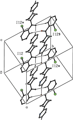
The hydrogen bonds give rise to a three-dimensional network structure of considerable complexity, but this is readily analysed in terms of three one-dimensional sub-structures (Fergusonet al., 1998a,b; Gregsonet al., 2000). The action of the two C—H O hydrogen bonds in combination links molecules related by inversion and translation into a chain of edge-fused rings running parallel to the [001] direc-tion (Fig. 2), in which R2
2(10) (Etter, 1990; Etteret al., 1990;
Bernstein et al., 1995) rings centred at (0.5, 0.5, n + 0.5) alternate with R2
4(28) rings centred at (0.5, 0.5, n), where n
represents an integer in each case.
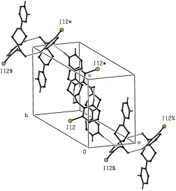
A second sub-structure can be identified in which the C— H (arene) hydrogen bond links molecules related by a 21
[image:2.610.44.298.74.214.2]screw axis to form a chain running parallel to the [010] direction (Fig. 3). The chains parallel to [010] and [001] each Table 1
Hydrogen-bond geometry (A˚ ,).
#Cg1 is the centroid of the C11–C16 ring.
D—H A D—H H A D A D—H A
C16—H16 O17i 0.95 2.50 3.395 (3) 157 C45—H45 O17ii 0.95 2.57 3.508 (3) 169
C46—H46 Cg1iii 0.95 2.72 3.596 (3) 153
Symmetry codes: (i)xþ1;yþ1;zþ1; (ii)x;y;zþ1; (iii)xþ1 2;yþ
1 2;zþ
[image:2.610.314.564.254.670.2]3 2.
Figure 2
Part of the crystal structure of compound (I) showing the formation of a hydrogen-bonded chain of edge-fused rings parallel to the [001] direction. Hydrogen bonds are drawn as dashed lines and, for the sake of clarity, the H atoms not involved in the motif shown have been omitted. The I atoms marked with an asterisk (*), a hash (#) or a dollar sign ($) are at the symmetry positions (1x, 1y, 1z), (x,y, 1 +z) and (1x, 1y, 2z), respectively.
Figure 1
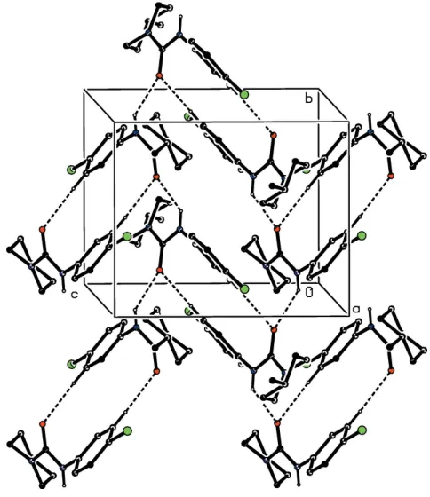
use only one type of hydrogen bond, but the alternating action
of the C—H O and C—H (arene) hydrogen bonds
involving atoms C16 and C46 as the donors (Table 1) links the molecules into a chain of rings running parallel to the [111] direction (Fig. 4). The combination of chains running parallel to [010], [001] and [111] suffices to generate a continuous three-dimensional network structure.
The formation of the hydrogen-bonded network is
augmented by two further intermolecular interactions, each of which involves inversion related pairs of molecules. The pyrimidine rings of the molecules at (x,y,z) and (1x, 1y, 2z), which are components of the hydrogen-bonded chain along [001], are strictly parallel with an interplanar spacing of 3.4295 (10) A˚ and a ring-centroid separation of 3.4924 (6) A˚, thus giving rise to a–stacking interaction (Fig. 5). Finally, we note a short intermolecular I N contact with geometrical parameters of I12 N41i= 3.168 (2) A˚ and C12—I12 N41i 174.83 (7)[symmetry code: (i)x, 1y, 1z], which can be
regarded as a halogen bond (Gildayet al., 2015; Cavalloet al., 2016).
4. Database survey
It is of interest briefly to compare the structure of compound (I) reported here with those of some related structures which have been recently reported. In
2-{4-[(1,3-benzodioxol-5-yl)methyl]piperazin-1-yl}pyrimidine (II), the molecules are linked into sheets by a combination of C—H (arene) and C—H (pyrimidine) hydrogen bonds (Wu et al., 2013). N-(4-Chlorophenyl)-4-(pyrimidin-2-yl)piperazine-1-carbox-amide (III) crystallizes withZ0 = 2 in space groupP2
1/c(Li,
2011b), and the molecules are linked into chains by two independent N—H O hydrogen bonds: these chains, parallel to [100], are of theC22(8) type rather than of theC(4) type as
originally reported. However, the original report overlooked
the presence of C—H O hydrogen bonds which, in
[image:3.610.314.566.380.654.2]combination with the N—H O hydrogen bond within the selected asymmetric unit, generates a second chain, this time running parallel to the [010] direction (Fig. 6), so that overall the supramolecular assembly takes the form of a sheet parallel to (001). In the simpler analogue N-(4-chlorophenyl)-4-methylpiperidine-1-carboxamide (IV), the assembly was reported (Li, 2011a) as consisting of simpleC(4) chains built from N—H O hydrogen bonds. However, the presence in (IV) of a C—H O hydrogen bond was overlooked, and the two hydrogen bonds together generate a complex sheet structure lying parallel to (100) (Fig. 7). Finally, we note also the structures of a number of salts of the 4-(pyrimidin-2-yl)piperazin-1-ium cation, including the chloride and nitrate
Figure 4
Part of the crystal structure of compound (I) showing the formation of a hydrogen-bonded chain of rings parallel to the [111] direction. Hydrogen bonds are drawn as dashed lines and, for the sake of clarity, the H atoms not involved in the motif shown have been omitted. The I atoms marked with an asterisk (*), a hash (#), a dollar sign ($), an ampersand (&) or a percent sign (%) are at the symmetry positions (1x, 1y, 1z), (1
2x, 1
2+y, 3 2z), (
1 2+x,
3 2y,
1 2+z), (
1 2+x,
1 2y,
1
2+z) and (1.5x, 1 2+y, 1
[image:3.610.68.272.407.664.2]2z) respectively.
Figure 3
Part of the crystal structure of compound (I) showing the formation of a hydrogen-bonded chain parallel to the [010] direction. Hydrogen bonds are drawn as dashed lines and, for the sake of clarity, the H atoms not involved in the motif shown have been omitted. The I atoms marked with an asterisk (*), a hash (#) or a dollar sign ($) are at the symmetry positions (1
2x, 1 2+y,
3 2z), (
1 2x,
1 2+y,
3
(Yamunaet al., 2014a), the hydrogenfumarate (Yamunaet al., 2014b) and the butanoate (Yamunaet al., 2014c).
5. Synthesis and crystallization
1-(2-Pyrimidyl)piperazine was purchased from Sigma– Aldrich. For the synthesis of compound (I), 1-(3-dimethyl-aminopropyl)-3-ethylcarbodimide (52 mg, 0.6 mmol), 1-hy-droxybenzotriazole (81 mg, 0.6 mmol) and triethylamine (0.5 ml, 1.8 mmol) were added to a solution of 2-iodobenzoic acid (0.6 mmol) in N,N-dimethylformamide (5 ml) and the resulting mixture stirred for 20 mins at 273 K. A solution of 1-(2-pyrimidyl)piperazine (100 mg, 0.6 mmol) in N,N-di-methylformamide (5 ml) was then added and stirring was continued overnight at ambient temperature. The reaction was confirmed to be complete using thin-layer chromatography, and the mixture was then quenched with water (10 ml) and extracted with ethyl acetate (20 ml). The organic layer was separated and washed successively with an aqueous hydro-chloric acid solution (1 mol dm3), a saturated solution of sodium hydrogencarbonate and then with brine. The organic phase was dried over anhydrous sodium sulfate and the solvent was removed under reduced pressure. Crystals suitable for single-crystal X-ray diffraction were grown by slow evaporation, at ambient temperature and in the presence of air, of a solution in methanol; m. p. 450–452 K.
Figure 6
[image:4.610.89.224.73.444.2]Part of the crystal structure of compound (III) showing the formation of a hydrogen-bonded chain parallel to [010]. The original atomic coordinates (Li, 2011b) have been used and, for the sake of clarity, the H atoms not involved in the motif shown have been omitted.
Figure 7
[image:4.610.318.560.413.693.2]Part of the crystal structure of compound (IV) showing the formation of a hydrogen-bonded sheet parallel to (100). The original atomic coordinates (Li, 2011a) have been used and, for the sake of clarity, the H atoms not involved in the motif shown have been omitted.
Figure 5
[image:4.610.45.302.508.698.2]6. Refinement
Crystal data, data collection and structure refinement details are summarized in Table 2. All H atoms were located in difference maps, and they were subsequently treated as riding atoms in geometrically idealized positions with C—H distances of 0.95 A˚ (aromatic) or 0.99 A˚ (CH2), and with
Uiso(H) = 1.2Ueq(C).
Acknowledgements
NM is grateful to the University of Mysore for research facilities.
Funding information
HSY is grateful to the UGC, New Delhi for the award of a BSR Faculty Fellowship for three years.
References
Abdel-Jalil, R. J., Aldoqum, H. M., Ayoub, M. T. & Voeller, W. (2005). Heterocycles,65, 2061–2070.
Amin, K. M., Hanna, M. M., Abo-Youssef, H. E. & George, R. F. (2009).Eur. J. Med. Chem.44, 4572–4584.
Bernstein, J., Davis, R. E., Shimoni, L. & Chang, N.-L. (1995).Angew. Chem. Int. Ed. Engl.34, 1555–1573.
Boeyens, J. C. A. (1978).J. Cryst. Mol. Struct.8, 317–320.
Bruker (2004).APEX2. Bruker AXS Inc., Madison, Wisconsin, USA. Bruker (2015).SADABSandSAINT. Bruker AXS Inc., Madison,
Wisconsin, USA.
Cavallo, G., Metrangolo, P., Milani, R., Pilati, T., Priimagi, A., Resnati, G. & Terraneo, G. (2016).Chem. Rev.116, 2478–2601. Etter, M. (1990).Acc. Chem. Res.23, 120–126.
Etter, M. C., MacDonald, J. C. & Bernstein, J. (1990).Acta Cryst.B46, 256–262.
Ferguson, G., Glidewell, C., Gregson, R. M. & Meehan, P. R. (1998a). Acta Cryst.B54, 129–138.
Ferguson, G., Glidewell, C., Gregson, R. M. & Meehan, P. R. (1998b). Acta Cryst.B54, 139–150.
Gilday, L. C., Robinson, S. W., Barendt, T. A., Langton, M. J., Mullaney, B. R. & Beer, P. D. (2015). Chem. Rev. 115, 7118– 7195.
Gregson, R. M., Glidewell, C., Ferguson, G. & Lough, A. J. (2000). Acta Cryst.B56, 39–57.
Ibrahim, D. A. & El-Metwally, A. M. (2010).Eur. J. Med. Chem.45, 1158–1166.
Kim, J. Y., Kim, D., Kang, S. Y., Park, W.-K., Kim, H. J., Jung, M. E., Son, E.-J., Pae, A. N., Kim, J. & Lee, J. (2010).Bioorg. Med. Chem. Lett.20, 6439–6442.
Kuyper, L. F., Garvey, J. M., Baccanari, D. P., Champness, J. N., Stammers, D. K. & Beddell, C. R. (1996).Bioorg. Med. Chem.4, 593–602.
Li, Y.-F. (2011a).Acta Cryst.E67, o1796. Li, Y.-F. (2011b).Acta Cryst.E67, o2575.
Padmaja, A., Payani, T., Reddy, G. D. & Padmavathi, V. (2009).Eur. J. Med. Chem.44, 4557–4566.
Sheldrick, G. M. (2015a).Acta Cryst.A71, 3–8. Sheldrick, G. M. (2015b).Acta Cryst.C71, 3–8. Spek, A. L. (2009).Acta Cryst.D65, 148–155.
Tollefson, G. D., Lancaster, S. P. & Montague-Clouse, J. (1991). Psychopharmacol. Bull.27, 163–170.
Wood, P. A., Allen, F. H. & Pidcock, E. (2009).CrystEngComm,11, 1563–1571.
Wu, C., Li, J., Wei, H., Hang, Y. & Jiang, Y. (2013).Acta Cryst.E69, o1140.
Yamuna, T. S., Jasinski, J. P., Kaur, M., Anderson, B. J. & Yathirajan, H. S. (2014a).Acta Cryst.E70, 203–206.
Yamuna, T. S., Jasinski, J. P., Kaur, M., Anderson, B. J. & Yathirajan, H. S. (2014c).Acta Cryst.E70, o1063–o1064.
[image:5.610.44.292.89.357.2]Yamuna, T. S., Kaur, M., Jasinski, J. P. & Yathirajan, H. S. (2014b). Acta Cryst.E70, o702–o703.
Table 2
Experimental details.
Crystal data
Chemical formula C15H15IN4O
Mr 394.21
Crystal system, space group Monoclinic,P21/n
Temperature (K) 173
a,b,c(A˚ ) 9.6417 (17), 13.604 (2), 12.174 (2)
() 105.155 (2)
V(A˚3) 1541.3 (4)
Z 4
Radiation type MoK
(mm1) 2.08
Crystal size (mm) 0.670.560.16
Data collection
Diffractometer Bruker APEXII CCD
Absorption correction Multi-scan (SADABS; Bruker, 2015)
Tmin,Tmax 0.345, 0.717
No. of measured, independent and observed [I> 2(I)] reflections
8152, 3452, 3188
Rint 0.067
(sin/)max(A˚
1) 0.652
Refinement
R[F2> 2(F2)],wR(F2),S 0.028, 0.075, 1.05
No. of reflections 3452
No. of parameters 190
H-atom treatment H-atom parameters constrained
max, min(e A˚
3) 0.96,0.74
Computer programs: APEX2 (Bruker, 2004), SAINT (Bruker, 2015), SHELXT
sup-1
Acta Cryst. (2019). E75, 129-133
supporting information
Acta Cryst. (2019). E75, 129-133 [https://doi.org/10.1107/S205698901801811X]
The crystal structure of 1-(2-iodobenzoyl)-4-(pyrimidin-2-yl)piperazine: a
three-dimensional hydrogen-bonded framework, augmented by
π
–
π
stacking
interactions and I
···
N halogen bonds
Ninganayaka Mahesha, Hemmige S. Yathirajan, Tetsundo Furuya, Takashiro Akitsu and
Christopher Glidewell
Computing details
Data collection: APEX2 (Bruker, 2004); cell refinement: SAINT (Bruker, 2015); data reduction: SAINT (Bruker, 2015); program(s) used to solve structure: SHELXT (Sheldrick, 2015a); program(s) used to refine structure: SHELXL2014
(Sheldrick, 2015b); molecular graphics: PLATON (Spek, 2009); software used to prepare material for publication:
SHELXL2014 (Sheldrick, 2015b) and PLATON (Spek, 2009).
1-(2-Iodobenzoyl)-4-(pyrimidin-2-yl)piperazine
Crystal data
C15H15IN4O Mr = 394.21
Monoclinic, P21/n a = 9.6417 (17) Å
b = 13.604 (2) Å
c = 12.174 (2) Å
β = 105.155 (2)°
V = 1541.3 (4) Å3
Z = 4
F(000) = 776
Dx = 1.699 Mg m−3
Mo Kα radiation, λ = 0.71073 Å Cell parameters from 3152 reflections
θ = 2.3–27.6°
µ = 2.08 mm−1 T = 173 K Plate, colour
0.67 × 0.56 × 0.16 mm
Data collection
Bruker APEXII CCD diffractometer
Radiation source: fine focus sealed tube Graphite monochromator
Detector resolution: 0.3333 pixels mm-1 φ and ω scans
Absorption correction: multi-scan
(SADABS; Bruker, 2015)
Tmin = 0.345, Tmax = 0.717
8152 measured reflections 3452 independent reflections 3188 reflections with I > 2σ(I)
Rint = 0.067
θmax = 27.6°, θmin = 2.3° h = −12→10
k = −17→16
l = −15→15
Refinement
Refinement on F2
Least-squares matrix: full
R[F2 > 2σ(F2)] = 0.028 wR(F2) = 0.075 S = 1.05
3452 reflections 190 parameters 0 restraints
sup-2
Acta Cryst. (2019). E75, 129-133
H-atom parameters constrained
w = 1/[σ2(F
o2) + (0.0235P)2 + 0.7279P]
where P = (Fo2 + 2Fc2)/3
(Δ/σ)max = 0.001
Δρmax = 0.96 e Å−3
Δρmin = −0.74 e Å−3
Special details
Geometry. All esds (except the esd in the dihedral angle between two l.s. planes) are estimated using the full covariance matrix. The cell esds are taken into account individually in the estimation of esds in distances, angles and torsion angles; correlations between esds in cell parameters are only used when they are defined by crystal symmetry. An approximate (isotropic) treatment of cell esds is used for estimating esds involving l.s. planes.
Fractional atomic coordinates and isotropic or equivalent isotropic displacement parameters (Å2)
x y z Uiso*/Ueq
N1 0.2388 (2) 0.44984 (14) 0.52539 (16) 0.0253 (4) C2 0.2263 (3) 0.36447 (15) 0.5950 (2) 0.0282 (6)
H2A 0.2567 0.3046 0.5613 0.034*
H2B 0.1248 0.3559 0.5964 0.034*
C3 0.3195 (3) 0.37777 (17) 0.7159 (2) 0.0290 (6)
H3A 0.3033 0.3225 0.7640 0.035*
H3B 0.4222 0.3777 0.7159 0.035*
N4 0.2839 (2) 0.47043 (14) 0.76229 (16) 0.0260 (4) C5 0.2977 (3) 0.55610 (16) 0.69257 (18) 0.0247 (5)
H5A 0.3991 0.5637 0.6904 0.030*
H5B 0.2689 0.6163 0.7265 0.030*
C6 0.2028 (3) 0.54263 (17) 0.57233 (19) 0.0273 (5)
H6A 0.1005 0.5421 0.5735 0.033*
H6B 0.2174 0.5980 0.5239 0.033*
C17 0.2929 (3) 0.45026 (16) 0.43404 (17) 0.0211 (4) O17 0.3118 (2) 0.52552 (12) 0.38448 (14) 0.0334 (4) C11 0.3328 (3) 0.35268 (15) 0.39109 (19) 0.0195 (5) C12 0.2434 (2) 0.30692 (15) 0.29625 (17) 0.0191 (4) I12 0.03709 (2) 0.36186 (2) 0.21850 (2) 0.02547 (8) C13 0.2883 (3) 0.22226 (16) 0.25098 (19) 0.0257 (5)
H13 0.2268 0.1909 0.1867 0.031*
C14 0.4237 (3) 0.18428 (17) 0.3008 (2) 0.0327 (6)
H14 0.4553 0.1269 0.2699 0.039*
C15 0.5130 (3) 0.22889 (19) 0.3947 (2) 0.0346 (6)
H15 0.6054 0.2021 0.4284 0.042*
C16 0.4677 (3) 0.31314 (18) 0.4401 (2) 0.0279 (5)
H16 0.5293 0.3438 0.5048 0.033*
N41 0.2769 (2) 0.57158 (16) 0.91400 (17) 0.0301 (5) C42 0.3025 (2) 0.48172 (17) 0.87784 (18) 0.0221 (4) N43 0.3414 (2) 0.40176 (15) 0.94349 (17) 0.0280 (4) C44 0.3528 (3) 0.4150 (2) 1.0545 (2) 0.0328 (6)
H44 0.3790 0.3601 1.1039 0.039*
C45 0.3288 (3) 0.5033 (2) 1.1012 (2) 0.0318 (6)
H45 0.3381 0.5108 1.1804 0.038*
C46 0.2902 (3) 0.5804 (2) 1.0258 (2) 0.0338 (6)
sup-3
Acta Cryst. (2019). E75, 129-133
Atomic displacement parameters (Å2)
U11 U22 U33 U12 U13 U23
N1 0.0418 (12) 0.0171 (8) 0.0180 (9) 0.0026 (9) 0.0095 (8) −0.0025 (7) C2 0.0455 (17) 0.0190 (11) 0.0247 (13) −0.0072 (10) 0.0172 (12) −0.0029 (8) C3 0.0496 (17) 0.0193 (10) 0.0211 (12) 0.0030 (12) 0.0149 (11) 0.0016 (9) N4 0.0421 (12) 0.0213 (9) 0.0161 (9) 0.0009 (10) 0.0101 (8) −0.0012 (7) C5 0.0377 (13) 0.0191 (10) 0.0182 (11) 0.0013 (11) 0.0088 (9) −0.0022 (8) C6 0.0411 (14) 0.0223 (10) 0.0181 (10) 0.0059 (11) 0.0069 (10) −0.0043 (9) C17 0.0304 (11) 0.0176 (9) 0.0124 (9) 0.0005 (10) 0.0007 (8) −0.0020 (8) O17 0.0646 (13) 0.0173 (7) 0.0196 (8) 0.0013 (9) 0.0133 (8) 0.0014 (6) C11 0.0302 (12) 0.0145 (9) 0.0150 (10) −0.0006 (9) 0.0079 (9) 0.0014 (7) C12 0.0263 (11) 0.0174 (9) 0.0153 (9) −0.0017 (9) 0.0084 (8) 0.0003 (8) I12 0.02670 (12) 0.02407 (11) 0.02409 (11) −0.00088 (5) 0.00387 (8) −0.00347 (5) C13 0.0421 (14) 0.0163 (10) 0.0218 (11) −0.0010 (11) 0.0141 (10) −0.0016 (8) C14 0.0491 (16) 0.0192 (11) 0.0374 (14) 0.0070 (12) 0.0250 (12) 0.0005 (10) C15 0.0345 (13) 0.0304 (13) 0.0408 (14) 0.0108 (13) 0.0132 (11) 0.0083 (11) C16 0.0313 (12) 0.0261 (11) 0.0238 (11) 0.0009 (11) 0.0031 (9) 0.0020 (9) N41 0.0378 (12) 0.0343 (11) 0.0186 (10) 0.0082 (10) 0.0081 (8) −0.0043 (8) C42 0.0214 (10) 0.0286 (11) 0.0174 (10) −0.0028 (10) 0.0070 (8) −0.0009 (9) N43 0.0368 (12) 0.0279 (10) 0.0214 (10) −0.0055 (10) 0.0115 (8) 0.0019 (8) C44 0.0384 (14) 0.0388 (14) 0.0224 (12) −0.0092 (13) 0.0099 (10) 0.0063 (10) C45 0.0351 (13) 0.0452 (14) 0.0169 (11) −0.0073 (13) 0.0098 (9) −0.0023 (10) C46 0.0400 (14) 0.0428 (14) 0.0199 (12) 0.0034 (13) 0.0102 (10) −0.0060 (11)
Geometric parameters (Å, º)
N1—C17 1.346 (3) C11—C12 1.394 (3)
N1—C2 1.461 (3) C12—C13 1.393 (3)
N1—C6 1.464 (3) C12—I12 2.104 (2)
C2—C3 1.521 (4) C13—C14 1.388 (4)
C2—H2A 0.9900 C13—H13 0.9500
C2—H2B 0.9900 C14—C15 1.379 (4)
C3—N4 1.459 (3) C14—H14 0.9500
C3—H3A 0.9900 C15—C16 1.391 (3)
C3—H3B 0.9900 C15—H15 0.9500
N4—C42 1.379 (3) C16—H16 0.9500
N4—C5 1.468 (3) N41—C46 1.339 (3)
C5—C6 1.521 (3) N41—C42 1.344 (3)
C5—H5A 0.9900 C42—N43 1.344 (3)
C5—H5B 0.9900 N43—C44 1.340 (3)
C6—H6A 0.9900 C44—C45 1.375 (4)
C6—H6B 0.9900 C44—H44 0.9500
C17—O17 1.226 (3) C45—C46 1.380 (4)
C17—C11 1.513 (3) C45—H45 0.9500
C11—C16 1.390 (3) C46—H46 0.9500
sup-4
Acta Cryst. (2019). E75, 129-133
C17—N1—C6 120.10 (19) C16—C11—C17 119.1 (2) C2—N1—C6 113.23 (17) C12—C11—C17 121.3 (2) N1—C2—C3 110.4 (2) C13—C12—C11 120.5 (2) N1—C2—H2A 109.6 C13—C12—I12 118.05 (18) C3—C2—H2A 109.6 C11—C12—I12 121.41 (16)
N1—C2—H2B 109.6 C14—C13—C12 119.2 (2)
C3—C2—H2B 109.6 C14—C13—H13 120.4
H2A—C2—H2B 108.1 C12—C13—H13 120.4
N4—C3—C2 109.7 (2) C15—C14—C13 120.7 (2)
N4—C3—H3A 109.7 C15—C14—H14 119.6
C2—C3—H3A 109.7 C13—C14—H14 119.6
N4—C3—H3B 109.7 C14—C15—C16 119.9 (2)
C2—C3—H3B 109.7 C14—C15—H15 120.0
H3A—C3—H3B 108.2 C16—C15—H15 120.0
C42—N4—C3 120.7 (2) C11—C16—C15 120.3 (2) C42—N4—C5 119.60 (18) C11—C16—H16 119.8 C3—N4—C5 113.33 (17) C15—C16—H16 119.8 N4—C5—C6 109.75 (19) C46—N41—C42 116.0 (2)
N4—C5—H5A 109.7 N41—C42—N43 126.0 (2)
C6—C5—H5A 109.7 N41—C42—N4 116.7 (2)
N4—C5—H5B 109.7 N43—C42—N4 117.3 (2)
C6—C5—H5B 109.7 C44—N43—C42 115.3 (2)
H5A—C5—H5B 108.2 N43—C44—C45 123.8 (2)
N1—C6—C5 109.6 (2) N43—C44—H44 118.1
N1—C6—H6A 109.8 C45—C44—H44 118.1
C5—C6—H6A 109.8 C44—C45—C46 115.8 (2)
N1—C6—H6B 109.8 C44—C45—H45 122.1
C5—C6—H6B 109.8 C46—C45—H45 122.1
H6A—C6—H6B 108.2 N41—C46—C45 123.1 (2) O17—C17—N1 123.3 (2) N41—C46—H46 118.5 O17—C17—C11 118.59 (19) C45—C46—H46 118.5 N1—C17—C11 118.07 (19)
sup-5
Acta Cryst. (2019). E75, 129-133
N1—C17—C11—C16 −85.3 (3) N4—C42—N43—C44 −177.5 (2) O17—C17—C11—C12 −78.9 (3) C42—N43—C44—C45 −0.7 (4) N1—C17—C11—C12 101.4 (3) N43—C44—C45—C46 0.4 (4) C16—C11—C12—C13 0.2 (3) C42—N41—C46—C45 0.5 (4) C17—C11—C12—C13 173.52 (19) C44—C45—C46—N41 −0.3 (4) C16—C11—C12—I12 179.44 (16)
Hydrogen-bond geometry (Å, º)
# Cg1 is the centroid of the C11–C16 ring.
D—H···A D—H H···A D···A D—H···A
C13—H13···O17i 0.95 2.40 3.160 (3) 136
C16—H16···O17ii 0.95 2.50 3.395 (3) 157
C45—H45···O17iii 0.95 2.57 3.508 (3) 169
C46—H46···Cg1iv 0.95 2.72 3.596 (3) 153