Received 14 November 2017 Accepted 5 March 2018
Edited by C. Massera, Universita` di Parma, Italy
Keywords:crystal structure; CoIIcomplex with imidazopyridine; noncovalent interactions; supramolecular assembly; Hirshfeld surface; fingerprint plot.
Supporting information:this article has supporting information at journals.iucr.org/e
Structural characterization and Hirshfeld surface
analysis of a Co
IIcomplex with imidazo[1,2-
a
]-pyridine
Saikat Kumar Seth*
Department of Physics, Jadavpur University, Kolkata 700 032, India. *Correspondence e-mail: [email protected]
A new mononuclear tetrahedral CoII complex, dichloridobis(imidazo[1,2-a ]-pyridine-N1)cobalt(II), [CoCl2(C7H6N2)2], has been synthesized using a bioactive imidazopyridine ligand. X-ray crystallography reveals that the solid-state structure of the title complex exhibits both C—H Cl and–stacking interactions in building supramolecular assemblies. Indeed, the molecules are linked by C—H Cl interactions into a two-dimensional framework, with finite zero-dimensional dimeric units as building blocks, whereas–stacking plays a crucial role in building a supramolecular layered network. An exhaustive investigation of the diverse intermolecular interactions via Hirshfeld surface analysis enables contributions to the crystal packing of the title complex to be quantified. The fingerprint plots associated with the Hirshfeld surface clearly display each significant interaction involved in the structure, by quantifying them in an effective visual manner.
1. Chemical context
In the realm of the synthesis of heterocyclic compounds, imidazopyridines have proven to be a most important class of molecules and have attracted significant interest because of their promising applications. They are biologically important and have shown a wide variety of pharmacological effects (Adib et al., 2011): anti-inflammatory (Rupert et al., 2003), antiviral (Puerstinger et al., 2007), antiulcer (Kaminski & Doweyko, 1997), antibacterial (Rival et al., 1992), antifungal (Rivalet al., 1991), antiprotozoal (Biftuet al., 2006; Ismailet al.
2008), antiherpes (Gudmundsson & Johns, 2007; Ve´ronet al., 2007), and for the treatment of hepatitis C (Braviet al., 2007), and HIV (Gudmundsson & Boggs, 2007). These medically relevant compounds exhibit a wide range of activities including anti-herpes, antiapoptotic, sedative, anxiolytic, anticonvulsant, muscle relaxant, analgesic, antituberculosis and anticancer actions (Dymin´ska, 2015; Bagdi et al., 2015). The core structure of imidazo[1,2-a]pyridine is present in several drugs, such as zolpidem, alpidem, zolimidine, olpri-none, GSK812397, saripidem, and necopidem (Gunja, 2013; Harrison & Keating, 2005; Bagdi et al., 2015). Besides, this heterocyclic scaffold has attracted tremendous attention from the synthetic community due to its prevalence in dyes, ligands for metal catalysts, and electronic materials (Enguehard-Gueiffier & (Enguehard-Gueiffier, 2007; Prostota et al., 2013; Ke et al., 2013).
Inspired by the manifold potential applications of imidazo[1,2-a]pyridine, we focused our attention on its
coordination behavior towards metal ions and to the structural features of the resulting complexes. Herein, the crystal and molecular structure of a new CoII complex with imidazo-[1,2-a]pyridine is described, along with an investigation of the intermolecular interactionsviaHirshfeld surface analysis.
2. Structural commentary
The molecular structure of the title complex is shown in Fig. 1. The CoIIion is located on a twofold axis, so that half of the complex is generated by symmetry. The metal center is coordinated to the nitrogen atoms of two imidazopyridine ligands and to two chlorine ions, and shows a tetrahedral geometry with angles ranging from 107.70 (5) to 112.44 (5). Selected geometric parameters around CoIIare reported in Table 1. The imidazopyridine moiety is planar, with a dihedral angle between the rings of 2.47 (3). In the imidazopyridine moiety, atoms C6 and C4 show the largest deviations in opposite directions [C6: +0.034 (1) and N1:0.037 (1)] from the least-squares mean plane through the atoms N1/C6/C7/N2/ C1–C5.
3. Supramolecular features
The title structure exhibits intermolecular C—H Cl and–
stacking interactions; the details are included in Tables 2 and 3, respectively. It is convenient to consider the ‘substructures’ generated by each interaction individually, and then combine these substructures to build up the supramolecular assembly.
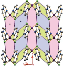
The first substructure is formed considering the pyridine ring carbon atom C5 in a general position, which acts as donor to the Cl1 atom at (x, y, 1 z). This C5—H5 Cl1 inter-action and its centrosymmetric analogue generate anR2
2(18) dimeric ring (M) centered at (0, 0, 1/2) (Fig. 2). A second substructure is formed via pairs of symmetry-related C7— H7 Cl1(x,1 +y,z) interactions, which generate a dimeric
R2
2(10) ring (N) (Fig. 2). The propagation of these dimers produces two infinite chains, the first running parallel to the (101) plane and the second running parallel to the [010] direction. The interconnection of the two chains leads to the generation of another tetramericR2
4(14) ring motif (P). Thus, the two types ofR22(18) andR
2
[image:2.610.313.562.93.134.2]4(14) rings are alternately linked into infinite MPMP. . . chains along the [010] direction
Figure 1
ORTEPview with atom-numbering scheme of the title complex with displacement ellipsoids drawn at the 30% probability level. The unlabeled counterpart is generated by the symmetry operationx+1
2, y,z+3
2.
Table 1
Selected geometric parameters (A˚ ,).
Co1—N1 2.0168 (4) Co1—Cl1 2.2556 (5)
N1—Co1—N1i 107.70 (5) N1i—Co1—Cl1 112.44 (5)
N1—Co1—Cl1 106.83 (1) Cl1—Co1—Cl1i 110.64 (4)
Symmetry code: (i)xþ1 2;y;zþ
[image:2.610.68.273.146.279.2]3 2.
Table 2
Hydrogen-bond geometry (A˚ ,).
D—H A D—H H A D A D—H A
C5—H5 Cl1ii 0.93 2.89 3.663 (1) 141
C7—H7 Cl1iii 0.93 2.88 3.734 (1) 153
[image:2.610.312.564.445.712.2]Symmetry codes: (ii)x;y;zþ1; (iii)x;y1;z.
Figure 2
[image:2.610.45.300.580.700.2]whereas theR22(10) andR 2
4(14) rings are linked alternately into an infiniteNPNP. . . chain parallel to the (101) plane (Fig. 2). Another substructure can be described considering that the molecules, because of their self-complementarity nature, are juxtaposed through – stacking interactions (Seth et al., 2011a, 2013; Mannaet al., 2013, 2014a). The molecular packing of the complex is such that the – stacking interactions between the pyridine rings, as well as between the imidazo rings, are optimized. The pyridine rings of the molecules at (x,y,z) and (x+ 1,y,z+ 1) are strictly parallel, with an interplanar spacing of 3.4671 (9) A˚ and a ring-centroid separation of 3.5293 (16) A˚ , corresponding to a ring offset of 0.659 A˚ . In addition, the imidazo rings at (x,y,z) and (x,y, z+ 1) are juxtaposed through face-to-face -stacking with an inter-centroid separation of 3.6414 (16) A˚ . Moreover, the imidazo and pyridine rings of the parent molecules are also involved into multi-stacking interactions with each other. In particular, the interplanar spacing between the imidazo ring in a general position and the pyridine rings at (x,y,z+ 1) and (x+ 1,y,z+ 1) are of 3.5303 (9) and 3.4625 (9) A˚ , respectively, while the relative ring-centroid separations are 3.9583 (16) and 3.8371 (16) A˚ . These–stacking interactions result in a two-dimensional supramolecular layered assembly parallel to the (010) plane (Fig. 3).
4. Hirshfeld surface analysis
Molecular Hirshfeld surfaces (Spackman & McKinnon, 2002) in the crystal structure are constructed considering the elec-tron distribution calculated as the sum of spherical atom electron densities (Spackman & Byrom, 1997; McKinnonet al., 1998). The normalized contact distance (dnorm) based on bothdeanddi, and the van der Waals (vdw) radii of the atom, given by the equation
dnorm¼
dir vdw i
rvdw i
þder vdw i
rvdw e
enable the identification of the regions of particular impor-tance to intermolecular interactions (McKinnonet al., 2007). The combination ofdeanddiin the form of a two-dimensional fingerprint plot (Rohlet al., 2008) provides a summary of the intermolecular contacts in the crystal (Spackman & McKinnon, 2002). The Hirshfeld surfaces are mapped with
dnorm, and the two-dimensional fingerprint plots presented in this work were generated usingCrystalExplorer 3.1(Wolffet al., 2012).
[image:3.610.44.565.136.194.2] [image:3.610.45.295.542.673.2]The pattern of the intermolecular interactions of the solid-state structure of the title complex prompted us to explore and quantify the contribution of the non-covalent interactions in the crystal packing, as well as the importance of the C— H Cl bonding in directing the organization of the extended supramolecular network (Seth et al., 2011a,b, Manna et al.,
Figure 3
Monomeric units linked through multi–stacking interactions leading to the formation of a supramolecular layered assembly. Color codes: the green and yellow dotted lines denote–stacking interactions between two pyridine rings and two imidazo rings, respectively, whereas –
stacking interactions between pyridine and imidazo rings are represented by pink dotted lines.
Table 3
Geometrical parameters (A˚ ,) for–stacking.
(a)Cg1 andCg2 are the centroids of the (N1/C1/N2/C6/C7) and (N2/C1–C5) rings, respectively; (b) centroid–centroid distance between ringiand ringj; (c) vertical distance from ring centroidito ringj; (d) vertical distance from ring centroidjto ringi; (e) dihedral angle between the first ring mean plane and the second ring mean plane of the partner molecule; (f) angle between the centroid of the first ring and the second ring; (g) angle between the centroid of the first ring and the normal to the mean plane of the second ring of the partner molecule.
Ringsi–ja
Rcb
R1vc
R2vd
e
f
g
Slippage
Cg1 Cg1ii 3.6414 (16) 3.4980 (8) 3.4980 (8) 0.0 16.13 16.13 1.012
Cg1 Cg2ii 3.9583 (16) 3.5303 (9) 3.5035 (9) 2.47 27.73 26.89 –
Cg1 Cg2iv 3.8371 (16) 3.4625 (9) 3.4846 (9) 2.47 24.75 25.53 –
Cg2 Cg2iv 3.5293 (16) 3.4671 (9) 3.4671 (9) 0.0 10.77 10.77 0.659
Symmetry codes: (ii)x,y,z+ 1; (iv)x+ 1,y,z+ 1.
Figure 4
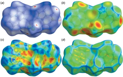
[image:3.610.314.564.562.719.2]2012; Seth, 2013; Mitraet al., 2014). In this present investiga-tion, the contacts responsible for building the supramolecular assembly were evaluated with respect to their contribution to the overall stability of the crystal structure. In this context, the Hirshfeld surface analysis (Spackman & McKinnon, 2002; Sethet al., 2011a,b,c,d; Mitraet al., 2013) of the title complex was performed and the results are illustrated in Fig. 4. The surfaces represented were mapped overdnorm,de, shape-index and curvedness in the ranges0.0620 to 0.9660 A˚ , 1.0626 to 2.4714 A˚ , 1.0000 to 1.0000 A˚ and 4.0000 to 0.4000 A˚ , respectively. The information regarding the intermolecular interactions summarized in Tables 2 and 3 are visible as spots on the Hirshfeld surfaces (Fig. 4). For instance, the distinct circular depressions (red spots) on thednormsurface (Fig. 4a) are due to the C—H Cl contacts, whereas other visible spots are due to H H contacts. From the Hirshfeld surfaces, it is also evident that the molecules are related to one another by
–stacking interactions, as can be inferred from inspection of the adjacent red and blue triangles (highlighted by yellow circles) on the shape-index surface (Fig. 4c). Indeed, the pattern of red and blue triangles in the same region of the shape-index surface is characteristic of – stacking inter-actions; the blue triangles represent convex regions resulting from the presence of ring carbon atoms of the molecule inside the surface, while the red triangles represent concave regions
caused by carbon atoms of the-stacked molecule above it. The presence of–stacking is also evident in the flat region toward the bottom of both sides of the molecules and is clearly visible on the curvedness surface (Fig. 4d): the shape of the blue outline on the curvedness surface unambiguously delineates the contacting patches of the molecules. On thede surface, this feature appears as a relatively flat green region where the contact distances are similar (Fig. 4b).
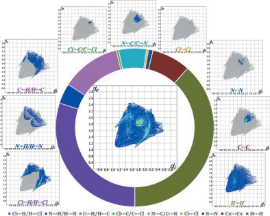
[image:4.610.313.565.93.160.2]The intermolecular interactions present in the structure are also visible on the two-dimensional fingerprint plot (Rohlet al., 2008; Samantaet al., 2014; Seth, 2014a,b,c), which can be decomposed to quantify the individual contributions of each intermolecular interaction involved in the structure (Mannaet al., 2014b). These complementary regions are visible in the fingerprint, where one molecule acts as donor (de>di) and the other as an acceptor (di>de). Table 4 contains the percentages
Figure 5
[image:4.610.116.510.398.713.2]Fingerprint plots: full (middle) and decomposed plots corresponding to all contacts involved in the structure [clockwise: from bottom left to bottom right]. The relative contributions of various intermolecular contacts to the Hirshfeld surface area of the title structure are displayed by the schematic illustration.
Table 4
Percentage contributions of interatomic contacts to the Hirshfeld surface.
Contact % contribution Contact % contribution
Cl H/H Cl 30.0 Cl Cl 0.4
N H/H N 4.1 N N 0.9
C H/H C 12.1 C C 7.9
Cl C/C Cl 0.5 H H 38.4
of contributions for a variety of contacts in the crystal struc-ture of the title compound.
The C—H Cl interactions appear as two distinct spikes in the fingerprint plot (Fig. 5) of the title complex, where Cl H interactions have a larger contribution (18.4%) than their H Cl counterparts (11.6%). Thus, the sum of Cl H/H Cl interactions comprises 30.0% of the total Hirshfeld surface area of the molecule (Table 4). The Cl H/H Cl inter-actions represented by the spikes in the bottom right and left region (de + di’ 2.77 A˚ ) indicate that the hydrogen atoms from the ligand moiety are in contact with the metal-coord-inated Cl atoms to build the two-dimensional supramolecular framework. The spoon-like tips in the region (de + di ’ 3.37 A˚ ) of the fingerprint plot (Fig. 5) represent a significant N H/H N contribution, covering 4.1% of the total Hirsh-feld surface of the molecules. The forceps-like tips in the region (de + di ’ 3.12 A˚ ) of the fingerprint plot (Fig. 5) represent the C H/H C contacts where the C H coun-terpart shows a larger contribution (7.6%) than the H C counterpart (4.5%). Overall, the C H/H C interactions account for 12.1% of the total Hirshfeld surface of the mol-ecules (Table 4), and the carbon atoms of the imidazopyridine moiety mainly act as donors in building the molecular assembly. The scattered points in the breakdown of the fingerprint plot show that the – stacking interactions comprise 7.9% of the total Hirshfeld surface of the molecule (Table 5) displayed as a region of blue/green color on the
diagonal at aroundde’di’1.743 A˚ . Another contribution comes from H H contacts (38.4%) represented by the scattered points in the fingerprint plots, and spread up only to
di=de= 1.092 A˚ (Fig. 5).
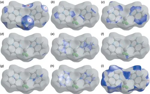
Finally, the short inter-atomic contacts of the structure (Table 5) of the type Cl C/ C Cl, N C/ C N, Cl Cl and N N are clearly visible as scattered points in the region
de+di’4.07 A˚ ,de+di’3.58 A˚ ,de+di’4.11 A˚ andde+di ’3.82 A˚ of the breakdown fingerprint plots (Fig. 5). They contribute 0.5%, 5.7%, 0.4% and 0.9%, respectively, to the total Hirshfeld area of the title complex (Table 4, see Fig. 6).
Figure 6
[image:5.610.312.566.93.197.2]Perspective view of the decomposeddnormsurfaces of the title structure corresponding to (a) Cl H/H Cl; (b) N H/H N; (c) C H/H C; (d) Cl C/C Cl; (e) N C/C N; (f) Cl Cl; (g) N N; (h) C C and (i) H H contacts.
Table 5
Summary of the short interatomic contacts (A˚ ).
Contact Distance Contact Distance
Cl1 H7v 2.883 C2 H6v 2.992
Cl1 C2i 3.613 (2) C2 C5iv 3.535 (3)
Cl1 H2i 2.932 C2 H3vii 3.021
Cl1 H5ii 2.893 C4 C4viii 3.525 (3)
Cl1 H3vi 3.055 C4 H4viii 2.834
N1 N1i 3.257 (2) C6 H2iii 3.050
C1 C4iv 3.482 (3) H2 H3vii 2.416
C1 C5iv 3.516 (3) H4 H4viii 2.309
C1 C6ii 3.518 (3) H6 H2iii 2.535
Symmetry codes: (i)x+1
2,y,z+32; (ii)x,y,z+ 1; (iii)x,y1,z; (iv)x+ 1,y,
z+ 1; (v)x,y+ 1,z; (vi)x1
2,y+ 1,z+12; (vii)x+ 1,y+ 1,z+ 1; (viii)x+12,y,
[image:5.610.52.552.408.721.2]The individual intermolecular interactions described above and the quantitative contributions included in Table 4 can be also visualized by the differentdnormsurfaces shown in Fig. 6, confirming that the Hirshfeld surface analysis provides a full understanding of the intermolecular interactions in a facile and immediate way.
5. Database survey
A search in the Cambridge Structural Database (Version 5.38, update May 2017; Groom et al., 2016) for structures of the general formula [ML2X2], whereMis any transition metal,L is the ligand imidazo[1,2-a]pyridine, and X any halogen, yielded no results. However, two related complexes exist, with ruthenium and tin, respectively: (i) dichloro-[2,20 -(pyridine-2,6-diyl)bis(imidazo[1,2-a ]pyridine)]triphenylphosphineruth-enium(II) (GULNEI; Li et al., 2015); (ii) dibromo-bis-(imidazo[1,2-a]pyridine)dimethyltin (NODREF; Agrawal et al., 2014). In both cases, the presence of the halogen atoms is relevant to the stabilization of the crystal structure. In the case of the ruthenium compound, the complex molecules are linked into discrete supramolecular dimers through pairs of C—H(imidazo) Cl interactions. On the other hand, the tin complex forms undulating sheets parallel to the (100) plane by means of C—H(pyridine) Br interactions in which both the Br ions and the ligands of one complex act as acceptor and donor, respectively.
6. Synthesis and crystallization
The title complex was prepared by simple hydrothermal reaction. CoCl26H2O (2.0 mmol, 0.476 g) was dissolved in water (20 ml) yielding a clear pink solution. A hot water– methanol (1:1) solution (20 ml) of imidazo[1,2-a]pyridine (1.0 mmol, 0.118 g) was added dropwise to the above solution under continuous stirring. The solution mixture thus obtained was further heated at 343 K for 2 h and then kept for crys-tallization at room temperature (303 K). The resulting solu-tion was allowed to evaporate slowly at room temperature for several weeks, yielding testable dark-pink crystals, which were collected by filtration, washed with water and dried in air.
7. Refinement details
Crystal data, data collection and structure refinement details are summarized in Table 6. The hydrogen atoms were located in the difference-Fourier map and refined as riding atoms, with C—H = 0.93 A˚ andUiso(H) = 1.2Ueq(C).
Funding information
The author is grateful to the Science and Engineering Research Board (SERB) –Department of Science and Tech-nology (DST), Govt. of India for a SERB Overseas Post-doctoral Fellowship (SB/OS/PDF-524/2015–16).
References
Adib, M., Sheikhi, E. & Rezaei, N. (2011).Tetrahedron Lett.52, 3191– 3194.
Agrawal, R., Goyal, V., Gupta, R., Pallepogu, R., Kotikalapudi, R., Jones, P. G. & Bansal, R. K. (2014).Polyhedron,70, 138–143. Bagdi, A. K., Santra, S., Monir, K. & Hajra, A. (2015). Chem.
Commun.51, 1555–1575.
Biftu, T., Feng, D., Fisher, M., Liang, G. B., Qian, X., Scribner, A., Dennis, R., Lee, S., Liberator, P. A., Brown, C., Gurnett, A., Leavitt, P. S., Thompson, D., Mathew, J., Misura, A., Samaras, S., Tamas, T., Sina, J. F., McNulty, K. A., McKnight, C. G., Schmatz, D. M. & Wyvratt, M. (2006).Bioorg. Med. Chem. Lett.16, 2479– 2483.
Brandenburg, K. (2006).DIAMOND. Crystal Impact GbR, Bonn, Germany.
Bravi, G., Cheasty, A. G., Corfield, J. A., Grimes, R. M., Harrison, D., Hartley, C. D., Howes, P. D., Medhurst, K. J., Meeson, M. L., Mordaunt, J. E.,et al.(2007). World patent WO 2,007,039,146. Bruker (2007).APEX2,SAINT,XPREPandSADABS. Bruker AXS
Inc., Madison, Wisconsin, USA.
Dymin´ska, L. (2015).Bioorg. Med. Chem.23, 6087–6099.
Enguehard-Gueiffier, C. & Gueiffier, A. (2007). Mini Rev. Med.
Chem.7, 888–899.
Farrugia, L. J. (2012).J. Appl. Cryst.45, 849–854.
Groom, C. R., Bruno, I. J., Lightfoot, M. P. & Ward, S. C. (2016).Acta Cryst.B72, 171–179.
Gudmundsson, K. S. & Boggs, S. D. (2007). World patent WO 2,007 027,999.
Gudmundsson, K. S. & Johns, B. A. (2007).Bioorg. Med. Chem. Lett.
17, 2735–2739.
Gunja, N. (2013).J. Med. Toxicol.9, 163–171.
Harrison, T. S. & Keating, G. M. (2005).CNS Drugs,19, 709–716. Ismail, M. A., Arafa, R. K., Wenzler, T., Brun, R., Tanious, F. A.,
[image:6.610.43.291.88.365.2]Wilson, W. D. & Boykin, D. W. (2008).Bioorg. Med. Chem.16, 683– 691.
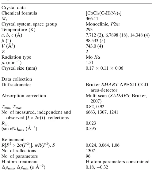
Table 6
Experimental details.
Crystal data
Chemical formula [CoCl2(C7H6N2)2]
Mr 366.11
Crystal system, space group Monoclinic,P2/n
Temperature (K) 293
a,b,c(A˚ ) 7.712 (2), 6.7898 (18), 14.348 (4)
() 98.533 (5)
V(A˚3) 743.0 (4)
Z 2
Radiation type MoK
(mm1) 1.51
Crystal size (mm) 0.170.110.06
Data collection
Diffractometer BrukerSMARTAPEXII CCD
area-detector
Absorption correction Multi-scan (SADABS; Bruker, 2007)
Tmin,Tmax 0.82, 0.92
No. of measured, independent and observed [I> 2(I)] reflections
6663, 1307, 1241
Rint 0.023
(sin /)max(A˚ 1
) 0.595
Refinement
R[F2> 2(F2)],wR(F2),S 0.024, 0.064, 1.06
No. of reflections 1307
No. of parameters 96
H-atom treatment H-atom parameters constrained
max,min(e A˚ 3
) 0.18,0.32
Kaminski, J. J. & Doweyko, A. M. (1997).J. Med. Chem. 40, 427– 436.
Ke, C.-H., Kuo, B.-C., Nandi, D. & Lee, H. M. (2013). Organome-tallics,32, 4775–4784.
Li, K., Niu, J.-L., Yang, M.-Z., Li, Z., Wu, L.-Y., Hao, X.-Q. & Song, M.-P. (2015).Organometallics,34, 1170–1176.
Macrae, C. F., Edgington, P. R., McCabe, P., Pidcock, E., Shields, G. P., Taylor, R., Towler, M. & van de Streek, J. (2006).J. Appl. Cryst.39, 453–457.
Manna, P., Ray Choudhury, S., Mitra, M., Kumar Seth, S., Helliwell, M., Bauza´, A., Frontera, A. & Mukhopadhyay, S. (2014b).J. Solid
State Chem.220, 149–156.
Manna, P., Seth, S. K., Das, A., Hemming, J., Prendergast, R., Helliwell, M., Choudhury, S. R., Frontera, A. & Mukhopadhyay, S. (2012).Inorg. Chem.51, 3557–3571.
Manna, P., Seth, S. K., Mitra, M., Choudhury, S. R., Bauza´, A., Frontera, A. & Mukhopadhyay, S. (2014a).Cryst. Growth Des.14, 5812–5821.
Manna, P., Seth, S. K., Mitra, M., Das, A., Singh, N. J., Choudhury, S. R., Kar, T. & Mukhopadhyay, S. (2013). CrystEngComm, 15, 7879–7886.
McKinnon, J. J., Jayatilaka, D. & Spackman, M. A. (2007). Chem.
Commun.pp. 3814–3816.
McKinnon, J. J., Mitchell, A. S. & Spackman, M. A. (1998).Chem. Eur. J.4, 2136–2141.
Mitra, M., Manna, P., Bauza´, A., Ballester, P., Seth, S. K., Ray Choudhury, S., Frontera, A. & Mukhopadhyay, S. (2014).J. Phys.
Chem. B,118, 14713–14726.
Mitra, M., Seth, S. K., Choudhury, S. R., Manna, P., Das, A., Helliwell, M., Bauza´, A., Frontera, A. & Mukhopadhyay, S. (2013).Eur. J.
Inorg. Chem.pp. 4679–4685.
Prostota, Y., Kachkovsky, O. D., Reis, L. V. & Santos, P. F. (2013).
Dyes Pigments,96, 554–562.
Puerstinger, G., Paeshuyse, J., De Clercq, E. & Neyts, J. (2007).
Bioorg. Med. Chem. Lett.17, 390–393.
Rival, Y., Grassy, G. & Michel, G. (1992). Chem. Pharm. Bull.40, 1170–1176.
Rival, Y., Grassy, G., Taudou, A. & Ecalle, R. (1991).Eur. J. Med.
Chem.26, 13–18.
Rohl, A. L., Moret, M., Kaminsky, W., Claborn, K., McKinnon, J. J. & Kahr, B. (2008).Cryst. Growth Des.8, 4517–4525.
Rupert, K. C., Henry, J. R., Dodd, J. H., Wadsworth, S. A., Cavender, D. E., Olini, G. C., Fahmy, B. & Siekierka, J. J. (2003).Bioorg. Med.
Chem. Lett.13, 347–350.
Samanta, T., Dey, L., Dinda, J., Chattopadhyay, S. K. & Seth, S. K. (2014).J. Mol. Struct.1068, 58–70.
Seth, S. K. (2013).CrystEngComm,15, 1772–1781. Seth, S. K. (2014a).J. Mol. Struct.1064, 70–75. Seth, S. K. (2014b).J. Mol. Struct.1070, 65–74. Seth, S. K. (2014c).Inorg. Chem. Commun.43, 60–63.
Seth, S. K., Manna, P., Singh, N. J., Mitra, M., Jana, A. D., Das, A., Choudhury, S. R., Kar, T., Mukhopadhyay, S. & Kim, K. S. (2013).
CrystEngComm,15, 1285–1288.
Seth, S. K., Saha, I., Estarellas, C., Frontera, A., Kar, T. & Mukhopadhyay, S. (2011b).Cryst. Growth Des.11, 3250–3265. Seth, S. K., Sarkar, D., Jana, A. D. & Kar, T. (2011d).Cryst. Growth
Des.11, 4837–4849.
Seth, S. K., Sarkar, D. & Kar, T. (2011a).CrystEngComm,13, 4528– 4535.
Seth, S. K., Sarkar, D., Roy, A. & Kar, T. (2011c).CrystEngComm,13, 6728–6741.
Sheldrick, G. M. (2015a).Acta Cryst.A71, 3–8. Sheldrick, G. M. (2015b).Acta Cryst.C71, 3–8.
Spackman, M. A. & Byrom, P. G. (1997).Chem. Phys. Lett.267, 215– 220.
Spackman, M. A. & McKinnon, J. J. (2002).CrystEngComm,4, 378– 392.
Spek, A. L. (2009).Acta Cryst.D65, 148–155.
Ve´ron, J. B., Enguehard-Gueiffier, C., Snoeck, R., Andrei, G., De Clercq, E. & Gueiffier, A. (2007).Bioorg. Med. Chem.15, 7209– 7219.
sup-1 Acta Cryst. (2018). E74, 600-606
supporting information
Acta Cryst. (2018). E74, 600-606 [https://doi.org/10.1107/S2056989018003857]
Structural characterization and Hirshfeld surface analysis of a Co
IIcomplex
with imidazo[1,2-
a
]pyridine
Saikat Kumar Seth
Computing details
Data collection: APEX2 (Bruker, 2007); cell refinement: APEX2 (Bruker, 2007) and SAINT (Bruker, 2007); data
reduction: SAINT (Bruker, 2007) and XPREP (Bruker, 2007); program(s) used to solve structure: SHELXT2014
(Sheldrick, 2015a); program(s) used to refine structure: SHELXL2018 (Sheldrick, 2015b); molecular graphics: ORTEP-3
for Windows (Farrugia, 2012), DIAMOND (Brandenburg, 2006) and Mercury (Macrae et al., 2006); software used to
prepare material for publication: PLATON (Spek, 2009).
Dichloridobis(imidazo[1,2-a]pyridine-κN1)cobalt(II)
Crystal data
[CoCl2(C7H6N2)2]
Mr = 366.11 Monoclinic, P2/n a = 7.712 (2) Å
b = 6.7898 (18) Å
c = 14.348 (4) Å
β = 98.533 (5)°
V = 743.0 (4) Å3
Z = 2
F(000) = 370
Dx = 1.636 Mg m−3
Mo Kα radiation, λ = 0.71073 Å Cell parameters from 647 reflections
θ = 1.5–25.0°
µ = 1.51 mm−1
T = 293 K Block, pink
0.17 × 0.11 × 0.06 mm
Data collection
Bruker SMART APEXII CCD area-detector diffractometer
Radiation source: fine-focus sealed tube Graphite monochromator
ω and φ scans
Absorption correction: multi-scan (SADABS; Bruker, 2007)
Tmin = 0.82, Tmax = 0.92
6663 measured reflections 1307 independent reflections 1241 reflections with I > 2σ(I)
Rint = 0.023
θmax = 25.0°, θmin = 2.8°
h = −8→9
k = −8→8
l = −17→17
Refinement
Refinement on F2 Least-squares matrix: full
R[F2 > 2σ(F2)] = 0.024
wR(F2) = 0.064
S = 1.06 1307 reflections 96 parameters 0 restraints
Primary atom site location: structure-invariant direct methods
Secondary atom site location: difference Fourier map
Hydrogen site location: inferred from neighbouring sites
sup-2 Acta Cryst. (2018). E74, 600-606
w = 1/[σ2(F
o2) + (0.0355P)2 + 0.2499P] where P = (Fo2 + 2Fc2)/3
(Δ/σ)max < 0.001
Δρmax = 0.18 e Å−3 Δρmin = −0.31 e Å−3
Special details
Geometry. All esds (except the esd in the dihedral angle between two l.s. planes) are estimated using the full covariance matrix. The cell esds are taken into account individually in the estimation of esds in distances, angles and torsion angles; correlations between esds in cell parameters are only used when they are defined by crystal symmetry. An approximate (isotropic) treatment of cell esds is used for estimating esds involving l.s. planes.
Refinement. Refinement of F2 against ALL reflections. The weighted R-factor wR and goodness of fit S are based on F2, conventional R-factors R are based on F, with F set to zero for negative F2. The threshold expression of F2 > 2sigma(F2) is used only for calculating R-factors(gt) etc. and is not relevant to the choice of reflections for refinement. R-factors based on F2 are statistically about twice as large as those based on F, and R- factors based on ALL data will be even larger.
Fractional atomic coordinates and isotropic or equivalent isotropic displacement parameters (Å2)
x y z Uiso*/Ueq
Co1 0.250000 0.21247 (5) 0.750000 0.03788 (13)
Cl1 0.00699 (6) 0.40150 (8) 0.73557 (4) 0.05261 (16)
N1 0.2270 (2) 0.0373 (2) 0.63532 (10) 0.0410 (3)
N2 0.2373 (2) −0.0539 (2) 0.48712 (11) 0.0452 (4)
C2 0.3250 (3) 0.2781 (3) 0.52292 (15) 0.0513 (5)
H2 0.341071 0.383348 0.564797 0.062*
C1 0.2664 (2) 0.0958 (3) 0.55145 (12) 0.0386 (4)
C6 0.1741 (3) −0.2106 (3) 0.53192 (17) 0.0554 (5)
H6 0.141218 −0.332623 0.505604 0.066*
C5 0.2770 (3) −0.0324 (4) 0.39615 (14) 0.0635 (6)
H5 0.261300 −0.136802 0.353786 0.076*
C7 0.1689 (3) −0.1537 (3) 0.62194 (16) 0.0503 (5)
H7 0.131285 −0.232355 0.668094 0.060*
C3 0.3582 (3) 0.2988 (4) 0.43289 (15) 0.0629 (6)
H3 0.394972 0.419967 0.412794 0.075*
C4 0.3379 (3) 0.1406 (5) 0.37060 (15) 0.0677 (7)
H4 0.367003 0.155512 0.310375 0.081*
Atomic displacement parameters (Å2)
U11 U22 U33 U12 U13 U23
Co1 0.0447 (2) 0.0343 (2) 0.0365 (2) 0.000 0.01225 (14) 0.000
Cl1 0.0507 (3) 0.0496 (3) 0.0578 (3) 0.0098 (2) 0.0093 (2) 0.0003 (2)
N1 0.0469 (8) 0.0342 (8) 0.0436 (8) −0.0029 (6) 0.0121 (7) −0.0024 (6)
N2 0.0415 (8) 0.0476 (9) 0.0451 (9) 0.0027 (7) 0.0022 (7) −0.0107 (7)
C2 0.0594 (12) 0.0493 (11) 0.0442 (11) −0.0098 (9) 0.0045 (9) 0.0033 (9)
C1 0.0383 (9) 0.0406 (10) 0.0367 (9) 0.0018 (7) 0.0049 (7) −0.0037 (7)
C6 0.0561 (12) 0.0403 (11) 0.0677 (14) −0.0017 (9) 0.0023 (10) −0.0165 (9)
C5 0.0534 (12) 0.0963 (18) 0.0387 (11) 0.0085 (13) −0.0003 (9) −0.0252 (12)
C7 0.0520 (11) 0.0353 (9) 0.0644 (12) −0.0050 (9) 0.0109 (9) 0.0038 (9)
C3 0.0607 (13) 0.0808 (16) 0.0454 (11) −0.0161 (12) 0.0019 (10) 0.0154 (11)
sup-3 Acta Cryst. (2018). E74, 600-606
Geometric parameters (Å, º)
Co1—N1 2.0168 (4) C2—C1 1.400 (3)
Co1—N1i 2.0169 (15) C2—H2 0.9300
Co1—Cl1 2.2556 (5) C6—C7 1.355 (3)
Co1—Cl1i 2.2556 (7) C6—H6 0.9300
N1—C1 1.344 (2) C5—C4 1.337 (4)
N1—C7 1.376 (2) C5—H5 0.9300
N2—C1 1.369 (2) C7—H7 0.9300
N2—C6 1.370 (3) C3—C4 1.391 (4)
N2—C5 1.392 (3) C3—H3 0.9300
C2—C3 1.361 (3) C4—H4 0.9300
N1—Co1—N1i 107.70 (5) N2—C1—C2 119.15 (17)
N1—Co1—Cl1 106.83 (1) C7—C6—N2 106.79 (17)
N1i—Co1—Cl1 112.44 (5) C7—C6—H6 126.6
N1—Co1—Cl1i 112.44 (5) N2—C6—H6 126.6
N1i—Co1—Cl1i 106.83 (5) C4—C5—N2 119.0 (2)
Cl1—Co1—Cl1i 110.64 (4) C4—C5—H5 120.5
C1—N1—C7 105.44 (16) N2—C5—H5 120.5
C1—N1—Co1 123.48 (12) C6—C7—N1 110.28 (19)
C7—N1—Co1 131.06 (14) C6—C7—H7 124.9
C1—N2—C6 107.13 (16) N1—C7—H7 124.9
C1—N2—C5 121.17 (19) C2—C3—C4 120.7 (2)
C6—N2—C5 131.65 (19) C2—C3—H3 119.6
C3—C2—C1 118.9 (2) C4—C3—H3 119.6
C3—C2—H2 120.5 C5—C4—C3 120.9 (2)
C1—C2—H2 120.5 C5—C4—H4 119.6
N1—C1—N2 110.35 (16) C3—C4—H4 119.6
N1—C1—C2 130.48 (17)
N1i—Co1—N1—C1 −157.31 (17) C5—N2—C1—C2 4.7 (3)
Cl1i—Co1—N1—C1 −39.88 (15) C3—C2—C1—N1 179.1 (2)
Cl1—Co1—N1—C1 81.68 (14) C3—C2—C1—N2 −2.6 (3)
N1i—Co1—N1—C7 24.35 (15) C1—N2—C6—C7 −0.9 (2)
Cl1i—Co1—N1—C7 141.78 (16) C5—N2—C6—C7 176.8 (2)
Cl1—Co1—N1—C7 −96.66 (17) C1—N2—C5—C4 −2.7 (3)
C7—N1—C1—N2 −1.1 (2) C6—N2—C5—C4 180.0 (2)
Co1—N1—C1—N2 −179.77 (11) N2—C6—C7—N1 0.2 (2)
C7—N1—C1—C2 177.4 (2) C1—N1—C7—C6 0.5 (2)
Co1—N1—C1—C2 −1.3 (3) Co1—N1—C7—C6 179.08 (14)
C6—N2—C1—N1 1.2 (2) C1—C2—C3—C4 −1.3 (3)
C5—N2—C1—N1 −176.69 (16) N2—C5—C4—C3 −1.3 (3)
C6—N2—C1—C2 −177.42 (18) C2—C3—C4—C5 3.4 (4)
sup-4 Acta Cryst. (2018). E74, 600-606
Hydrogen-bond geometry (Å, º)
D—H···A D—H H···A D···A D—H···A
C5—H5···Cl1ii 0.93 2.89 3.663 (1) 141
C7—H7···Cl1iii 0.93 2.88 3.734 (1) 153