Title: Restoring Mucosal Barrier Function and Modifying Macrophage Phenotype with an Extracellular Matrix Hydrogel: Potential Therapy for Ulcerative Colitis
Authors: Timothy J. Keane1,2, Jenna Dziki1,2, Eric Sobieski1,2, Adam Smoulder1,2, Arthur
Castleton1, Neill Turner1,3, Lisa J. White1,4, Stephen F. Badylak1,2,3,#
Affiliations: 1 McGowan Institute for Regenerative Medicine, Pittsburgh, PA 15219, USA 2 Dept. of Bioengineering, University of Pittsburgh, Pittsburgh, PA 15213, USA 3 Dept. of Surgery, University of Pittsburgh, Pittsburgh, PA 15219, USA 4 School of Pharmacy, University of Nottingham, Nottingham, NG7 2RD, UK
Short title: ECM Hydrogel for UC Therapy
# Corresponding Author: Stephen F. Badylak, DVM, Ph.D, MD
Professor, Department of Surgery and Bioengineering, University of Pittsburgh Deputy Director, McGowan Institute for Regenerative Medicine
450 Technology Drive, Suite 300, Pittsburgh, PA, 15219, USA. Tel: 412-624-5253
Email: [email protected]
Funding: This material is based upon work supported by the NSF Graduate Research Fellowship under Grant No. DGE-1247842 (to T.J.K. and J.D.) and by Asana Medical, Inc. The sponsors had no role in data collection or analysis.
Author Contributions: TJK and SFB developed study concept and design. TJK, JD, ES, AS, AC, NT, and LJW contributed to data acquisition. TJK, JD, and SFB analyzed and interpreted data. All authors critically reviewed and approved final version of the manuscript.
Abstract
Background & Aims: Despite advances in therapeutic options, more than half of all patients with ulcerative colitis (UC) do not achieve long-term remission, many require colectomy, and the disease still has a marked negative impact on quality of life. Extracellular matrix (ECM)
bioscaffolds facilitate the functional repair of many soft tissues by mechanisms that include mitigation of pro-inflammatory macrophage phenotype and mobilization of endogenous stem/progenitor cells. The aim of the present study was to determine if an ECM hydrogel therapy could influence outcomes in an inducible rodent model of UC.
Methods: The dextran sodium sulfate (DSS)-colitis model was used in male Sprague Dawley rats. Animals were treated via enema with an ECM hydrogel and the severity of colitis was determined by clinical and histologic criteria. Lamina propria cells were isolated and the production of inflammatory mediators was quantified. Mucosal permeability was assessed in-vivo by administering TRITC-dextran and in-vitro using transepithelial electrical resistance (TEER).
Results: ECM hydrogel therapy accelerated healing and improved outcome. The hydrogel was adhesive to colonic tissue, which allowed for targeted delivery of the therapy, and resulted in a reduction in clinical and histologic signs of disease. ECM hydrogel facilitated functional
improvement of colonic epithelial barrier function and the resolution of the pro-inflammatory state of tissue macrophages.
Conclusions: The present study shows that a nonsurgical and nonpharmacologic ECM-based therapy can abate DSS-colitis not by immunosuppression but by promoting phenotypic change in local macrophage phenotype and rapid replacement of the colonic mucosal barrier.
1. INTRODUCTION
Ulcerative colitis (UC) is one of the most common forms of inflammatory bowel disease, and represents a significant global health problem[1]. Since the 1930s, the fundamental approach to treatment has been pharmacologic (e.g., 5-amino salicylic acid, immunosuppressive therapy) and/or surgical intervention (e.g., colectomy). Nearly a century later, the basic tenets of patient care remain unchanged despite inadequate and less than acceptable results. Each year more than 50% of UC patients suffer from active flares and associated systemic effects. Overall, greater than 20 percent of patients diagnosed with UC will eventually require radical tissue resection (i.e. colectomy)—an alarming incidence that has remained unchanged over the last 50
years[2].
UC is a chronic relapsing disease consisting of acute flares followed by periods of remission and healing[3]. Active disease is characterized by chronic inflammation of the colon and defects in intestinal epithelial cell (IEC) barrier function[4]. Based upon observations that bioscaffolds composed of extracellular matrix (ECM) were shown to mitigate inflammation and support functional reconstruction of tissues including the gastrointestinal tract [5, 6, 7, 8], we
hypothesized that a similar approach to UC therapy will (1) abate inflammatory flares not by immune suppression but rather by promoting alternative activation of the local immune cell population, and (2) induce rapid restoration of the colonic mucosal barrier function not by simply providing a physical barrier between colonic submucosa and luminal contents but rather by promoting proliferation and replacement of the colonic mucosal epithelium. This two-pronged approach was tested by local delivery (enema) of an ECM hydrogel in a rodent model of UC.
cryptic peptide motifs released or exposed during in-vivo degradation of the ECM material, combined with the secreted products of ECM-exposed alternatively activated macrophages, promote stem/progenitor cell chemotaxis, proliferation, and differentiation [11, 12]. The objective of the present study was to determine if the above-mentioned ECM-induced biologic effects could influence outcomes in an inducible rodent model of UC.
2. MATERIALS AND METHODS 2.1. Experimental design
Ulcerative colitis was induced in male Sprague Dawley rats and treated with a daily enema of ECMH or vehicle (pepsin buffer) only for 7 days to determine the effect of an extracellular matrix hydrogel (ECMH) on colonic inflammation and barrier function. Animals were sacrificed at 7 days and 14 days post-DSS to evaluate the temporal response (n = 14 per time point per treatment) as shown in Supplemental Figure 1. Healthy control rats, which did not receive DSS, were included for comparison at both 7 and 14 days (n = 6 per time point). The study endpoints included clinical response, histologic scores of colon pathology, characterization of the
inflammatory response, and barrier function. The effect of ECMH on cell phenotype and epithelial barrier function was also measured in-vitro with lamina propria mononuclear cells (LPMCs) and intestinal epithelial cells, respectively.
2.2. ECM hydrogel preparation and formulations
rinses. SIS-ECM was frozen, lyophilized, and comminuted with a Wiley Mill using a #60-mesh screen, and digested at 10 mg/mL dry weight with 1 mg/mL pepsin (Sigma, St. Louis, MO) in 0.01N HCl while stirring for 20-26 h at 21-23°C. Digest was stored in aliquots at -20°C and pH neutralized with 0.1M NaOH prior to use. Hydrogel formation was induced by the neutralization step and an accompanying temperature increase to approximately 37°C following administration of the enema. All in-vivo studies used an ECM hydrogel (ECMH) concentration of 8 mg/mL and all in-vitro studies used an ECMH concentration of 500 μg/mL. The choice of 8 mg/mL ECMH
concentration was based on a hydrogel concentration that supported robust gel formation within 10 minutes of placement within the colon, would adhere to the ulcerated colonic wall for greater than 12 hours, and did not exhibit time-dependent viscosity as was observed in the 12 mg/mL ECMH (Supplemental Figure 2). To examine the effects of ECMH on cell in-vitro, the ECMH concentration had to be reduced to prevent gelation within the cell culture and 500 μg/mL was
used based on previous studies [10, 14].
14C-labeled ECMH was prepared as stated above with the intestines of pigs that were
injected with 14C-tagged proline, as previously described [15]. FITC-labeled ECMH was
prepared with a protein labeling kit (Thermo PierceNet) per manufacturer’s instructions[11].
2.3. ECMH adhesion testing
for 1 hour. After incubation, the upper washer was slowly withdrawn at a constant speed of 5 mm/min until a failure occurred between the surfaces.
2.4. Animals and husbandry
All procedures and animal studies were approved and conducted in compliance with the University of Pittsburgh Radiation Safety Committee and the Institutional Animal Care and Use Committee. Male Sprague Dawley rats, 8-12 weeks of age, were obtained from standard vendor (Harlan) and were housed and environmentally acclimated for 7-10 days. Animals were housed in standard laboratory conditions with a temperature of 21-23 °C and 12 hr dark/light cycles. Rats were allowed ad libitum access to food and water throughout the study.
2.5. Disease induction and monitoring
Five percent dextran sulfate sodium (DSS) salt (36,000-50,000 MW; MP Biomedical) was prepared daily in deionized water and administered to rats by ad libitum drinking for 7 days and the animals were monitored daily. Animal weight and consumption of food and water were tracked for each animal. Disease activity (i.e., stool consistency, presence of blood in stool, and weight loss) was measured every other day (i.e., days 1, 3, 5, 7, etc.) and scored on a range of 0 to 4. Stool was scored for consistency (0=normal, 2=loose, 4=diarrhea) and presence of blood (0=none, 2=occult, 4=gross bleeding). Stool was tested for the presence of blood using
ColoScreen ES Lab Pack Fecal Occult Tests. Weight loss compared to baseline was scored as follows: 0=none, 1= 1-5%, 2= 5-10%, 3= 10-20%, and 4= >20%.
2.6. ECMH retention studies with FITC- and 14C-ECMH
To determine hydrogel retention time, rats were administered FITC-labeled or 14C-labeled
ECMH via enema following disease induction. Eighteen rats were divided into 2 groups based on ECMH formulation (FITC- and 14C-ECMH) and sacrificed at 2 hr, 12 hr, and 24 hr post
enema (n = 3 per time point per ECMH formulation). Explanted colons from FITC-ECMH treated rats were processed to be optically clear such that the luminal contents were visible by
prevent photo bleaching of the FITC conjugate. Optical clearing of the colons was initiated by incubating in Dent’s fixative (1:4 dimethyl sulfoxide (DMSO): acetone) for 2 hours. Colons were then permeabilized and bleached in Dent’s bleach (1:4:1 DMSO: acetone: H2O2) for 1 hour.
Optically cleared colons were then imaged on a Fluorescent gel imager (Chemidoc Touch, Biorad). Exposure time was set to a control sample of FITC-ECMH and kept constant for all subsequent images.
For 14C measurements, the entire colon of each rat was individually flash frozen in liquid
nitrogen and homogenized. The frozen tissue was ground with mortar and pestle and mixed until homogenous. Approximately 40 mg of tissue samples was analyzed by accelerated mass spectrometry (AMS). Non-treated controls were used to subtract the background 14C levels in
native tissue.
2.7. Explanting and scoring of colonic tissue
Animals were sacrificed at predetermined time points as described previously. Euthanasia was achieved by CO2 inhalation and subsequent cervical dislocation in accordance with the
guidelines of the American Veterinary Medical Association (AVMA). Following euthanasia, the colon was resected following a ventral abdominal midline incision. A continuous colon segment was collected, spanning from the rectum to the cecum, and photographed. Colon length was measured as an indicator of disease activity. The colon was opened longitudinally and assessed grossly by investigators blinded to the treatment group for damage according to the metrics outlined in Supplemental Table 1.
The distal region of colon, 9 cm in length, was cut into thirds and opened longitudinally. Specimens were then collected for histologic examination, ex-vivo organ culture, and myeloperoxidase measurement. The colon specimens were paraffin embedded and tissue sections (5 μm) that were obtained from 2 to 8 cm from distal to proximal colon were stained
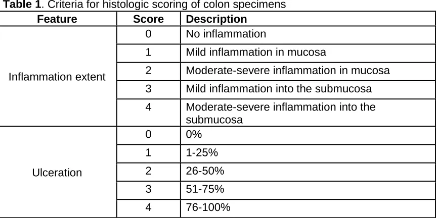
tissue sections were separated onto 2 slides and histologic scoring was performed according to Table 1 by six blinded investigators.
2.8. TRITC-Dextran permeability assay
Colonic mucosal permeability was assessed by enteral administration of TRITC-dextran (molecular mass 4.4 kDa; Sigma). Rats were administered TRITC-dextran (1mL, 10 mg/mL) enema 4 h before sacrifice. Whole blood was obtained at the time of sacrifice in serum collection tubes and allowed to clot undisturbed at room temperature for at least 30 minutes. The clot was removed by centrifuging at 1,000 x g for 10 minutes at 4°C. TRITC-dextran concentration in the serum were determined in triplicate on a SpectraMax plate reader (Molecular Devices), with serial dilutions of TRITC-dextran used as a standard curve.
2.9. Organ cultures
Full thickness biopsies were obtained following sacrifice from the explanted colon of each experimental and control animal at day 7 and day 14 using a 3 mm dermal punch as described previously [17]. Tissue specimens were cultured at 37°C with 5% CO2 for 48 h. The supernatants
were then harvested and stored at -80°C until the amount of TNFα and PGE2 was measured
using ELISA assays.
2.10. LPMC isolation and culture
Lamina propria mononuclear cells (LPMCs) were isolated from rats following colitis induction with DSS as described above. The colon was explanted, cleared of mesenteric fat tissue, and regions of Peyer’s patches were excised. The colon was then split in half longitudinally, cut into
pieces, and dissociated into single cell suspensions using a lamina propria dissociation kit (Miltenyi) according to manufacturer’s instructions. The suspension was then separated along a
40/70% Percoll gradient. The cells were suspended in RPMI 1640 containing 10% FBS and 100 U/mL penicillin and streptomycin, and then placed in 96-well plates at 2×10⁵ cells per well with or
2.11. IEC culture
For in-vitro barrier function assays, IECs (Caco-2, passages 24-28, ATCC) were cultured to approximately 80% confluence in MEM containing non-essential amino acids, 1mM sodium pyruvate, and 20% FBS. The functional response of IECs to ECMH was evaluated using rapid differentiation system (Corning Biocoat HTS Caco-2 Assay) per manufacturer’s instructions.
Confluent and differentiated cell monolayers were challenged with 100 ng/mL LPS for 2 hours and then treated with ECMH for 48 hours.
2.12. Transepithelial electrical resistance (TEER) measurement
TEER of Caco-2 monolayers was measured with an Epithelial Voltohmmeter (EVOM2, World Precision Instruments). Before seeding Caco-2 cells, electrical resistance of the supporting filter and buffer medium was measured and subtracted from the total electrical resistance determined with the monolayer to calculate the TEER of the monolayer. Only differentiated monolayers with TEER values greater than 300 Ω×cm2 were used in the study.
2.13. Immunolabeling
To determine the macrophage response following ECMH treatment, paraffin embedded histologic sections were deparaffinized and immunolabeled for a pan-macrophage marker (CD68)
and indicators of the M1-like (TNF) and M2-like (CD206) macrophage phenotypes. All primary
antibodies were confirmed to cross-react with rat epitopes. For visualizing the presence of Caco-2 adhesion proteins, trans-well inserts were fixed in 4% paraformaldehyde and immunolabeled for epithelial cadherin (E-cad). Sections were imaged at five random fields per tissue section. Quantification of localized staining was achieved using a custom image analysis algorithm developed using CellProfiler Image Analysis Software.
2.14. Statistical analysis
blinded to the experimental groupings. Quantitative outcomes were compared with a one-way or two-way analysis of variance (ANOVA) and post-hoc Tukey test to determine differences between groups. All statistical analysis was performed using SPSS Statistical Analysis Software (SPSS, IBM). Data are reported as mean ± standard deviation unless otherwise stated.
3. RESULTS
3.1. ECMH is Adhesive to Colon Tissue
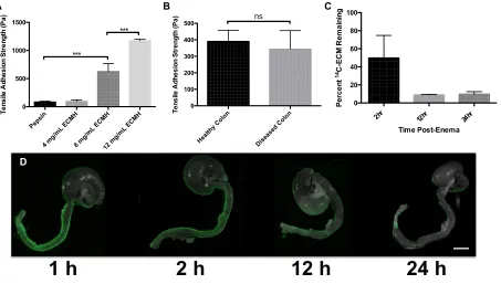
The therapeutic efficacy of ECMH is reliant upon its ability to adhere to the colon wall and interface with the resident cells. ECMH has the distinctive property of reverse thermal gelation and the hydrogel properties are dependent upon material characteristics (Supplemental Fig 3). Results of adhesion testing show that ECMH is mucoadhesive, with a dose-dependent increase in adhesion strength when tested on healthy colon (Fig 1A). Importantly, the 8 mg/mL ECMH dose used in the present study maintains equivalent adhesion strength in colitic rat colon when compared to healthy tissue (Fig 1B). It is noteworthy that mucosal adherence is not simply a property of thermoreversible gels. For example, Pluronic F-127 (20%; Sigma) did not show adhesion strength greater than the negative control (data not shown). When delivered via enema to colitic rats, the residence time of the ECMH is greater than 24 hours. Two hours after administering the enema, about 50% of the 14C-ECMH remains attached to the colon wall and
3.2. ECMH Treatment Mitigates Disease State
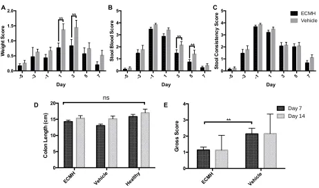
The DSS experimental model is a well-accepted UC-like self-limiting colitis phenotype with epithelial barrier defects[18]. Clinical signs of colitis (e.g., weight loss, stool blood, and stool consistency) were present following 3 days of exposure to 5% DSS in drinking water and reach their peak following 6 days (Supplemental Fig 4). ECMH treatment diminished clinical symptoms of UC in this rodent model. ECMH treated animals did not lose as much weight (at days 1 and 3) and had less blood in stool (at days 3 and 5) compared to the vehicle control. The shortening of the colon that was present at day 0 (Supp. Fig 3D) was no longer evident by days 7 and 14 across all groups (Fig 2D). ECMH treatment resulted in a reduction in the gross score compared to the vehicle at day 7 (Fig 2E). Histomorphologic analysis also showed that ECMH is
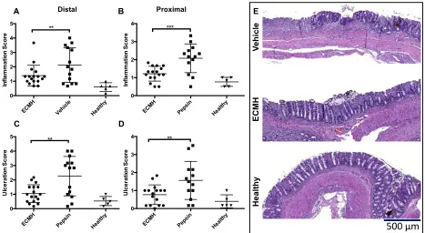
therapeutic in the present model and resulted in diminished signs of inflammation and a lower the degree of ulceration at 7 and 14 days in both distal and proximal tissue sections (Fig 3 and Supplemental Fig 5).
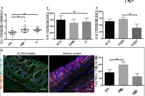
3.3. ECMH Restores Epithelial Barrier Function
3.4. ECMH Mitigates the Inflammatory Response
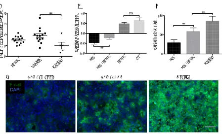
Recognized inflammatory mediators of IBD (i.e., TNFα and PGE2) were measured in the
present study. LPMCs isolated from colitic rats were plated and exposed to ECMH. The ECMH treatment resulted in a substantial reduction in the production of TNFα (Fig 5A) by the LPMCs
but had no effect on PGE2 production (Fig 5B). Organ cultures of biopsies collected from rats following ECMH or vehicle control treatment showed secreted PGE2 was similar to healthy controls in the ECMH treated animals while the vehicle controls had significantly elevated levels of mucosal PGE2 (Fig 5C). Secreted levels of TNFα were below detection in the organ cultures
regardless of experimental condition at the time points studied.
The effect of ECMH on macrophage phenotype in DSS-colitis was evaluated by quantifying the number of CD68+ macrophages in the colon that co-express TNFα or CD206. Interestingly,
the absolute number of individually labeled CD68+, CD206+, and TNFα+ cells was the same
across all treatment groups (data not shown) but ECMH treatment resulted in a reduction in the number of co-labeled CD68+/TNFα+ inflammatory macrophages at day 7 (Fig 5D). The fact that
ECMH did not affect the amount of global TNFα+ cells but did reduce the number of
CD68+/TNFα+ cells suggests a direct role for ECMH in modulating the macrophage response by
reducing the number of inflammatory macrophages present in the colonic tissue.
4. DISCUSSION
The present study shows that an ECM hydrogel composed of ECM mitigates the
proinflammatory macrophage phenotype and restores barrier function in a rodent model of UC. It is noteworthy that the total number of macrophages was not changed by ECMH treatment, but rather the phenotype of this cell population was changed. In addition, the barrier function was not restored by the physical presence of the hydrogel but rather by the restoration of an effective mucosal epithelium. These effects are a distinct departure from the
humans. More than half of all patients with UC do not achieve long-term remission and many require colectomy.
UC is a complex immune-mediated disease characterized by diffuse inflammation confined to the mucosa and submucosa of the colon and rectum. The inflammatory infiltrate consists of neutrophils, lymphocytes, and macrophages that penetrate the epithelium and lead to ulceration and crypt abscessation [20]. Macrophages, dependent upon their phenotype, are important effector cells of both the initiation/maintenance of inflammatory response and the
resolution/regeneration processes. Regulatory M2-like macrophages have been associated with mucosal healing outcomes[21, 22] in preclinical models of IBD. Similarly, the restoration of functional tissues via ECM bioscaffold mediated events in a variety of regenerative medicine applications has been shown to be either associated with, or dependent upon a timely shift in macrophage phenotype toward M2 predominance[23, 24, 25].
The findings of the present study are consistent with, and analogous to, the known
mechanisms by which ECM-based approaches facilitate the constructive remodeling of injured tissues in other anatomic locations[5, 26, 27, 28, 29, 30]. Specifically, ECM materials derived via decellularization of a variety of allogeneic or xenogeneic source tissues have been shown to induce a phenotypic transition from the proinflammatory macrophage and lymphocyte
phenotype toward a regulatory, “anti-inflammatory” and healing phenotype [9, 31, 32, 33], and to
promote endogenous stem/progenitor cell activation and recruitment [11, 29, 34, 35]. Furthermore, the findings in the present study are similar to the results in patients with
esophageal adenocarcinoma who were subjected to complete surgical removal of long segment esophageal mucosa and placement of a solid (i.e., not hydrogel) form of an ECM bioscaffold. These patients showed a rapid restoration of the esophageal mucosa and esophageal function without recalcitrant stricture.
current therapeutic strategies for UC, which are focused upon immune suppression (e.g., corticosteroids and anti-TNFα compounds) with the associated local and systemic effects.
Immune suppression is clearly different than maintaining complete functionality of the immune system while redirecting its’ biologic objectives. In fact, a robust immune system is necessary
for a healthy, functional gastrointestinal tract [36, 37, 38, 39].
The use of immunosuppressive and/or anti-inflammatory compounds has a limited ability to facilitate mucosal healing [40, 41], and yet the disrupted mucosal barrier integrity is a key component in the pathogenesis of UC. Barrier dysfunction enables the ingress of luminal antigens and pathogens, and the continuous activation of a proinflammatory immune response in the lamina propria and the associated chronic inflammation that is the hallmark of UC.
Restoration of barrier function is therefore an important therapeutic target in UC. In the present study, ECMH therapy had a protective downstream effect on the epithelial cells of the colonic mucosa. Results show that ECMH facilitated functional improvement of the epithelial barrier function and suggests that ECMH acts therapeutically either by limiting epithelial cell damage and/or by actively promoting mucosal integrity.
Recognition that effector cells of the immune system, such as the macrophages and T-helper cells, not only promote classic inflammatory processes but also orchestrate the temporal inhibition of inflammation and initiation of functional tissue remodeling [25, 31, 42, 43, 44, 45] provides the opportunity to re-examine immunosuppressive strategies for treatment of diseases such as UC. Although the signaling molecules that influence macrophage and lymphocyte phenotype transition are not fully understood, there is suggestive evidence that at least some of these regulators reside within the ECM [9, 10, 25]. Results of the present study suggest that ECMH modulates the innate immune response not by directly promoting an M2-like
macrophage phenotype but rather by reducing the number of TNF expressing M1-like
carcinogenesis and a sustained M2-like phenotype may not be optimal for UC therapies. However, the tumor associated macrophage (TAM) M2-like phenotype differs from the regulatory M2-like phenotype associated with tissue restoration [46, 47]. The present study showed the ECMH-mediated macrophage response to be transient and associated with a
reduction of TNF, a suspected participant in inflammation-mediated colon carcinogenesis[48].
[49].
Limitations of the present study include the use of one animal model and only 2 surface markers for macrophage phenotype. The DSS-colitis model was chosen because the model features innate immunity and epithelial barrier defects that are central to the present study’s
hypothesis. The outcomes in the DSS-colitis model can be effective in predicting clinical treatment of IBD [18]. It is well established that when DSS administration is halted, the animals will begin to spontaneously recover [50] and therefore differences among groups requires examining the temporal response. While only two markers for macrophage phenotype were used for immunolabeling, we chose the most representative marker of UC-like inflammation
(TNF) and for M2-like macrophages (CD206). Macrophages are a heterogeneous and plastic
cell population and the use of a single marker to delineate phenotype can result in ambiguity.
TNF, in particular, was chosen for the present study because of its integral role in the
pathogenesis of UC [51], however it is logical and plausible that the use of other markers could provide additional insight into the effects of ECMH treatment upon the resident macrophage population in the colitic microenvironment.
addition to the therapeutic efficacy, is that the well-accepted safety profile of ECM products may allow for accelerated transition to the clinic.
ACKNOWLEDGEMENTS
The authors would like to thank Lori Walton of the McGowan Histology Center for histologic section preparation; Andrew Lesniac for slide scanning; the Purdue Rare Isotope Measurement Laboratory at Purdue University for AMS processing of 14C samples; Janet Reing and Li Zhang
REFERENCES
1 Kaplan GG. The global burden of IBD: from 2015 to 2025. Nat Rev Gastroenterol Hepatol 2015;12:720-7.
2 Danese S, Fiocchi C. Ulcerative colitis. N Engl J Med 2011;365:1713-25.
3 Molodecky NA, Soon IS, Rabi DM, Ghali WA, Ferris M, Chernoff G, et al. Increasing incidence and prevalence of the inflammatory bowel diseases with time, based on systematic review. Gastroenterology 2012;142:46-54 e42; quiz e30.
4 de Souza HS, Fiocchi C. Immunopathogenesis of IBD: current state of the art. Nat Rev Gastroenterol Hepatol 2016;13:13-27.
5 Badylak SF, Hoppo T, Nieponice A, Gilbert TW, Davison JM, Jobe BA. Esophageal preservation in five male patients after endoscopic inner-layer circumferential resection in the setting of superficial cancer: a regenerative medicine approach with a biologic scaffold. Tissue Eng Part A 2011;17:1643-50.
6 Badylak SF, Vorp DA, Spievack AR, Simmons-Byrd A, Hanke J, Freytes DO, et al.
Esophageal reconstruction with ECM and muscle tissue in a dog model. Journal of Surgical Research 2005;128:87-97.
7 Chen MK, Badylak SF. Small bowel tissue engineering using small intestinal submucosa as a scaffold. Journal of Surgical Research 2001;99:352-8.
8 Hoppo T, Badylak SF, Jobe BA. A novel esophageal-preserving approach to treat high-grade dysplasia and superficial adenocarcinoma in the presence of chronic gastroesophageal reflux disease. World J Surg 2012;36:2390-3.
9 Sicari BM, Dziki JL, Siu BF, Medberry CJ, Dearth CL, Badylak SF. The promotion of a constructive macrophage phenotype by solubilized extracellular matrix. Biomaterials
2014;35:8605-12.
10 Slivka PF, Dearth CL, Keane TJ, Meng FW, Medberry CJ, Riggio RT, et al. Fractionation of an ECM hydrogel into structural and soluble components reveals distinctive roles in
regulating macrophage behavior. Biomaterials Science 2014;2:1521.
11 Agrawal V, Johnson SA, Reing J, Zhang L, Tottey S, Wang G, et al. Epimorphic regeneration approach to tissue replacement in adult mammals. Proc Natl Acad Sci U S A 2010;107:3351-5.
12 Cortiella J, Niles J, Cantu A, Brettler A, Pham A, Vargas G, et al. Influence of acellular natural lung matrix on murine embryonic stem cell differentiation and tissue formation. Tissue engineering Part A 2010;16:2565-80.
13 Badylak SF, Lantz GC, Coffey A, Geddes LA. Small intestinal submucosa as a large diameter vascular graft in the dog. J Surg Res 1989;47:74-80.
14 Dearth CL, Slivka PF, Stewart SA, Keane TJ, Tay JK, Londono R, et al. Inhibition of COX1/2 alters the host response and reduces ECM scaffold mediated constructive tissue remodeling in a rodent model of skeletal muscle injury. Acta Biomater 2016;31:50-60. 15 Gilbert TW, Stewart-Akers AM, Badylak SF. A quantitative method for evaluating the degradation of biologic scaffold materials. Biomaterials 2007;28:147-50.
16 Kammer HW. Adhesion between Polymers - Review. Acta Polymerica 1983;34:112-8. 17 Dieleman LA, Ridwan BU, Tennyson GS, Beagley KW, Elson CO. Dextran Sodium-Sulfate (Dss)-Induced Colitis Occurs in Severe Combined Immunodeficient (Scid) Mice. Gastroenterology 1993;104:A692-A.
18 Valatas V, Vakas M, Kolios G. The value of experimental models of colitis in predicting efficacy of biological therapies for inflammatory bowel diseases. Am J Physiol Gastrointest Liver Physiol 2013;305:G763-85.
20 Cottone M, Scimeca D, Mocciaro F, Civitavecchia G, Perricone G, Orlando A. Clinical course of ulcerative colitis. Dig Liver Dis 2008;40 Suppl 2:S247-52.
21 Hunter MM, Wang A, Parhar KS, Johnston MJ, Van Rooijen N, Beck PL, et al. In vitro-derived alternatively activated macrophages reduce colonic inflammation in mice.
Gastroenterology 2010;138:1395-405.
22 Vos AC, Wildenberg ME, Arijs I, Duijvestein M, Verhaar AP, de Hertogh G, et al.
Regulatory macrophages induced by infliximab are involved in healing in vivo and in vitro. Inflamm Bowel Dis 2012;18:401-8.
23 Brown BN, Badylak SF. Expanded applications, shifting paradigms and an improved understanding of host-biomaterial interactions. Acta Biomater 2013;9:4948-55.
24 Brown BN, Ratner BD, Goodman SB, Amar S, Badylak SF. Macrophage polarization: an opportunity for improved outcomes in biomaterials and regenerative medicine. Biomaterials 2012;33:3792-802.
25 Sadtler K, Estrellas K, Allen BW, Wolf MT, Fan H, Tam AJ, et al. Developing a pro-regenerative biomaterial scaffold microenvironment requires T helper 2 cells. Science 2016;352:366-70.
26 Sicari BM, Rubin JP, Dearth CL, Wolf MT, Ambrosio F, Boninger M, et al. An acellular biologic scaffold promotes skeletal muscle formation in mice and humans with volumetric muscle loss. Sci Transl Med 2014;6:234ra58.
27 Meng F, Modo M, Badylak SF. Biologic scaffold for CNS repair. Regen Med 2014;9:367-83.
28 Wainwright JM, Hashizume R, Fujimoto KL, Remlinger NT, Pesyna C, Wagner WR, et al. Right ventricular outflow tract repair with a cardiac biologic scaffold. Cells Tissues Organs 2012;195:159-70.
29 Agrawal V, Kelly J, Tottey S, Daly KA, Johnson SA, Siu BF, et al. An isolated cryptic peptide influences osteogenesis and bone remodeling in an adult mammalian model of digit amputation. Tissue Eng Part A 2011;17:3033-44.
30 Seif-Naraghi SB, Singelyn JM, Salvatore MA, Osborn KG, Wang JJ, Sampat U, et al.
Safety and efficacy of an injectable extracellular matrix hydrogel for treating myocardial infarction. Sci Transl Med 2013;5:173ra25.
31 Brown BN, Londono R, Tottey S, Zhang L, Kukla KA, Wolf MT, et al. Macrophage phenotype as a predictor of constructive remodeling following the implantation of biologically derived surgical mesh materials. Acta Biomater 2012;8:978-87.
32 Badylak SF, Valentin JE, Ravindra AK, McCabe GP, Stewart-Akers AM. Macrophage phenotype as a determinant of biologic scaffold remodeling. Tissue Eng Part A 2008;14:1835-42.
33 Fishman JM, Lowdell MW, Urbani L, Ansari T, Burns AJ, Turmaine M, et al.
Immunomodulatory effect of a decellularized skeletal muscle scaffold in a discordant xenotransplantation model. Proc Natl Acad Sci U S A 2013;110:14360-5.
34 Vorotnikova E, McIntosh D, Dewilde A, Zhang J, Reing JE, Zhang L, et al. Extracellular matrix-derived products modulate endothelial and progenitor cell migration and proliferation in vitro and stimulate regenerative healing in vivo. Matrix Biol 2010;29:690-700.
35 Reing JE, Zhang L, Myers-Irvin J, Cordero KE, Freytes DO, Heber-Katz E, et al.
Degradation products of extracellular matrix affect cell migration and proliferation. Tissue Eng Part A 2009;15:605-14.
36 Khamsi R. A gut feeling about immunity. Nat Med 2015;21:674-6.
37 Mantovani A, Marchesi F. IL-10 and macrophages orchestrate gut homeostasis. Immunity 2014;40:637-9.
39 Kayama H, Takeda K. Functions of innate immune cells and commensal bacteria in gut homeostasis. J Biochem 2016;159:141-9.
40 Magro F, Rodrigues-Pinto E, Coelho R, Andrade P, Santos-Antunes J, Lopes S, et al. Is it possible to change phenotype progression in Crohn's disease in the era of
immunomodulators? Predictive factors of phenotype progression. Am J Gastroenterol 2014;109:1026-36.
41 Neurath MF, Travis SP. Mucosal healing in inflammatory bowel diseases: a systematic review. Gut 2012;61:1619-35.
42 Mills CD, Kincaid K, Alt JM, Heilman MJ, Hill AM. M-1/M-2 macrophages and the Th1/Th2 paradigm. J Immunol 2000;164:6166-73.
43 Chazaud B. Macrophages: supportive cells for tissue repair and regeneration. Immunobiology 2014;219:172-8.
44 Novak ML, Koh TJ. Phenotypic transitions of macrophages orchestrate tissue repair. Am J Pathol 2013;183:1352-63.
45 Weidenbusch M, Anders HJ. Tissue microenvironments define and get reinforced by macrophage phenotypes in homeostasis or during inflammation, repair and fibrosis. J Innate Immun 2012;4:463-77.
46 Sica A, Schioppa T, Mantovani A, Allavena P. Tumour-associated macrophages are a distinct M2 polarised population promoting tumour progression: potential targets of anti-cancer therapy. Eur J Cancer 2006;42:717-27.
47 Mantovani A, Sozzani S, Locati M, Allavena P, Sica A. Macrophage polarization: tumor-associated macrophages as a paradigm for polarized M2 mononuclear phagocytes. Trends in Immunology 2002;23:549-55.
48 Popivanova BK, Kitamura K, Wu Y, Kondo T, Kagaya T, Kaneko S, et al. Blocking TNF-alpha in mice reduces colorectal carcinogenesis associated with chronic colitis. J Clin Invest 2008;118:560-70.
49 Itzkowitz SH, Yio X. Inflammation and cancer IV. Colorectal cancer in inflammatory bowel disease: the role of inflammation. Am J Physiol Gastrointest Liver Physiol 2004;287:G7-17.
50 Yan Y, Kolachala V, Dalmasso G, Nguyen H, Laroui H, Sitaraman SV, et al. Temporal and spatial analysis of clinical and molecular parameters in dextran sodium sulfate induced colitis. PLoS One 2009;4:e6073.
FIGURE LEGENDS
Figure 1: ECMH is Mucoadhesive. Tensile tests show dose-dependent increase in adhesion
strength of ECMH to healthy colon (A) and equivalent adhesion in healthy vs. diseased colon (B). The resident time of ECMH following enema delivery was tested with 14C (C) and FITC-labeled
ECMH (D). Scale bar = 1 cm.
Figure 2: ECMH Treatment Reduces Disease Activity. The effect of ECMH treatment on clinical
symptoms (A-C), colon length (D), and gross score at explant (E) was tracked and compared to the vehicle (pepsin) alone.
Figure 3: ECMH Treatment Lowers Histologic Score. Distal and proximal tissue sections stained
with hematoxylin and eosin were scored by blinded investigators and compared with vehicle/pepsin buffer. The extent of inflammation (A, B) and degree of ulceration (C,D) were quantified at 7 days. Representative images used for inflammation scoring are shown in panel E.
Figure 4: ECM Restores Barrier Function. TRITC-Dextran permeability assay showed that the
barrier function of ECMH-treated animals was similar to healthy animals while the colonic epithelial barrier in the vehicle-treated control group remained impaired compared to the healthy control (A). Differentiated and LPS-damaged monolayers of IECs responded to ECMH treatment in-vitro with functional recovery as shown by TEER readings (B). The increased barrier function was associated with an increased presence of E-cadherin compared with negative controls (C-D). Scale bar = 100 μm.
Figure 5: ECMH Mediates Inflammation. Lamina propria cells exposed to ECMH results in
TABLES
Table 1. Criteria for histologic scoring of colon specimens Feature Score Description
Inflammation extent
0 No inflammation
1 Mild inflammation in mucosa
2 Moderate-severe inflammation in mucosa 3 Mild inflammation into the submucosa 4 Moderate-severe inflammation into the
submucosa
Ulceration
0 0%
1 1-25%
2 26-50%
3 51-75%
FIGURES
2 hr 12 hr
24 hr
0 20 40 60 80 100 Time Post-Enema P e rc e n t 14C -E C M R e m a in in g Heal thy Colo n Dise ased Col on 0 100 200 300 400 500 T e n s ile A d h e s io n S tr e n g th ( P a ) ns Peps in
4 m g/m
L EC MH
8 m g/m
L EC MH
12 m g/m
L EC MH 0 500 1000 1500 T e n s ile A d h e s io n S tr e n g th (P a ) *** ***
A B C
D
[image:23.612.84.537.216.473.2]-5 -3 -1 1 3 5 7 0.0 0.5 1.0 1.5 2.0 Day W e ig h t S c o re ECMH Vehicle ** **
-5 -3 -1 1 3 5 7
0 1 2 3 4 5 Day S to o l B lo o d S c o re ECMH Pepsin ** **
A B
D E
-5 -3 -1 1 3 5 7
[image:24.612.77.539.137.410.2]Distal Proximal
A B
C D
LPS Damaged LPS +ECMH Healthy
E-cad DAPI
A C
D B LPS LPS +ECM H Heal thy 0 50 100 P e rc e n t E -c a d
+ C
[image:26.612.98.547.142.414.2]ECM H
Vehi
cle NT
0 1000 2000 3000 4000 P G E 2 C o n c e n tr a tio n (n g /m l) ns
A B C
ECM H Vehi cle Heal thy 0 100 200 300 400 P G E 2 C o n c e n tr a tio n (n g /m l) ** D
ECMH Vehicle
Healt hy 0 50 100 150 200 C D 6 8 +/T N F -a
+ C
e lls p e r F ie ld ** C D 6 8 / T N Fα / C D 2 0 6 / D A P I
ECMH treated Vehicle control
Fig 5
ECMH Vehicle NT
-2 0 2 4 6 T N F a C o n c e n tr a tio n R e la ti v e to N T ***
[image:27.612.92.560.97.406.2]