Progress and problems in the biology,
diagnostics, and therapeutics of prion diseases
Adriano Aguzzi, … , Mathias Heikenwalder, Gino Miele
J Clin Invest. 2004;
114(2)
:153-160.
https://doi.org/10.1172/JCI22438
.
The term “prion” was introduced by Stanley Prusiner in 1982 to describe the atypical
infectious agent that causes transmissible spongiform encephalopathies, a group of
infectious neurodegenerative diseases that include scrapie in sheep, Creutzfeldt-Jakob
disease in humans, chronic wasting disease in cervids, and bovine spongiform
encephalopathy in cattle. Over the past twenty years, the word “prion” has been taken to
signify various subtly different concepts. In this article, we refer to the prion as the
transmissible principle underlying prion diseases, without necessarily implying any specific
biochemical or structural identity. When Prusiner started his seminal work, the study of
transmissible spongiform encephalopathies was undertaken by only a handful of scientists.
Since that time, the “mad cow” crisis has put prion diseases on the agenda of both
politicians and the media. Significant progress has been made in prion disease research,
and many aspects of prion pathogenesis are now understood. And yet the diagnostic
procedures available for prion diseases are not nearly as sensitive as they ought to be, and
no therapeutic intervention has been shown to reliably affect the course of the diseases.
This article reviews recent progress in the areas of pathogenesis of, diagnostics of, and
therapy for prion diseases and highlights some conspicuous problems that remain to be
addressed in each of these fields.
Science in Medicine
Find the latest version:
Progress and problems in the biology,
diagnostics, and therapeutics of prion diseases
Adriano Aguzzi, Mathias Heikenwalder, and Gino Miele
Institute of Neuropathology, University Hospital Zurich, Zurich, Switzerland.
The term “prion” was introduced by Stanley Prusiner in 1982 to describe the atypical
infec-tious agent that causes transmissible spongiform encephalopathies, a group of infecinfec-tious
neurodegenerative diseases that include scrapie in sheep, Creutzfeldt-Jakob disease in humans,
chronic wasting disease in cervids, and bovine spongiform encephalopathy in cattle. Over the
past twenty years, the word “prion” has been taken to signify various subtly different concepts.
In this article, we refer to the prion as the transmissible principle underlying prion diseases,
with-out necessarily implying any specific biochemical or structural identity. When Prusiner started
his seminal work, the study of transmissible spongiform encephalopathies was undertaken by only a handful of
scientists. Since that time, the “mad cow” crisis has put prion diseases on the agenda of both politicians and the
media. Significant progress has been made in prion disease research, and many aspects of prion pathogenesis are
now understood. And yet the diagnostic procedures available for prion diseases are not nearly as sensitive as they
ought to be, and no therapeutic intervention has been shown to reliably affect the course of the diseases. This
article reviews recent progress in the areas of pathogenesis of, diagnostics of, and therapy for prion diseases and
highlights some conspicuous problems that remain to be addressed in each of these fields.
Prion pathogenesis, diagnostics, and therapy: where do we stand?
Prion diseases, also known as transmissible spongiform encephalopathies (TSEs), are invariably fatal neurodegenerative disorders affecting a broad spectrum of host species and arise via genetic, infectious, or sporadic mechanisms (Table 1). In humans, prion diseases result from infectious modes of transmission (vari-ant Creutzfeldt-Jakob disease [vCJD], iatrogenic CJD, Kuru); inherited modes of transmission in which there is nonconservative germ line mutation of the PRNP gene open reading frame (famil-ial CJD, Gerstmann-Sträussler-Scheinker Syndrome, Fatal Famil-ial Insomnia) (1, 2); and modes of transmission that have as yet been neither determined nor understood (sporadic CJD [sCJD]). The clinical symptoms associated with each of the human prion disease forms vary dramatically (2).
Nomenclature applied to prion biology continues to be complex and confusing to nonspecialists. Here we utilize the term “prion” to denote the causative agent of prion diseases, without implying associated structural properties. We refer to the disease-associated prion protein (PrPSc), a disease-specific isoform of the
host-encod-ed cellular prion protein (PrPC), which accumulates in
individu-als affected with most forms of TSE (Figure 1) (3). While PrPSc is
classically defined as partially protease-resistant, aggregated PrP, it has recently been shown that PrPC may undergo disease-associated
structural modifications that do not impart properties of inherent protease resistance (4). In light of this, it is advisable that PrPSc be
defined on the basis of disease-associated structural modifications rather than properties of protease resistance.
Prion diseases are conceptually recent; the first cases of Creutzfeldt-Jakob disease were described eight decades ago (5, 6), yet the protein-only theory of prion infection was originally for-mulated in 1967 (7) and later refined and the term “prion” coined in 1982 (8). The precise physical nature of the prion agent is still the subject of intense scientific controversy. PrPSc may or may not
be congruent with the infectious agent. It remains to be formally proven whether the infectious unit consists primarily or exclu-sively of: (a) a subspecies of PrPSc; (b) an intermediate form of PrP
(PrP*) (9); (c) other host-derived proteins (10); or (d) nonprotein compounds (which may include glycosaminoglycans and maybe even nucleic acids) (11). We still do not know, therefore, whether the prion hypothesis is correct in its entirety.
As with any other disease, a thorough mechanistic understand-ing of pathogenesis is the best foundation for devisunderstand-ing sensitive predictive diagnostics and efficacious therapeutic regimens.
The purpose of the present article is to discuss some aspects of the state of the art in prion science and their impact on prion diagnostics, primarily with respect to peripherally acquired prion disease. As of now, no causal therapies can be offered to prion dis-ease victims. Yet we are witnessing the emergence of an impressive wealth of therapeutic approaches, some of which certainly deserve to be tested for their validity.
Progress in understanding prion pathogenesis
Prion pathogenesis is a dynamic process that can be broken down into spatially and temporally distinct phases: (a) infection and
The Science in medicine series is supported in part by a generous grant from the Doris Duke Charitable Foundation.
Nonstandard abbreviations used: bovine spongiform encephalopathy (BSE); cellular prion protein (PrPC); Creutzfeldt-Jakob disease (CJD); C-terminal transmembrane PrP (CtmPrP); CXC chemokine receptor 5 (CXCR5); cytidyl-guanyl oligodeoxynucleotide (CpG-ODN); days post-inoculation (dpi); disease-associated prion protein (PrPSc); erythroid differentiation–related factor (EDRF); follicular dendritic cell (FDC); LTβ receptor immunoglobulin fusion protein (LTβR-Ig); lym-phoreticular system (LRS); lymphotoxin (LT); Prnp knockout (Prnpo/o); Rocky Moun-tain Laboratory (RML); sporadic Creutzfeldt-Jakob disease (sCJD); toll-like receptor 9 (TLR9); transmissible spongiform encephalopathy (TSE); tyrosine-tyrosine-arginine (YYR); variant Creutzfeldt-Jakob disease (vCJD).
Conflict of interest: The authors have declared that no conflict of interest exists.
science in medicine
peripheral replication; (b) transmigration from the periphery to the CNS (also termed “neuroinvasion”); and (c) neurodegeneration. But what are the mechanisms underlying neuroinvasion, and which cellular compartments are involved in replication and neu-roinvasion of prions?
Peripheral replication
Cell tropism of prions varies dramatically among animal species and is also in part dependent on the particular strain of prion agent. For example, prions are lymphotropic in sheep scrapie and vCJD (12) but less so in sCJD (13) and bovine spongiform encephalopathy (BSE). Different prion strains can lead to differ-ent routes of peripheral
repli-cation in experimental models of scrapie (14, 15), and, there-fore, strain-encoded properties might also determine the route of peripheral replication. With respect to peripheral patho-genesis of prion diseases, it is well established that replica-tion of the prion agent occurs in high titers in lymphoid tis-sues such as spleen and lymph nodes well before neuroinva-sion and subsequent detection in the CNS (16).
Upon oral challenge, an early rise in prion infectiv-ity is observed in the distal ileum of infected organisms. This applies to several spe-cies but has been most exten-sively investigated in sheep, and Western blot analyses and bioassays have shown that Peyer’s patches accumulate PrPSc and contain high titers
of prion infectivity. This is true also in the mouse model of scrapie, where
administra-tion of mouse-adapted scrapie prions (Rocky Mountain Labora-tory [RML] strain) induces a surge in intestinal prion infectivity as early as a few days after inoculation (17, 18). Indeed, immune cells are crucially involved in the process of neuroinvasion after oral application: mature follicular dendritic cells (FDCs), located in Peyer’s patches, may be critical for the transmission of scrapie from the gastrointestinal tract (16, 18).
Neuroinvasion
The resistance to prions of mice that lack expression of PrPC,
[image:3.585.54.528.113.265.2]encoded by Prnp (a single-copy gene located on chromosome 2 in mice and 20 in humans), is well documented (19, 20). While the
Table 1
Spectrum of prion diseases of humans and animals
Prion disease Natural host species Etiology
sCJD Humans Unknown (somatic PRNP mutation?)
Familial Creutzfeldt-Jakob disease (fCJD) Humans Familial (germ line PRNP mutation) Iatrogenic Creutzfeldt-Jakob disease (iCJD) Humans Surgical procedures (infection)
vCJD Humans Ingestion of BSE-contaminated food; transfusion medicine (infection)
Kuru Humans Ingestion, ritualistic cannibalism (infection)
Fatal Familial Insomnia (FFI) Humans Familial (germ line PRNP mutation)
Gerstmann-Sträussler-Scheinker Syndrome Humans Familial (germ line PRNP mutation)
Scrapie Sheep, goats Infection, natural; mode of transmission unclear
Chronic Wasting Disease (CWD) Deer, Elk Infection; mode of transmission unclear
BSE Cattle Ingestion of BSE-contaminated feed (infection)
[image:3.585.172.543.447.638.2]Transmissible mink encephalopathy Mink Ingestion (infection); Origin unclear Feline spongiform encephalopathy Cats Ingestion of BSE-contaminated feed (infection) Spongiform encephalopathy of zoo animals Zoologic bovids, primates Ingestion of BSE-contaminated feed (infection)
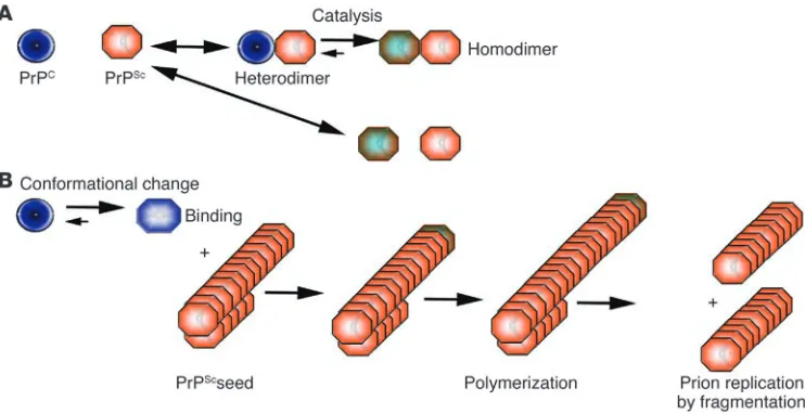
Figure 1
Models of PrPC to PrPSc conversion. (A) The heterodimer model proposes that upon infection of an
appro-priate host cell, the incoming PrPSc (orange) starts a catalytic cascade using PrPC (blue) or a partially
unfolded intermediate arising from stochastic fluctuations in PrPC conformations as a substrate, converting
it by a conformational change into a new β-sheet–rich protein. The newly formed PrPSc (green-orange) will
in turn convert new PrPC molecules. (B) The noncatalytic nucleated polymerization model proposes that
the conformational change of PrPC into PrPSc is thermodynamically controlled: the conversion of PrPC to
PrPSc is a reversible process but at equilibrium strongly favors the conformation of PrPC. Converted PrPSc is
established only when it adds onto a fibril-like seed or aggregate of PrPSc. Once a seed is present, further
precise physiological function of PrPC is unclear, expression of it
is absolutely required for transportation of the infectious agent both from the peripheral sites to the CNS (21) and within the CNS (22). However, reconstitution of Prnp knockout (Prnpo/o) mice with
WT bone marrow is insufficient to restore neuroinvasion in Prnpo/o
mice (21). Hence one could argue that the elemental compartment required for prion neuroinvasion is stromal and must express PrPC. Nevertheless, in adoptive transfer experiments on Prnpo/o
mice with WT bone marrow, the capability of the spleen to accu-mulate prions of the RML strain is restored (21, 23). This suggests that hematopoietic cells transport prions from the entry site to the lymphoreticular system (LRS), which accumulates and replicates prions. B lymphocytes (not necessarily expressing PrPC) are crucial
for peripheral prion spread and neuroinvasion (24–26).
This dependence of FDCs on lymphotoxin (LT) signaling by B cells likely may explain — at least in part — the apparent requirement for B cells in peripheral pathogenesis: FDCs have been reported to accumulate PrPSc following scrapie infection (27). Indeed, blockade
of LT signaling via administration of a soluble, dimeric LTβ recep-tor immunoglobulin fusion protein (LTβR-Ig) ablates mature FDCs and significantly impairs peripheral prion pathogenesis (28, 29).
FDCs are bifunctional cells: they support the formation and maintenance of the lymphoid microarchitecture but also play a
role in antigen trapping — capturing immune complexes by Fcγ
receptors and binding opsonized antigens to the CD21/CD35 complement receptors. Two studies have demonstrated that the complement system is relevant to prion pathogenesis. Mice geneti-cally engineered to lack complement factors (30) or mice depleted of the C3 complement component (31) exhibited enhanced resis-tance to peripheral prion inoculation. However, FDCs are most likely immobile cells and therefore unlikely to be responsible for prion transport into the CNS.
But just which cell types are involved in neuroinvasion? The innervation pattern of lymphoid organs is primarily sympathetic (32). Sympathectomy delays the onset of scrapie, while sympa-thetic hyperinnervation enhances splenic prion replication and neuroinvasion, which suggests that innervation of secondary lym-phoid organs is the rate-limiting step to neuroinvasion (33). How-ever, there is no physical synapse between FDCs and sympathetic nerve endings (34). So how can prions transmigrate from FDCs to sympathetic nerve fibers? A series of recent experiments (discussed below) may go some way toward providing answers.
FDC positioning is a primary determinant of velocity of neuroinvasion
We investigated how the distance between FDCs and splenic nerves affects the velocity of neuroinvasion, utilizing mice deficient in the CXC chemokine receptor 5 (CXCR5), which directs lymphocytes toward specific microcompartments (35). While density, distri-bution, and branching patterns of sympathetic nerve processes in CXCR5–/– spleens are normal, the distance between FDCs and
nerve endings is greatly reduced (36).
After peripheral administration of high doses of prions, velocity of pathogenesis was similar in CXCR5–/– and WT mice; however,
delivery of smaller inocula resulted in a dose-dependent increase in incubation periods in WT mice that was not evident in CXCR5–/–
mice. Peripheral prion pathogenesis in CXCR5–/– mice is therefore
more efficient upon incremental reduction of the inoculum. What is the basis of this reduced incubation period? Kinetics measurements of prion infectivity titers in the thoracic spinal cord provided the answer: following peripheral administration, only traces of infectivity were found in WT spinal cords at 80 days post-inoculation (dpi), whereas infectivity rose to measurable levels in the spinal cords of CXCR5–/– mice already at 60 dpi. This suggests
that increased velocity of prion entry into the CNS in CXCR5–/–
mice is due to the repositioning of FDCs near highly innervated, splenic arterioles (Figure 2). This was validated by the finding that incubation periods were prolonged in CXCR5–/– mice treated with
soluble LTβR-Ig to deplete mature FDCs.
Hence topographical relationships within lymphoid organs con-tribute to prion neuroinvasion. However, it remains to be deter-mined whether this results from passive diffusion of prions or whether mobile cells (e.g., germinal center B cells) are involved in an active transport process.
This study also raises the possibility that spread of infection to peripheral nerves occurs more rapidly when FDCs are in close proximity to nerves in lymphoid tissue other than spleen, such as Peyer’s patches. Indeed, FDCs are crucial to disease progres-sion but only during a short window of time following oral scrapie challenge (17). This implicates the efficiency of neuroim-mune transfer of prions as a primary determinant of neuroinva-sion. The detection of PrPSc in spleens of sCJD patients (12)
[image:4.585.50.280.79.318.2]sug-gests that the interface between cells of the immune system and
Figure 2
Positioning of FDCs in spleens of WT and CXCR5–/– mice. (A and B)
Diagrammatic representation of white pulp follicles in prion-infected CXCR5–/– and WT mice. Anti-CD21 immunostaining was performed to
visualize the lymphoid white pulp follicle microarchitecture. (C) Atypical-ly localized perivascular FDCs in Atypical-lymph follicles in CXCR5–/– mice.
Sym-pathetic nerves, visualized with antibodies to tyrosine hydroxylase (TH), are in close vicinity to FDCs (visualized by FDC-M1 immunostaining) (D). Scale bar: 50 mm. In contrast, sympathetic nerves in WT FDCs are localized in B cell areas at the periphery of the follicles (E and F). Arrowheads indicate TH and FDC-M1 positive areas. (G) Sympathetic nerves lining the thoracic spinal cord connect lymphoid organs and the CNS. (H) Shortened prion disease incubation period in CXCR5–/– mice
science in medicine
peripheral nerves (the neuroimmune connection) might also be of relevance in sporadic prion disease.
The neurodegeneration issue
There has certainly been progress in understanding the events underlying peripheral prion pathogenesis and neuroinvasion (37). However, prions exert their destructive effects exclusively within the CNS. The precise cause of neurodegeneration remains poorly understood and is a point of contention among prionologists. It seems unlikely that PrPSc is directly toxic, since tissue devoid of
PrPC that subsequently accumulates PrPSc remains healthy and
free of pathology (20, 38). During the conversion process, PrPC
lev-els may be depleted, yet this is also an unlikely cause of pathology, since ablation of PrPC does not result in scrapie-like symptoms
(39), even when ablated postnatally (40).
Lindquist and colleagues have suggested a mechanism that may account for prion-associated toxicity: (a) expression of a PrP variant resident in the cytosol was strongly neurotoxic in cultured cells and transgenic mice, which suggests a common framework for diverse PrP neurodegenerative disorders (41); and (b) PrP, retrogradely transported out of the endoplasmic reticulum, produced amorphous aggregates of PrP possess-ing partial proteinase K resistance in the cytosol. Once conver-sion occurred, it was self-sustaining (42). It will be interesting to determine whether the disease generated in these mice is, in some way, transmissible. However, while the results obtained here are certainly intriguing, it should be noted that reports else-where, although not refuting these observations, argue against the contribution of such potential neurotoxic PrP species (43,
44). Similarly, it has been reported that PrPC in some forms of
prion disease assumes a transmembrane topology, C-terminal transmembrane PrP (CtmPrP), and that the extent of neurotoxicity
is a result of concentration of CtmPrP, thereby arguing that CtmPrP may represent a major toxic moiety (45, 46). However,
while we still do not understand the biochemical events involved in cytosolic or CtmPrP-induced neurotoxicity, elucidation of this
may aid in the much-needed identification of therapeutic tar-gets. Additionally, in-depth characterization of transgenic mice expressing amino-terminally truncated PrPC (47), in which
cere-bellar neurodegeneration occurs, may not only aid in the elucida-tion of the molecular events responsible for potentially common neurodegenerative processes but perhaps also provide clues to the physiological function of PrPC itself.
Prion diagnostics
The ability to secure early diagnosis is vital for therapeutic inter-ventions to be of real value. With respect to animals destined for the human food chain, there is the additional demand to deter-mine presence of the prion agent in tissues in asymptomatic organisms, well before the appearance of any clinical symptoms. This applies equally to the detection of prions in humans, who may participate in tissue donation programs.
[image:5.585.53.533.114.379.2]Prions were transmitted via blood transfusion in sheep using blood obtained from infected animals prior to the onset of clinical symptoms (48, 49). If the same route applies to humans, this could represent a nightmare scenario for the blood transfusion services (50). A transfusion recipient received blood from an individual harboring the vCJD agent 3.5 years prior to the development of
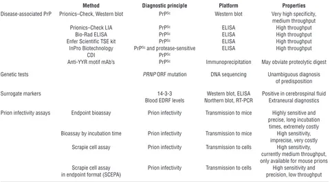
Table 2
Molecular diagnosis of prion disease and prion infectivity
Method Diagnostic principle Platform Properties Disease-associated PrP Prionics–Check, Western blot PrPSc Western blot Very high specificity,
medium throughput
Prionics–Check LIA PrPSc ELISA High throughput
Bio-Rad ELISA PrPSc ELISA High throughput
Enfer Scientific TSE kit PrPSc ELISA High throughput
InPro Biotechnology PrPSc and protease-sensitive ELISA High throughput
CDI PrPSc
Anti-YYR motif mAb’s PrPSc Immunoprecipitation May obviate proteolytic digest
Genetic tests PRNP ORF mutation DNA sequencing Unambiguous diagnosis
of predisposition
Surrogate markers 14-3-3 Western blot, ELISA Positive in cerebrospinal fluid
Blood EDRF levels Northern blot, RT-PCR Extraneural diagnostics
Prion infectivity assays Endpoint bioassay Prion infectivity Transmission to mice Highly sensitive and precise, long incubation
times, extremely costly Bioassay by incubation time Prion infectivity Transmission to mice High sensitivity,
imprecise, very costly Scrapie cell assay Prion infectivity Transmission to cells High sensitivity,
currently medium throughput, only available for mouse prions Scrapie cell assay Prion infectivity Transmission to cells High sensitivity and
in endpoint format (SCEPA) precision, low throughput
All currently reported methods of assessing the presence of prion disease are listed, including validated methods and nonvalidated experimental approach-es. Most commercial assays rely on the direct detection of disease-associated PrPSc. LIA, luminescence immunoassay; CDI, conformation-dependent
any clinical signs of prion disease in the donor. The unfortunate recipient developed disease 6.5 years after the transfusion.
Detection of PrPSc
To be truly useful, prion diagnostics should identify “suspect” cases as early during pathogenesis as possible. However, the currently available methods are quite insensitive when compared with those available for other infectious diseases. PrPSc represents the only
dis-ease-specific macromolecule identified to date, and all approved commercial testing procedures are based on the immunological detection of PrPSc. While around 50 companies are reported to be
developing prion diagnostic assays, all commercial test kits vali-dated for use by the European Union rely on proteolytic removal of endogenous PrPC prior to detection of PrPSc (Table 2). In addition,
the conformation-dependent immunoassay (4) utilizes the differ-ential binding of antibodies to native or denatured PrPSc.
Circumvention of the protease digestion step might theoreti-cally yield increased sensitivity of PrPSc-based detection methods
and make these methods more amenable to high-throughput technologies. However, it has proved difficult to discriminate between PrPC and PrPSc with antibodies, despite some early reports
(51). Interestingly, tyrosine-tyrosine-arginine (YYR) motifs (52) were reasoned to be more solvent-accessible in the pathological isoform of PrP, and a monoclonal antibody directed against these motifs was reported to be capable of selectively detecting PrPSc
across a variety of platforms. However, YYR motifs are certainly not unique to pathological prion proteins, and it remains to be determined whether this reagent can really improve the sensitivity of detection of prion infections.
Deposition of PrPSc in lymphoid tissues of human prion disease
patients has long been believed to be restricted to vCJD. However, recent results (12) imply that PrPSc is present in spleens and muscle
tissue from as much as one third of patients with sCJD. It is pres-ently unclear whether the patients with extraneural PrP represent a specific subset of CJD patients or whether the extraneural-negative patients may simply deposit PrPSc in muscle and spleen at levels
that are below the detectability threshold of the assay. If the lat-ter scenario proves true, and if the assay sensitivity can be raised, minimally invasive muscle biopsies may replace brain biopsy in clinical CJD diagnostics.
Surrogate markers and prion infectivity
While presence of PrPSc secures diagnostic association with the
presence of prion disease, PrPSc is not always easily detectable in
several forms of prion disease (53–55). In order to enhance the safe-ty of the blood supply and of products of bovine origin, absolute specificity in securing diagnosis of asymptomatic prion disease may not be required. Instead, it may be prudent to accommodate less than 100% specificity with a panel of surrogate markers capa-ble of identifying donated blood units from “suspect” individuals rather than requiring definitive diagnosis. It could be envisaged that wide-scale primary screens accommodate a certain degree of loss of specificity to identify samples to be re-tested in a secondary screen utilizing more specific (and likely labor-intensive) criteria.
Several research efforts have been directed at identifying tran-scripts and proteins differentially expressed in tissues of prion-infected animals relative to disease-free controls (56–58). However, these have mostly focused primarily either on prion-infected neural cell lines or on CNS tissue, frequently with emphasis on late-stage disease. Ideally, these surrogate markers should be detectable (and
differentially expressed) in easily accessible body fluids, such as blood or urine. At present, only one extraneural gene was reported to be differentially expressed during prion infection (59): erythroid differentiation–related factor (EDRF; also known as erythroid-associated factor) levels were progressively reduced in spleens of prion-infected mice throughout pathogenesis and also in blood of experimentally infected mice, cattle with BSE, and sheep with clinically manifest scrapie.
Assessment of the levels of surrogate markers in healthy indi-viduals is crucial in order to define the normal range of expression (according to age, sex, etc.) in order to determine what represents abnormal levels. In this respect, it is worth noting that determina-tion of normal expression range must utilize appropriate controls. For example, EDRF transcript levels have recently been reported to show a broad range of variation in healthy human subjects (60). However, since EDRF is an erythroid-specific transcript, it would be imperative to utilize other erythroid transcripts as internal con-trols to normalize for variations in numbers of circulating cells in which the transcript under study is expressed relative to total cells. More searches for surrogate markers would certainly be useful, and it is likely that surrogate markers of prion disease, particularly if they are detectable in body fluids, will expand the panel of tools available for screening for prion infections.
It is also worth noting here that recent advances in neuroimaging techniques, particularly with respect to MRI, may lead to clinically useful methods of assessment of prion disease in humans, perhaps even the ability to distinguish between sCJD and vCJD (61). For example, in vCJD the pulvinar sign (a high T2 MRI signal in the posterior thalamus) has been suggested to be relatively specific for vCJD, being present in approximately 75% of vCJD patients tested (62). In sCJD, fluid-attenuated inversion recovery and diffusion-weighted MRI sequences appear to be associated with high sensi-tivity and specificity. MRI imaging techniques such as these may therefore represent a relatively noninvasive method to corroborate suspicion of clinical presentation of human prion disease.
While surrogate markers such as S-100, neuron-specific enolase, and 14-3-3 protein have been suggested as potential biomarkers of prion disease using body fluids such as cerebrospinal fluid (CSF) (63, 64), it is worth remembering that these are clearly sur-rogate markers of general neurodegenerative disease and are not therefore predictive for human prion disease. For example, one study reported false positives of 14-3-3 detection in CSF samples of patients with herpes simplex encephalitis, hypoxic brain dam-age, atypical encephalitis, intracerebral metastases of a bronchial carcinoma, and metabolic encephalopathy (65).
It should not be forgotten that there is no ultimate consen-sus on the nature of the prion: PrPSc itself might represent a
sur-rogate marker of prion disease (66). The real gold standard of prion diagnostics is the detection of prion infectivity (whether or not PrPSc is present). Until recently, the only method available
science in medicine
the prion agent. However, it should be noted that these cell lines are currently reported only to be permissive to murine prions. It is to be expected that the spectrum of prion strains that can be assayed using this technology will expand.
Prion therapy
For all the promising approaches that are being explored (Table 3), no therapy for prion diseases is available as of yet. Many substances appear to possess prion-curing properties in vitro, including Congo red (68), amphotericin B, anthracyclines (69), sulfated polyanions (70), porphyrins (71), branched polyamines (72), �-sheet breakers (73), the spice curcumin (74), and recently even small interfering RNAs (75). The majority of these molecules exert their biological effects by directly or indirectly interfering with conversion of PrPC to
PrPSc, thereby also aiding clearance of PrPSc. Yet none of these
com-pounds have proved very effective for actual therapy.
In a recent report, results obtained in mice have led to the theory that administration of cytidyl-guanyl oligodeoxynucleotides (CpG-ODNs), which stimulate the innate immune system via toll-like receptor 9 (TLR9) signaling receptors on a variety of immune cells, may represent an applicable treatment regimen to delay prion dis-ease in humans (76). Here it was shown that the incubation period of prion disease was extended in mice multidose treated with CpG-ODNs for twenty days. It was concluded that stimulation of innate immunity accounts for this apparent anti-prion effect, possibly through induction of anti-PrP antibodies. However, this is diffi-cult to reconcile with several studies indicating that immune defi-ciencies of various sorts inhibit prion pathogenesis (24, 25, 30, 77). In addition, prion pathogenesis is unhampered in MyD88–/– mice,
in which there is impaired TLR9 signaling (78). In fact, repeated CpG-ODN treatment has severe side effects, ranging from lym-phoid follicle destruction and impaired antibody class switch to the development of ascites, hepatotoxicity, and thrombocytopenia (79). In addition, anti-PrP antibodies are not detectable in CpG-ODN–treated mice (79). It is likely therefore that the anti-prion effects of repeated CpG-ODN treatment arise via indiscriminate and undesirable follicular destruction.
Is vaccination against prion disease possible?
Anti-PrP antibodies (30) and F(ab) fragments to PrP (80, 81) can suppress prion replication in cultured cells. However, the mamma-lian immune system is essentially tolerant to PrPC (82). Ablation
of Prnp (39) renders mice highly susceptible to immunization with prions (22). Tolerance can be circumvented by transgenic expres-sion of an immunoglobulin µ chain containing the epitope-inter-acting region of a high-affinity anti-PrP monoclonal antibody. This sufficed to block prion pathogenesis upon intraperitoneal prion inoculation (83). Passive immunization may be a useful strategy for prophylaxis of prion diseases, since it has been shown that passive transfer of anti-PrP monoclonal antibodies prior to the onset of clinical symptoms is able to delay the onset of prion disease in mice inoculated intraperitoneally (84). Unfortunately, several efforts aimed at active immunization strategies have met with little success due to the robust immune tolerance to PrPC. In
this respect, it is certainly worth noting that extensive neuronal apoptosis in hippocampus and cerebellum has been shown follow-ing intracranial delivery of monoclonal antibodies reactive against a subset of PrP epitopes (85). The implications here are obvious; clearly, exhaustive in-vivo safety trials must be performed prior to the utilization of such strategies in humans.
Soluble prionostatic candidates
Do any serious candidates for prion therapeutic strategies exist? It is well established that expression of two PrPC moieties that differ
subtly from each other are able to inhibit prion replication (10). For example, humans heterozygous for a common PRNP polymor-phism at codon 129 are largely protected from CJD (86). Although the precise molecular basis for this effect is unclear, it is possible that heterologous PrPC may exert inhibitory action on prion
rep-lication by sequestration. This has been addressed directly by fusion of PrPC to an immunoglobulin Fcγ domain (87), allowing
for ligand dimerization, expression of the molecule as a soluble moiety, and also, therefore, increased overall stability in body flu-ids. Transgenetic expression of this PrP-Fc2 fusion protein resulted
in significant prolongation of incubation period upon prion
inoc-Table 3
Approaches to prion therapy
Therapeutic approach Target Properties References
Polyanions Possibly membrane-resident PrPSc Efficient in cultured cells; (70, 88)
relatively toxic in vivo
Curcumin Unknown Efficacy in vivo unproven (74)
Soluble lymphotoxin receptor FDCs Effective in vivo, but only on peripheral (28, 29) pathogenesis; moderate untoward effects
CpG oligodeoxynucleotides FDCs, DCs; B cells and macrophages Severely immunoclastic at doses effective in vivo (76, 79) Anti-PrP antibodies PrPC Effective in vivo only if administered in massive doses (80, 83, 84)
Amyloidotropic intercalators PrPSc Toxic; questionable efficacy in vivo (89, 90)
(e.g., Congo red)
Chemical or immunological Peripheral nerves involved Very efficacious, but unacceptable (33)
sympathectomy in neuroinvasion toxicity in vivo
Polyene antibiotics Possibly membrane-resident PrPSc Low efficacy in vivo (91, 92)
Chlorpromazine and quinacrine Unknown Questionable efficacy in vivo; hepatotoxicity (93–95)
Soluble dimeric PrPC PrPSc Effective as transgene, but efficacy (87)
immunoadhesin upon injection unproven
ulation via competition with PrPSc. It remains to be established
whether PrP-Fc2 is acting as an anti-prion compound when
deliv-ered exogenously. If so, soluble prion protein mutants may well represent anti-prion compounds.
An outlook for prion therapy
Prion diseases continue to present a diagnostic and therapeu-tic challenge to clinicians and researchers worldwide. There are many aspects of prion biology that remain unclear; we still do not know the precise physical nature of the infectious agent, the molecular and biochemical mechanisms underlying associated neurodegeneration, or the physiological function of PrPC. The
diagnostic tools currently available for prion diseases are
signifi-cantly less sensitive and satisfactory than those available for other infectious diseases. Additionally, there is a dearth of therapeutic intervention strategies available for these diseases. However, that said, the last decade or so of prion research has witnessed astound-ing advances in our knowledge and understandastound-ing of basic prion biology, and the field has attracted increasing numbers of research-ers from divresearch-erse disciplines. Undoubtedly, this trend will trigger further important advances in prion science.
Address correspondence to: Adriano Aguzzi, Institut für Neuro-pathologie, UniversitätsSpital Zürich, Schmelzbergstrasse 12, CH-8091 Zurich, Switzerland. Phone: 2107; Fax: 41-1-255-4402; E-mail: [email protected].
1. Glatzel, M., et al. 2003. Human prion diseases: epi-demiology and integrated risk assessment. Lancet Neurol.2:757–763.
2. Collins, P.S., Lawson, V.A., and Masters, P.C. 2004. Transmissible spongiform encephalopathies.
Lancet.363:51–61.
3. Bolton, D.C., McKinley, M.P., and Prusiner, S.B. 1982. Identification of a protein that purifies with the scrapie prion. Science.218:1309–1311. 4. Safar, J.G., et al. 2002. Measuring prions causing
bovine spongiform encephalopathy or chronic wasting disease by immunoassays and transgenic mice. Nat. Biotechnol.20:1147–1150.
5. Creutzfeldt, H.G. 1920. Über eine eigenartige herd-förmige Erkrankung des Zentralnervensystems.
Zeitschrift für die gesamte Neurologie und Psychiatrie.
57:1–19.
6. Jakob, A. 1921. Über eigenartige Erkrankungen des Zentralnervensystems mit bemerkenswertem anatomischem Befunde. (Spastische Pseudoskle-rose-Encephalomyelopathie mit disseminierten Degenerationsherden). Zeitschrift für die gesamte Neurologie und Psychiatrie.64:147–228.
7. Griffith, J.S. 1967. Self-replication and scrapie.
Nature.215:1043–1044.
8. Prusiner, S.B. 1982. Novel proteinaceous infectious particles cause scrapie. Science.216:136–144. 9. Weissmann, C. 1991. A ‘unified theory’ of prion
propagation. Nature.352:679–683.
10. Telling, G.C., et al. 1995. Prion propagation in mice expressing human and chimeric PrP transgenes implicates the interaction of cellular PrP with another protein. Cell.83:79–90.
11. Priola, S.A., Chesebro, B., and Caughey, B. 2003. Biomedicine. A view from the top--prion diseases from 10,000 feet. Science.300:917–919.
12. Glatzel, M., Abela, E., Maissen, M., and Aguzzi, A. 2003. Extraneural pathologic prion protein in sporadic Creutzfeldt-Jakob disease. N. Engl. J. Med.
349:1812–1820.
13. Hill, A.F., et al. 2003. Molecular classification of sporadic Creutzfeldt-Jakob disease. Brain.
126:1333–1346.
14. Aguzzi, A., Montrasio, F., and Kaeser, P.S. 2001. Pri-ons: health scare and biological challenge. Nat. Rev. Mol. Cell Biol.2:118–126.
15. Brown, K.L., et al. 1999. Scrapie replication in lym-phoid tissues depends on prion protein- expressing follicular dendritic cells. Nat. Med.5:1308–1312. 16. Aguzzi, A. 2003. Prions and the immune system:
a journey through gut, spleen, and nerves. Adv. Immunol.81:123–171.
17. Mabbott, N.A., Young, J., McConnell, I., and Bruce, M.E. 2003. Follicular dendritic cell dedifferentiation by treatment with an inhibitor of the lymphotoxin pathway dramatically reduces scrapie susceptibility. J. Virol.77:6845–6854. 18. Prinz, M., et al. 2003. Oral prion infection requires
normal numbers of Peyer’s patches but not of enteric lymphocytes. Am. J. Pathol.162:1103–1111.
19. Büeler, H.R., et al. 1993. Mice devoid of PrP are resistant to scrapie. Cell.73:1339–1347.
20. Brandner, S., et al. 1996. Normal host prion pro-tein necessary for scrapie-induced neurotoxicity.
Nature.379:339–343.
21. Blättler, T., et al. 1997. PrP-expressing tissue required for transfer of scrapie infectivity from spleen to brain. Nature.389:69–73.
22. Brandner, S., et al. 1996. Normal host prion pro-tein (PrPC) is required for scrapie spread within the central nervous system. Proc. Natl. Acad. Sci. U. S. A.
93:13148–13151.
23. Kaeser, P.S., Klein, M.A., Schwarz, P., and Aguzzi, A. 2001. Efficient lymphoreticular prion propagation requires prp(c) in stromal and hematopoietic cells.
J. Virol.75:7097–7106.
24. Klein, M.A., et al. 1997. A crucial role for B cells in neuroinvasive scrapie. Nature.390:687–690. 25. Klein, M.A., et al. 1998. PrP expression in B
lym-phocytes is not required for prion neuroinvasion.
Nat. Med.4:1429–1433.
26. Montrasio, F., et al. 2001. B lymphocyte-restricted expression of prion protein does not enable prion replication in prion protein knockout mice. Proc. Natl. Acad. Sci. U. S. A.98:4034–4037.
27. Kitamoto, T., Muramoto, T., Mohri, S., Doh-Ura, K., and Tateishi, J. 1991. Abnormal isoform of prion protein accumulates in follicular dendritic cells in mice with Creutzfeldt-Jakob disease. J. Virol.
65:6292–6295.
28. Montrasio, F., et al. 2000. Impaired prion replica-tion in spleens of mice lacking funcreplica-tional follicular dendritic cells. Science.288:1257–1259.
29. Mabbott, N.A., Mackay, F., Minns, F., and Bruce, M.E. 2000. Temporary inactivation of follicular dendritic cells delays neuroinvasion of scrapie [let-ter]. Nat. Med.6:719–720.
30. Klein, M.A., et al. 2001. Complement facilitates early prion pathogenesis. Nat. Med.7:488–492. 31. Mabbott, N.A., Bruce, M.E., Botto, M., Walport,
M.J., and Pepys, M.B. 2001. Temporary depletion of complement component C3 or genetic deficiency of C1q significantly delays onset of scrapie. Nat. Med.7:485–487.
32. Felten, D.L., and Felten, S.Y. 1988. Sympathetic noradrenergic innervation of immune organs.
Brain Behav. Immun.2:293–300.
33. Glatzel, M., Heppner, F.L., Albers, K.M., and Aguzzi, A. 2001. Sympathetic innervation of lymphoreticu-lar organs is rate limiting for prion neuroinvasion.
Neuron.31:25–34.
34. Heinen, E., Bosseloir, A., and Bouzahzah, F. 1995. Follicular dendritic cells: origin and function. Curr. Top. Microbiol. Immunol.201:15–47.
35. Forster, R., et al. 1996. A putative chemokine recep-tor, BLR1, directs B cell migration to defined lym-phoid organs and specific anatomic compartments of the spleen. Cell.87:1037–1047.
36. Prinz, M., et al. 2003. Positioning of follicular dendritic cells within the spleen controls prion
neuroinvasion. Nature.425:957–962.
37. Aguzzi, A., and Polymenidou, M. 2004. Mamma-lian prion biology. One century of evolving con-cepts. Cell.116:313–327.
38. Mallucci, G., et al. 2003. Depleting neuronal PrP in prion infection prevents disease and reverses spon-giosis. Science.302:871–874.
39. Büeler, H.R., et al. 1992. Normal development and behaviour of mice lacking the neuronal cell-surface PrP protein. Nature.356:577–582.
40. Mallucci, G.R., et al. 2002. Post-natal knockout of prion protein alters hippocampal CA1 properties, but does not result in neurodegeneration. EMBO J.
21:202–210.
41. Ma, J., Wollmann, R., and Lindquist, S. 2002. Neurotoxicity and neurodegeneration when PrP accumulates in the cytosol. Science.298:1781–1785. 42. Ma, J., and Lindquist, S. 2002. Conversion of PrP to
a self-perpetuating PrPSc-like conformation in the cytosol. Science.298:1785–1788.
43. Drisaldi, B., et al. 2003. Mutant PrP is delayed in its exit from the endoplasmic reticulum, but neither wild-type nor mutant PrP undergoes retrotrans-location prior to proteasomal degradation. J. Biol. Chem. 278:21732–21743.
44. Roucou, X., Guo, Q., Zhang, Y., Goodyer, C.G., and LeBlanc, A.C. 2003. Cytosolic prion protein is not toxic and protects against Bax-mediated cell death in human primary neurons. J. Biol. Chem.
278:40877–40881.
45. Hegde, R.S., et al. 1998. A transmembrane form of the prion protein in neurodegenerative disease.
Science.279:827–834.
46. Hegde, R.S., et al. 1999. Transmissible and genet-ic prion diseases share a common pathway of neurodegeneration. Nature.402:822–826. 47. Shmerling, D., et al. 1998. Expression of
amino-ter-minally truncated PrP in the mouse leading to atax-ia and specific cerebellar lesions. Cell.93:203–214. 48. Houston, F., Foster, J.D., Chong, A., Hunter, N.,
and Bostock, C.J. 2000. Transmission of BSE by blood transfusion in sheep. Lancet.356:999–1000. 49. Hunter, N., et al. 2002. Transmission of prion
diseas-es by blood transfusion. J. Gen. Virol.83:2897–2905. 50. Aguzzi, A. 2000. Prion diseases, blood and
the immune system: concerns and reality.
Haematologica.85:3–10.
51. Korth, C., et al. 1997. Prion (PrPSc)-specific epitope defined by a monoclonal antibody. Nature.
390:74–77.
52. Paramithiotis, E., et al. 2003. A prion protein epitope selective for the pathologically misfolded conformation. Nat. Med.9:893–899.
53. Hsiao, K.K., et al. 1994. Serial transmission in rodents of neurodegeneration from transgenic mice expressing mutant prion protein. Proc. Natl. Acad. Sci. U. S. A.91:9126–9130.
science in medicine
55. Tagliavini, F., et al. 1994. Amyloid fibrils in Gerst-mann-Straussler-Scheinker disease (Indiana and Swedish kindreds) express only PrP peptides encoded by the mutant allele. Cell.79:695–703. 56. Duguid, J.R., and Dinauer, M.C. 1990. Library
subtraction of in vitro cDNA libraries to identify differentially expressed genes in scrapie infection.
Nucleic Acids Res.18:2789–2792.
57. Duguid, J., and Trzepacz, C. 1993. Major histocom-patibility complex genes have an increased brain expression after scrapie infection. Proc. Natl. Acad. Sci. U. S. A.90:114–117.
58. Dandoy-Dron, F., et al. 1998. Gene expression in scrapie. Cloning of a new scrapie-responsive gene and the identification of increased levels of seven other mRNA transcripts. J. Biol. Chem.
273:7691–7697.
59. Miele, G., Manson, J., and Clinton, M. 2001. A novel erythroid-specific marker of transmissible spongi-form encephalopathies. Nat. Med.7:361–364. 60. Glock, B., et al. 2003. Transcript level of erythroid
differentiation-related factor, a candidate surro-gate marker for transmissible spongiform enceph-alopathy diseases in blood, shows a broad range of variation in healthy individuals. Transfusion.
43:1706–1710.
61. Tribl, G.G., et al. 2002. Sequential MRI in a case of Creutzfeldt-Jakob disease. Neuroradiology.
44:223–226.
62. Zeidler, M., Collie, D.A., Macleod, M.A., Sellar, R.J., and Knight, R. 2001. FLAIR MRI in sporadic Creutzfeldt-Jakob disease. Neurology.56:282. 63. Hsich, G., Kinney, K., Gibbs, C.J., Lee, K.H., and
Harrington, M.G. 1996. The 14-3-3 brain protein in cerebrospinal fluid as a marker for transmis-sible spongiform encephalopathies. N. Engl. J. Med.
335:924–930.
64. Beaudry, P., et al. 1999. 14-3-3 protein, neuron-spe-cific enolase, and S-100 protein in cerebrospinal fluid of patients with Creutzfeldt-Jakob disease.
Dement. Geriatr. Cogn. Disord.10:40–46.
65. Zerr, I., et al. 1998. Detection of 14-3-3 protein in the cerebrospinal fluid supports the diagnosis of Creutzfeldt-Jakob disease. Ann. Neurol.43:32–40. 66. Aguzzi, A., and Weissmann, C. 1997. Prion research:
the next frontiers. Nature.389:795–798.
67. Klohn, P.C., Stoltze, L., Flechsig, E., Enari, M., and
Weissmann, C. 2003. A quantitative, highly sensitive cell-based infectivity assay for mouse scrapie prions.
Proc. Natl. Acad. Sci. U. S. A.100:11666–11671. 68. Caughey, B., and Race, R.E. 1992. Potent inhibition
of scrapie-associated PrP accumulation by congo red. J. Neurochem.59:768–771.
69. Tagliavini, F., et al. 1997. Effectiveness of anthracycline against experimental prion disease in Syrian hamsters. Science.276:1119–1122. 70. Caughey, B., and Raymond, G.J. 1993. Sulfated
polyanion inhibition of scrapie-associated PrP accu-mulation in cultured cells. J. Virol.67:643–650. 71. Priola, S.A., Raines, A., and Caughey, W.S. 2000.
Porphyrin and phthalocyanine antiscrapie com-pounds. Science.287:1503–1506.
72. Supattapone, S., et al. 2001. Branched polyamines cure prion-infected neuroblastoma cells. J. Virol.
75:3453–3461.
73. Soto, C., et al. 2000. Reversion of prion protein conformational changes by synthetic beta- sheet breaker peptides. Lancet.355:192–197.
74. Caughey, B., et al. 2003. Inhibition of protease-resistant prion protein accumulation in vitro by curcumin. J. Virol.77:5499–5502.
75. Daude, N., Marella, M., and Chabry, J. 2003. Spe-cific inhibition of pathological prion protein accu-mulation by small interfering RNAs. J. Cell. Sci.
116:2775–2779.
76. Sethi, S., Lipford, G., Wagner, H., and Kretzschmar, H. 2002. Postexposure prophylaxis against prion disease with a stimulator of innate immunity.
Lancet.360:229–230.
77. Frigg, R., Klein, M.A., Hegyi, I., Zinkernagel, R.M., and Aguzzi, A. 1999. Scrapie pathogenesis in sub-clinically infected B-cell-deficient mice. J. Virol.
73:9584–9588.
78. Prinz, M., et al. 2003. Prion pathogenesis in the absence of Toll-like receptor signalling. EMBO Rep.
4:195–199.
79. Heikenwalder, M., et al. 2004. Lymphoid follicle destruction and immunosuppression after repeat-ed CpG oligodeoxynucleotide administration. Nat. Med.10:187–192.
80. Peretz, D., et al. 2001. Antibodies inhibit prion propagation and clear cell cultures of prion infec-tivity. Nature.412:739–743.
81. Enari, M., Flechsig, E., and Weissmann, C. 2001.
Scrapie prion protein accumulation by scrapie-infected neuroblastoma cells abrogated by expo-sure to a prion protein antibody. Proc. Natl. Acad. Sci. U. S. A.98:9295–9299.
82. Souan, L., et al. 2001. Modulation of proteinase-K resistant prion protein by prion peptide immuniza-tion. Eur. J. Immunol.31:2338–2346.
83. Heppner, F.L., et al. 2001. Prevention of scrapie pathogenesis by transgenic expression of anti-prion protein antibodies. Science.294:178–182. 84. White, A.R., et al. 2003. Monoclonal antibodies
inhibit prion replication and delay the develop-ment of prion disease. Nature.422:80–83. 85. Solforosi, L., et al. 2004. Cross-linking cellular
prion protein triggers neuronal apoptosis in vivo.
Science. 303:1514–1516.
86. Mead, S., et al. 2003. Balancing selection at the prion protein gene consistent with prehistoric kurulike epidemics. Science.300:640–643. 87. Meier, P., et al. 2003. Soluble dimeric prion protein
binds PrP(Sc) in vivo and antagonizes prion dis-ease. Cell.113:49–60.
88. Ladogana, A., et al. 1992. Sulphate polyanions prolong the incubation period of scrapie-infected hamsters. J. Gen. Virol.73:661–665.
89. Prusiner, S.B., et al. 1983. Scrapie prions aggre-gate to form amyloid-like birefringent rods. Cell.
35:349–358.
90. Caughey, B., Ernst, D., and Race, R.E. 1993. Congo red inhibition of scrapie agent replication. J. Virol.
67:6270–6272.
91. Pocchiari, M., Schmittinger, S., and Masullo, C. 1987. Amphotericin B delays the incubation period of scrapie in intracerebrally inoculated hamsters.
J. Gen. Virol.68:219–223.
92. Mange, A., et al. 2000. Amphotericin B inhibits the generation of the scrapie isoform of the prion pro-tein in infected cultures. J. Virol.74:3135–3140. 93. Korth, C., May, B.C., Cohen, F.E., and Prusiner, S.B.
2001. Acridine and phenothiazine derivatives as pharmacotherapeutics for prion disease. Proc. Natl. Acad. Sci. U. S. A.98:9836–9841.