© 2016, IRJET | Impact Factor value: 4.45 | ISO 9001:2008 Certified Journal
| Page 2439
A Modified Approach to FECG Extraction using Sequential and Parallel
Kalman Filter
Rohit K. Paithane
1, Faisal I. Shaikh
21
M.E. Student Electronics & Telecommunication ,J.N.E.C.
Aurangabad, Maharashtra, India.
2
Professor,
Electronics & Telecommunication ,J.N.E.C.
Aurangabad, Maharashtra, India.
---***---Abstract -
The extraction of Fetal Electrocardiogram (FECG) is very important to know the health of fetus .It is also useful for doctors to get complete well being of fetus and decide to take required decision regarding delivery. The extraction of fetal electrocardiogram (FECG) is from maternal ECG signal obtained from the abdominal lead.The FECG contains activity of electrical depolarization and repolarization of fetal heart. This paper suggests extended Kalman filter (EKF) for the extraction of Fetal ECG (FECG) from Mother’s ECG (MECG) and filtering of noisy FECG signals. An extended Kalman filtering is used for extracting electrocardiograms (ECGs) from a single channel as encountered in the fetal and maternal ECG extraction from abdominal sensor is done. The proposed system facilitates the prenatal procedures for monitoring of the cardiac condition of both the mother and the fetus. The database used is taken from physionet.org. i.e. direct fetal electrocardiogram.Key Words: Abdominal ECG (AECG), Extended Kalman
filtering (EKF), Fetal electrocardiogram (FECG) extraction, Mother’s ECG (MECG), Parallel EKF (par-EKF), Sequential EKF (seq-EKF), Linear quadratic estimation (LQE).
1. INTRODUCTION
The Fetal ECG (FECG) signal is obtained from the Abdominal ECG (AECG) of a pregnant woman. This FECG is an effective diagnostic tool for determining the general condition of the fetus. The fetal contribution to the AECG is very small, so it is common to record a very degraded signal from which even the fetal heart rate (FHR) can hardly be detected. Hence we have to eliminate MECG and noise signals to extract FECG.
Adaptive filtering is a common approach for removing MECG and extracting FECG. The basic adaptive filtering is based on adaptive filter for either removing the MECG using one or several maternal reference channels or directly using the filter for extracting the fetal QRS waves. However, existing adaptive filtering methods for MECG artifact removal either require a reference MECG channel
that is morphologically similar to the contaminating waveform or require several linearly independent channels to roughly reconstruct any morphologic shape from the references. Both of these approaches are practically inconvenient and with limiting performance, because their morphology.
The Kalman filter, also known as linear quadratic estimation (LQE) an algorithm that uses a series of measurements observed over time, containing noise (random variations) and other inaccuracies, and produces estimates of unknown variables that tend to be more precise than those based on a single measurement alone. The Kalman filter has numerous applications in technology. Furthermore, the Kalman filter is a widely applied concept in time series analysis used in fields such as signal processing and econometrics. he algorithm works in a two-step process. In the prediction step, the Kalman filter produces estimates of the current state variables, along with their uncertainties. Once the outcome of the next measurement (necessarily corrupted with some amount of error, including random noise) is observed, these estimates are updated using a weighted average, with more weight being given to estimates with higher certainty. Because of the algorithm's recursive nature, it can run in real time using only the present input measurements and the previously calculated state and its uncertainty matrix; no additional past information is required. It is true that premature standardization of technique is not necessarily advantageous.
© 2016, IRJET | Impact Factor value: 4.45 | ISO 9001:2008 Certified Journal
| Page 2440
and FECG. However, as mentioned in, the filter fails to discriminate between the maternal and fetal components when the MECG and FECG waves fully overlap in time. The reason is that when MECG is being estimated, FECG and other components are supposed to be Gaussian noises.
However, this assumption is not true, especially when MECG and FECG waves fully overlap in time it is difficult for the filter to follow desired ECG. Clinical monitoring of fetal cardiac activity is usually based on a small number of electrodes located on mother’s abdomen, and on a sound sensitive sensor. In such a context, in this study, we wonder what performance can be obtained with only one electrode, by using a refined model of the signal recorded on the unique electrode: the model will explicitly take into account that the signal is the superposition of a few ECG signals. The rest of this paper is organized as follows. In Section II, equations and theory supporting our proposed method including the Bayesian filtering theory and dynamic ECG model are described. In Section III, results of the proposed method applied on different data and discussion about the results are presented. Finally, our conclusion is stated in Section IV.
1.1
METHOD
A. Sequential EKF
The goal of KF is to estimate the state of a discrete-time controlled process. Consider a state vector xk+1 governed by
a nonlinear stochastic difference equation with measurement vector yk+1at time instant k + 1:
Xk+1 = f (Xk, Wk, k+1)
Yk+1 = h (Xk+1, Vk+1, k+1) (1)
where the random variables Wk and Vk represent the process and measurement noises, with associated covariance matrices Qk = E {Wk,WkT} and Rk = E{Vk,VkT} . The extended Kalman filter (EKF) is an extension of the standard KF to nonlinear systems, which linearize about the current mean and covariance. In this study, a synthetic dynamic ECG model is used to extract FECG from mixture of an MECG, one (or more) FECG(s), and other signals considered as noises. In polar coordinates, one ECG signal can be expressed as the sum of five Gaussian functions defined by their peak amplitude, width, and center, denoted αi , bi , and ψi, respectively. From the ECG, one can define the observed phase φk by a linear time wrapping of the R–R time intervals into [0, 2π][1].
The ECGs composing the observed mixture can be estimated by applying the described EKF: at each step, one ECG is extracted according to a procedure. In case of a mixture of MECG and one FECG, the first step extracts, from the raw recording, the dominant ECG (often the MECG) considering the concurrent ECG (respectively, FECG) and other noises as a unique Gaussian noise. After subtracting the dominant ECG from the original signal, the second step is the extraction of FECG from the residual signal. This procedure is referred to as sequential EKF (seq-EKF). In this extraction, during the first step, the concurrent MECG and additional noise are modeled by Gaussian noises. In fact, although this assumption may be acceptable when there are not strong artifacts interfering with the ECG, it is no longer accurate when other ECG artifacts are considerable (i.e., at the first step) since the noise is no longer normally distributed.
B. Parallel EKF
The extended state Kalman filtering procedure is referred to as parallel EKF or EKS (par-EKF). The related extended state vector xk=[θ(1)k , z(1)k , . . . , θ (N )k , z(N )k ]T is thus defined by where each [θ(i)k , z(i)k ]Tis related to one of the ECGs. Finally, themeasurement process leads to express the measurement vectoryk+1 = [φ(1)k+1, . . . , φ(N )k+1, sk+1]T[1]. This par-EKF is more accurate to extract FECG from abdominal sensors than the seq-EKF. Indeed, in the proposed method, all ECGs are jointly modeled by dynamic states so that only the state and measurement noise vectors are assumed to be normally distributed.
Moreover, the extended state par-EKF fully models overlapping waves of several ECGs. Finally, the state and observation noises, ηnk and vnk , respectively, allow the filter to fit some variability of the ECG shapes. Although the model does not fit too large variations inspection of the residue will reveal these abnormal beats.
C. Model Parameters Estimation
© 2016, IRJET | Impact Factor value: 4.45 | ISO 9001:2008 Certified Journal
| Page 2441
rough estimation of FECG is obtained by using the seq-EKF algorithm, which now allows us to detect easily the fetal R-peaks. Then, for each ECG, each beat (defined by the signals between two consecutive R peaks) is time wrapped into [0,2π]. The average of the ECG waveform is obtained by the mean of all time-wrapped beats, for all phases between 0 and 2π. Finally, by using a nonlinear least-squares approach, the best estimate of the parameters in the minimum mean square error (MMSE) sense is found.
1.2 RESULTS AND DISCUSSIONS
Synthetic and actual data have been used to study performance of the proposed method. In Subsection III-A, the synthetic mixed ECG is considered and performance of the method has been studied. In Subsection III-B, the effectiveness of the method on actual data has been examined.
A. Experimental Performance on Synthetic Data
In realistic synthetic mixtures of MECG and FECG with white Gaussian noise have been generated for different situations, and the proposed method has been applied on them to extract MECG and FECG. Synthetic MECG and FECG used in this study are based on single dipole vector of the heart, proposed in and inspired by the single-channel ECG dynamic model presented in. Sampling frequency is set to 500 Hz and signals include 1000 samples. The main parameters that can affect the mixtures are input noise power, ratio between amplitudes of FECG and MECG, and ratio between fetal and maternal heart rates. In order to investigate the performance of the proposed method, several trials were carried out under each value of these parameters. In the output, estimated MECG and FECG signals, ˆsm and ˆsf , are assumed to be the sum of MECG, FECG, and noise, such that
ˆsm = a1sm + a2sf + a3n
ˆsf = b1sm + b2sf + b3n (2)
where coefficients a1 , a2 , a3 , b1 , b2 , and b3 have to be estimated and sm, sf , and n denote MECG, FECG, and noise, respectively[1].
[image:3.595.310.559.189.255.2]Fig 1: Mixture synthesized ECG
[image:3.595.308.560.285.349.2]Fig 2: Mixture synthesized ECG R-R peak detection
Fig 3: Maternal separation from mixture using par-EKF
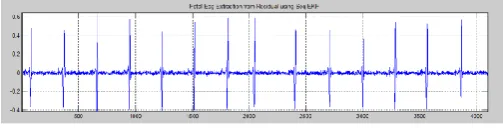
Fig 4: Filtered Fetal-ECG from residual ECG using seq-EKF
B. Fetal ECG Extraction on Actual (Physionet) Data
Abdominal Direct FECG Database: This database consists ofa series of abdominal FECG recordings, taken from a single subject between 21 and 40 weeks of pregnancy.
The recordings include direct abdominal signal. The signals were recorded at 1 kHz. Fig. shows results of Seq-EKS and par-EKS using single channel of the first 10s of namely the r01_direct dataset. To show the effectiveness of the proposed method in extraction of the FECG at different periods of pregnancy, and from channel locations, the first 10s of the mixtures and fetal par-EKS outputs of the datasets r04, r07, r08, r010, are plotted in Figures [7]
.
0 1000 2000 3000 4000 5000 6000 7000 8000 9000 10000 -2000
-1500 -1000 -500 0 500 1000
1500 Loaded Signal
Samples
Vo
lta
ge
(m
icr
o V
)
[image:3.595.329.537.576.635.2]Noisy ECG Signal
Fig 5: Direct abdominal ECG (r01) from Physionet
0 500 1000 1500 2000 2500 3000 3500 4000 -1500
-1000 -500 0 500 1000 1500
Samples
Vo
lta
ge
(m
icr
o
V)
Phase Assignement using R-detection
ECG Signal R-waves
© 2016, IRJET | Impact Factor value: 4.45 | ISO 9001:2008 Certified Journal
| Page 2442
Fig 5: Maternal separation from mixture using par-EKF
0 1000 2000 3000 4000 5000 6000 7000 8000 9000 10000 -30
-20 -10 0 10 20 30
Samples
Vo
lta
ge
(m
V)
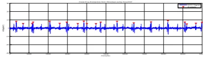
Fetal Ecg Extraction from Residual using Seq-EKF
ECG Signal R-waves
Fig 6: Filtered Fetal-ECG from residual ECG using seq-EKF
C. Analytical Results
Applying Kalman Filter on the 8 data sets, the FECG signals were extracted from 8 of them, and with an average percentage of 90.0 % of FECG R peaks extraction as calculated in Table I. Figures above shows the comparison between abdominal signal and extracted FECG signal. Looking at the figures in earlier points, maternal and extracted fetal ECG two different signals can be seen. Maternal ECG signal was removed followed by fetal ECG. Applying the peak detection algorithm to extracted FECG signals, the number of detected fetal R peaks was
calculated. Table I shows the number of peaks of the direct FECG from scalp and the extracted ones in addition to the percentage of extraction for the different 8 sets and the total average percentage after applying the technique[19].
Table -1: THE PERCENTAGE OF THE R PEAKS EXTRACTION OF EACH DATA SET AFTER APPLYING KALMAN FILTER
Data Set NO.
Peak numbers of Direct FECG
Peak numbers of
Extracted FECG
% of Extraction
DATA
Set 1 20 17 85
DATA
Set 2 20 20 100
DATA
Set 3 20 17 85
DATA
Set 4 20 20 100
DATA
Set 5 21 21 100
DATA
Set 6 18 17 94.44444444
DATA
Set 7 18 20 111.1111111
DATA
Set 8 18 12 66.66666667
Average of % Extraction 90
3. CONCLUSIONS
In this paper, a synthetic ECG has been used for Kalman Filter. The same concept is used for actual data taken from Physionet i.e. direct Fetal ECG from abdomen 2. Here the actual data is downloaded and filtered followed by peak detection, i.e. R-R interval. After the peaks are detected then the aligned with phase and Maternal ECG is extracted by using Par-EKF and residual signal remains which consist Fetal ECG. To detect the peaks of Fetal from the residual signal the Seq-EKF is used extract desired FECGs from that mixture. This proposed method only uses a single channel to separate different ECGs, i.e. Maternal and Fetal ECGs. Synthetic data and illustrated on actual data (single channel fetal ECG). The comparison of the extraction is done by counting the number of peaks of Direct FECG captured from the scalp with the signal acquired from the various leads of abdomen of the same pregnant women. Hence the comparison is done by counting the number of peaks as shown in the table.
ACKNOWLEDGEMENT
I express my sincere gratitude to my M. E. guide, Prof. F. I. Shaikh, Associate Professor, Electronics and Telecommication Department, MGM’s Jawaharlal Nehru Engineering College, Aurangabad, for writing this paper and his guidance throughout my dissertation.
REFERENCES
1] Mohammad, Niknazar, Bertrand Rivet, and Christian Jutten, “Fetal ECG Extraction by Extended State Kalman Filtering Based on Single-Channel Recordings” IEEE TRANSACTIONS ON BIOMEDICAL ENGINEERING, VOL. 60, NO. 5, MAY 2013. [2] M. Vennila and Sikkandar Mohamed Yacin,“A Modified Approach on Fetal ECG Extraction Using Extended State Kalman Filtering” International Conference on Green Computing, Communication and Electrical Engineering (ICGCCEE’14). [3] Mahsa Akhbari, Mohammad Niknazar, Christian Jutten, Mohammad Bagher, Shamsollahi, Bertrand Rivet “Fetal Electrocardiogram R-peak Detection using Robust Tensor Decomposition and Extended Kalman Filtering.”
[image:4.595.59.266.197.257.2]© 2016, IRJET | Impact Factor value: 4.45 | ISO 9001:2008 Certified Journal
| Page 2443
Hagen Malberg “Maternal Signal Estimation by Kalman Filtering and Template Adaptation for Fetal Heart Rate Extraction”, Computing in Cardiology 2013; 40:193-196, ISSN 2325-8861. [5] Reza Sameni, M.B. Shamsollahi, Christian Jutten,
Massoud Babaie-Zadeh.“Filtering Noisy ECG Signals Using the Extended Kalman Filter Based on a Dynamic ECG Model.” Proc.Of the 32nd Annual International Conference on Computers in Cardiology, Lyon, France, Sep.25-28, 2005, Sep 2005, Lyon, France. 32, pp.1017-1020. <hal-00174343>. [6] Reza Sameni, M.B. Shamsollahi, Christian Jutten, “Filtering Electrocardiogram Signals Using the Extended Kalman Filter.”, 27th Annual International Conference of the IEEE Engineering in Medicine and Biology Society (EMBS), Sep 2005, Shanghai, China. pp.5639-5642. <hal-00174347> [7] Physionet:
https://www.physionet.org/cgi-bin/atm/ATM/database [8] H. M. Jenkins, “Technical progress in fetal
electrocardiography—A review,”J. Perinat. Med., vol. 14, no. 6, pp. 365–370, 1986. [9] G. Camps, M. Martinez, and E. Soria, “Fetal ECG
extraction using an fir neural network,” in Proc. Comput. Cardiol., 2001, pp. 249–252. [10] R. Sameni and G. D. Clifford, “A review of fetal ECG
signal processing; Issues and promising directions,” Open Pacing, Electrophysiol. Ther. J.,vol.3, pp. 4–20, 2010. [11] A. Khamene and S. Negahdaripour, “A new method for the extraction of fetal ECG from the composite abdominal signal,” IEEE Trans. Biomed.Eng., vol. 47, no. 4, pp. 507–
516, Apr. 2000. [12] P.P. Kanjilal, S. Palit, and G. Saha, “Fetal ECG
extraction from singlechannelmaternal ECG using singular value decomposition,” IEEE Trans.Biomed. Eng., vol. 44, no. 1, pp. 51–59, Jan. 1997. [13] J.-F. Cardoso, “Multidimensional independent
component analysis,” in Proc. IEEE Int. Conf. Acoust.,
Speech, Signal Process., May 1998, vol. 4, pp. 1941–1944. [14] L. de Lathauwer, B. de Moor, and J. Vandewalle, “Fetal
electrocardiogramextraction by blind source subspace separation,” IEEE Trans. Biomed.Eng., vol. 47, no. 5, pp.
567–572, May 2000. [15] R. Sameni, C. Jutten, and M. B. Shamsollahi,
“Multichannel electrocardiogram decomposition using periodic component analysis,” IEEE Trans.Biomed. Eng., vol. 55, no. 8, pp. 1935–1940, Aug. 2008. [16] J. L. Camargo-Olivares, R. Marti-Clemente, S. Hornillo-Mellado, M. M. Elena, and I. Roman, “The maternal abdominal ECG as input to MICA in the fetal ECG
extraction problem,” IEEE Signal Process. Lett., vol. 18, no. 3, pp. 161–164, Mar. 2011. [17] Vijay K. Madisetti, “The Digital signal Processing
Handbook”, Second Edition [18] Paulo. S.R.Diniz, “Adaptive Filtering”, Algorithms and Practical implementation, Second Edition. [19] Dina Shehada and Ahsan H. Khandoker,
“Non-Invasive Extraction of Fetal Electrocardiogram Using Fast Independent Component Analysis Technique” Middle East Conference on Biomedical Engineering (MECBME), 978-1-4799-4799-7/14,February 17-20, 2014 [20] V. Vigneron, A. Paraschiv-Ionescu, A. Azancot, O.