www.impactjournals.com/oncotarget/ Oncotarget, Vol. 6, No. 36
PPE26 induces TLR2-dependent activation of macrophages and
drives Th1-type T-cell immunity by triggering the cross-talk of
multiple pathways involved in the host response
Haibo Su1, Cong Kong1, Lin Zhu1, Qi Huang1, Liulin Luo1,2, Honghai Wang1 and Ying Xu1
1 State Key Laboratory of Genetic Engineering, Institute of Genetics, School of Life Science, Fudan University, Shanghai, China
2 Department of Clinical Laboratory Medicine, Shanghai Pulmonary Hospital, Tongji University School of Medicine, Shanghai, China
Correspondence to: Ying Xu, email: [email protected] Correspondence to: Honghai Wang, email: [email protected]
Keywords: PPE26, PE/PPE, mycobacterium tuberculosis, TLR2, proteomics, Immunology and Microbiology Section, Immune re-sponse and Immunity
Received: June 21, 2015 Accepted: September 12, 2015 Published: October 02, 2015
This is an open-access article distributed under the terms of the Creative Commons Attribution License, which permits unrestricted use, distribution, and reproduction in any medium, provided the original author and source are credited.
ABSTRACT
The pathophysiological functions and the underlying molecular basis of PE / PPE proteins of M. tuberculosis remain largely unknown. In this study, we focused on the link between PPE26 and host response. We demonstrated that PPE26 can induce extensive inflammatory responses in macrophages through triggering the cross-talk of multiple pathways involved in the host response, as revealed by iTRAQ-based subcellular quantitative proteomics. We observed that PPE26 is able to specifically bind to TLR2 leading to the subsequent activation of MAPKs and NF-κB signaling. PPE26 functionally stimulates macrophage activation by augmenting pro-inflammatory cytokine production (TNF-α, IL-6 and IL-12 p40) and the expression of cell surface markers (CD80, CD86, MHC class I and II). We observed that PPE26-treated macrophages effectively polarizes naïve CD4+ T cells to up-regulate CXCR3
expression, and to secrete IFN-γ and IL-2, indicating PPE26 contributes to the Th1 polarization during the immune response. Importantly, rBCG::PPE26 induces stronger antigen-specific TNF-а and IFN-γ activity, and higher levels of the Th1 cytokines TNF-а and IFN-γ comparable to BCG. Moreover, PPE26 effectively induces the reciprocal expansion of effector/memory CD4+/CD8+ CD44highCD62Llow T cells in the spleens of
mice immunized with this strain. These results suggest that PPE26 may be a TLR2 agonist that stimulates innate immunity and adaptive immunity, indicating that PPE26 is a potential antigen for the rational design of an efficient vaccine against M. tuberculosis.
INTRODUCTION
Mycobacterium tuberculosis is considered to be one of the most successful and widespread intracellular pathogens, with approximately one-third of the world’s population infected and 1.5 million deaths annually [1, 2]. Almost a century after its introduction, Bacille Calmette-Guérin (BCG) is still the only current widely used vaccine for protection against TB [3]. However, the major
tuberculosis antigen is reasonably essential to understand the connection between the host and the pathogen, and also can facilitate the development of prospective vaccines candidates [7-9].
Macrophages in the alveoli are thought to serve as the main effector cells during the early stages of infection with M. tuberculosis. Once the pathogen enters the lung via small aerosolized droplets exhaled by infected individuals, macrophages phagocytose the bacilli, transport it into deeper tissues and limit bacterial survival and proliferation [10]. However, M. tuberculosis can use multiple and even overlapping strategies to hide and replicate within permissive macrophages recruited to the lung. These strategies include blockage of phagocytosis, attenuation of macrophage antigen presentation,
interference with cellular trafficking and immune
recognition, and manipulation of autophagy [11-15]. TLR2 on APCs initiates innate immune responses and modulates adaptive immune responses through the recognition of microbial molecules, which restricts
M. tuberculosis replication and eventually leads to its elimination. TLR2 is reported to be recognized by M. tuberculosis components such as lipoproteins [16-21], peptidoglycan [22-24] and PE/PPE proteins [25-30]. Rv1818c was shown to interact directly with TLR2, thereby increasing the Th1 cytokine production [31]. Lipomannan (LM) from several mycobacterial species
was found to activate macrophage characterized by TNF-α
and nitric oxide secretion through TLR2 [32]. The binding of M. tuberculosis antigens to TLR2 through Toll/IL-1R homology domains results in recruitment of the adaptor molecules MyD88 and/or TRIF, ultimately leading to the activation of MAPKs and transcription factors (i.e.,
NF-κB and IRFs) [21, 33-35]. TLR2-dependent activation of
macrophages/DC can up-regulate the expression of surface molecules (i.e., CD80, CD86, MHC I and MHC II),
and induce the secretion of pro-inflammatory cytokines (i.e., TNF-α, IL-6 and IL-12) [36, 37]. Together, TLR2
engagement on APC or T cells can drive Th1 polarization and enhance effector functions or protective responses against M. tuberculosis [8, 30, 38].
PE (Pro-Glu) and PPE (Pro-Pro-Glu) are two gene families that account for almost 10% of the M. tuberculosis genome and include more than 160 members in M. tuberculosis [39]. PE and PPE family proteins are named for the presence of multiple repeats at their N-terminal domain, which are particularly critical for generating antigenic variation and evading host immune responses [40]. PE/PPE proteins have been linked to
the induction of pro- or anti-inflammatory response by
modulating the activation of macrophages. PPE18 was
observed to induce anti-inflammatory response to suppress
macrophage innate-effector functions [27]. PPE57 could
trigger pro-inflammatory programming and drive Th1-type
cytokine secretion in macrophages [29]. To determine the effects of PE/PPE proteins on macrophages gives insights
into the pathogenesis associated with TB and may provide new strategies for protection against M. tuberculosis.
PPE26 (Rv1789) is one of the ESX-5-encoded PE/PPE proteins and has been found in the membrane fraction of M. tuberculosis [41]. PPE26 is a non-essential gene for the in vitro growth of H37Rv [42] and
caused no significant difference in the growth rate of
the rBCG::PPE26 strain compared with the BCG strain (Supplementary Figure S1B). Although comparative analyses suggest that PPE26 may be associated with pathogenesis [43, 44], the immunological function of this protein is not fully understood, especially with respect to its role in innate and adaptive immunity. Here, we attempted to clarify the precise mechanism by which PPE26 triggered the Th1-type immune response via macrophages activation. The iTRAQ-based subcellular quantitative proteomic changes and concurrent biological validations revealed that PPE26 induced macrophage activation by triggering TLR2-dependent cross-talk of multiple pathways involved in the host response. PPE26 could up-regulate macrophages function and induce the Th1 immune response. Moreover, immunization with rBCG::PPE26 effectively polarized T cells towards Th1 phenotype and promoted the proliferation of effector/ memory CD4+/CD8+CD44highCD62Llow T cells. Deep
comprehension of the PPE26 immunological functions in the host immune response may be useful for understanding host-pathogen interactions and the development of more effective vaccines.
RESULTS
PPE26 triggered the cross-talk among multiple signaling pathways downstream of TLR2, as revealed by iTRAQ-based subcellular quantitative proteomic
We designed an iTRAQ-based subcellular quantitative proteomic approach to identify proteins associated with PPE26 functions in macrophages (Supplementary Figure S2). As a result, 352 up-regulated or 214 down-up-regulated were found in the cytoplasmic fraction. Meanwhile, 281 up-regulated or 205 down-regulated were detected in the nuclear fraction (Supplementary Table 2-5, Supplementary Figure S3). The proteins that were differentially expressed in the cytosol and nucleus were associated with signal transduction, immunology and defense, response to tress and apoptosis (Supplementary Figure S4 and Figure S5). It is inferred
that the PPE26 could trigger extensive inflammatory
response in macrophages by eliciting a series of intracellular signaling cascades such as TLR signaling and
NF-κB-regulated signaling.
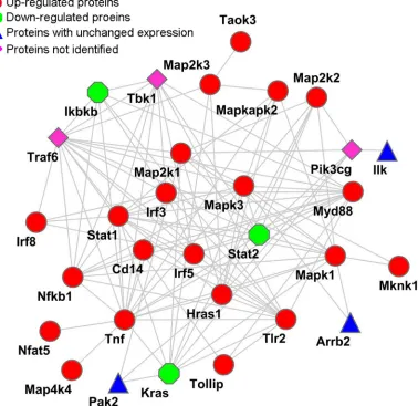
with macrophages. PANTHER was used to determine the differentially expressed proteins engaged in the TLR2,
MAPKs, NF-κB, and IRF signaling pathways (Table
1). Subsequently, STRING was used to reconstruct an interaction network. As shown in Figure 1. A possible cross-talk connection involving the key components of
MAPKs, NF-κB, and IRF signaling may coordinate the modulation of inflammatory factors for the PPE26-induced
TLR2-mediated early response.
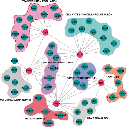
The 120 transcription factors or cofactors
(Supplementary Table 6) indentified in the nuclear
fraction were mainly involved in transcription, immune system processes, signaling transduction and response
to interferon-γ (Supplementary Figure S6). The global
functional maps generated from data-dependent bioinformatics analysis illustrated how the TFs regulatory network operated in conjunction with upstream signal cascades to generate the response following PPE26 stimulation (Figure 2 and Supplementary Figure S7).
Together, we hypothesized that PPE26 could bind to the cell surface receptor TLR2, thereby activating MAPKs,
NF-κB and IRFs pathways (Supplementary Figure S8), and finally initiating pro-inflammatory programming in
macrophages.
PPE26 induces pro-inflammatory cytokine production through TLR2
To validate whether PPE26 induced macrophage
activation and promoted pro-inflammatory cytokine
production in macrophages, we measured cytokine levels in the culture supernatants of RAW264.7 cells treated with PPE26, LPS, Pam3CSK4 or proteinase K for 24 h
by ELISA. As shown in Figure 3A, PPE26 significantly increased the production of TNF-α, IL-6, and IL-12 p40 in
a dose-dependent manner. PPE26 also produced a similar
increase in the relative expression of TNF-α, IL-6, and
[image:3.612.147.525.305.672.2]IL-12 p40 mRNA (Figure 3B). In contrast, proteinase K
Figure 1: Reconstructed networks involving the NF-κB, MAPK, IRF, and TLR signaling pathway responses to PPE26
stimulation. The key components (Table 1), such as NF-κB, MAPK, IRF, and TLR2, were acquired by PANTHER (http://www.pantherdb. org/) and submitted to STRING (http://string.embl.de/) for network construction. The network was modified using the Cytoscape 3.1.0
treatment abolished the PPE26-induced increase in TNF-α,
IL-6, and IL-12 p40 production, indicating that PPE26
specifically induced macrophages to secrete cytokines.
In MS data, TLR2 were increased by 54% in macrophages infected with rBCG::PPE26 compared with the control group, indicating that TLR2 might be the interacting partner of PPE26. To clarify this, we
examined whether PPE26 interacted with TLR2 using confocal microscopy. As shown in Figure 3C, PPE26 was observed to preferentially bind to TLR2 but not TLR4.
Additionally, immunoprecipitation further confirmed this observation (Figure 3D). Next, we measured TNF-α,
[image:4.612.61.552.61.610.2]IL-6, and IL-12 p40 levels in the supernatants of PPE26-treated macrophages from WT and TLR2-/- mice. We
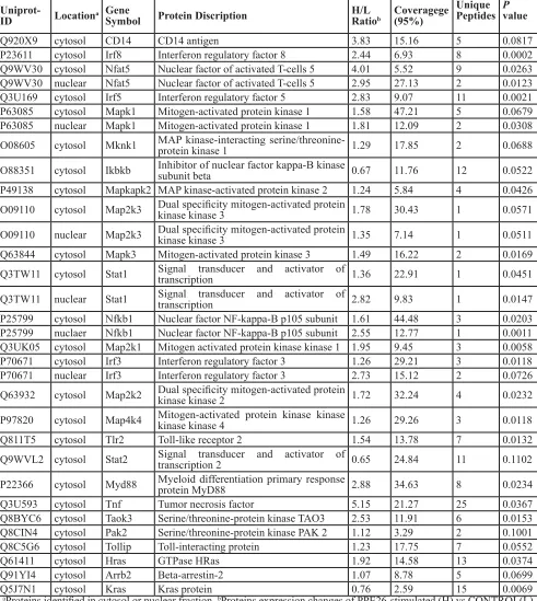
Table 1: Quantified Proteins Involved in TLR2, NF-κB, MAPK and IRF Signaling Pathway
Uniprot-ID Locationa GeneSymbol Protein Discription H/L Ratiob Coveragege(95%)
Unique Peptides Pvalue
Q920X9 cytosol CD14 CD14 antigen 3.83 15.16 5 0.0817
P23611 cytosol Irf8 Interferon regulatory factor 8 2.44 6.93 8 0.0002
Q9WV30 cytosol Nfat5 Nuclear factor of activated T-cells 5 4.01 5.52 9 0.0263 Q9WV30 nuclear Nfat5 Nuclear factor of activated T-cells 5 2.95 27.13 2 0.0123
Q3U169 cytosol Irf5 Interferon regulatory factor 5 2.83 9.07 11 0.0021
P63085 cytosol Mapk1 Mitogen-activated protein kinase 1 1.58 47.21 5 0.0679 P63085 nuclear Mapk1 Mitogen-activated protein kinase 1 1.81 12.09 2 0.0308 O08605 cytosol Mknk1 MAP kinase-interacting serine/threonine-protein kinase 1 1.29 17.85 2 0.0688
O88351 cytosol Ikbkb Inhibitor of nuclear factor kappa-B kinase subunit beta 0.67 11.76 12 0.0522 P49138 cytosol Mapkapk2 MAP kinase-activated protein kinase 2 1.24 5.84 4 0.0426 O09110 cytosol Map2k3 Dual specificity mitogen-activated protein kinase kinase 3 1.78 30.43 1 0.0571
O09110 nuclear Map2k3 Dual specificity mitogen-activated protein kinase kinase 3 1.35 7.14 1 0.0511 Q63844 cytosol Mapk3 Mitogen-activated protein kinase 3 1.49 16.22 2 0.0169 Q3TW11 cytosol Stat1 Signal transducer and activator of transcription 1.36 22.91 1 0.0451
Q3TW11 nuclear Stat1 Signal transducer and activator of transcription 2.82 9.83 1 0.0147 P25799 cytosol Nfkb1 Nuclear factor NF-kappa-B p105 subunit 1.61 44.48 3 0.0203 P25799 nuclaer Nfkb1 Nuclear factor NF-kappa-B p105 subunit 2.55 12.77 1 0.0011 Q3UK05 cytosol Map2k1 Mitogen activated protein kinase kinase 1 1.95 9.45 3 0.0058
P70671 cytosol Irf3 Interferon regulatory factor 3 1.26 29.21 3 0.0118
P70671 nuclear Irf3 Interferon regulatory factor 3 2.73 15.12 2 0.0726
Q63932 cytosol Map2k2 Dual specificity mitogen-activated protein kinase kinase 2 1.72 32.24 4 0.0232
P97820 cytosol Map4k4 Mitogen-activated protein kinase kinase kinase kinase 4 1.26 29.26 3 0.0118
Q811T5 cytosol Tlr2 Toll-like receptor 2 1.54 13.78 7 0.0132
Q9WVL2 cytosol Stat2 Signal transducer and activator of transcription 2 0.65 24.84 11 0.1102
P22366 cytosol Myd88 Myeloid differentiation primary response protein MyD88 2.88 34.63 8 0.0234
Q3U593 cytosol Tnf Tumor necrosis factor 5.15 21.27 25 0.0367
Q8BYC6 cytosol Taok3 Serine/threonine-protein kinase TAO3 2.53 11.91 6 0.0153 Q8CIN4 cytosol Pak2 Serine/threonine-protein kinase PAK 2 1.12 3.29 2 0.1001
Q8C5G6 cytosol Tollip Toll-interacting protein 1.23 17.75 7 0.0552
Q61411 cytosol Hras GTPase HRas 1.92 14.58 13 0.0374
Q91YI4 cytosol Arrb2 Beta-arrestin-2 1.07 8.78 5 0.0699
Q5J7N1 cytosol Kras Kras protein 0.76 2.59 15 0.0069
aProteins identified in cytosol or nuclear fraction. bProteins expression changes of PPE26-stimulated (H) vs CONTROL(L)
demonstrated that PPE26-induced cytokine production
was significantly decreased in macrophages from TLR2
-/- mice compared with WT mice (Figure 3E). Our results
suggested that PPE26 can stimulate macrophage activation
and induce the production of pro-inflammatory cytokines
through TLR2.
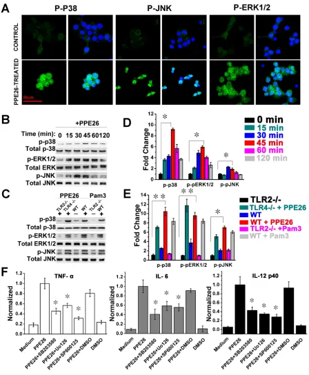
PPE26-induced cytokines production is involved in the activation of MAPKs pathway
In our MS data, we found that Mitogen-activated protein kinase kinase 1 (MAP2K1), which can phosphorylate and activate ERK1 and ERK2, was increased by 95% in the cytosol. MAP kinase kinase kinase kinase 4 (MAP4K4) acts as a link to the JNK and was
increased by 26% in the cytosol. We also found that MAP kinase kinase 3 (MAP2K3), an upstream kinase related to p38 activation, was increased by 78% in the cytosol and 35% in the nucleus. The evidence indicated that the ERK, JNK, and p38 cascades were activated following PPE26 stimulation. To clarify this, we examined the effect of PPE26 on MAPK activation by confocal microscopy and western blotting analysis. As shown in Figure 4A, PPE26 produced strong phosphorylation of p38, JNK, and ERK1/2. Next, western blot analysis also showed that the levels of the phosphorylation of MAPKs were obviously up-regulated, and the peak phosphorylation occurred within 45 min following PPE26 stimulation (Figure 4B and 4D). Moreover, PPE26-induced phosphorylation of
p38, JNK and ERK1/2 were significantly attenuated in
[image:5.612.104.519.260.666.2]macrophages from WT and TLR4-/- mice compared to
Figure 3: PPE26 induces cytokine production by mouse macrophages through TLR2. A.. RAW264.7 cells were incubated
with various concentrations of PPE26 (0.1-10μg/ml), proteinase K (PK; 50 mg/ml), PPE26 (10μg/ml) + PK (50 mg/ml), Pam3CSK4 (5 mg/ ml) (Pam3 = Pam3CSK4) or LPS (1μg/ml). After 24 h of incubation, supernatants were collected, and TNF-a, IL-6, and IL-12p40 levels were measured by ELISA. B.. Semiquantitative and quantitative RT-PCR analysis of mRNA levels for TNF-a, IL-6, and IL-12p40 in total RNA that was extracted from RAW264.7 cells incubated in medium alone, Pam3CSK4(5 mg/ml) or PPE26 (10μg/ml). The mRNA levels were normalized to the β-actin mRNA level. C.. Macrophages derived from WT, TLR2-/-, and TLR4-/- mice were incubated with PPE26-His (10μg/ml) for 1 h. After washing and staining, the cells were fixed and photographed by confocal microscopy. Scale bar, 50μm. D.. Macrophages derived from WT, TLR2-/-, and TLR4-/- mice were treated with PPE26 (10μg/ml) for 6 h, cell lysates were immunoprecipitated with anti-rat IgG, anti-mouse IgG, anti-His, anti-TLR2, or anti-TLR4; then, proteins were visualized by immunoblotting with the anti-His,
anti-TLR2, or anti-TLR4 Abs. Total cell lysate was used as an input control. E.. Macrophages derived from TLR2-/- and WT mice were treated with medium, PPE26 (10μg/ml) or Pam3CSK4(5 mg/ml). TNF-α, IL-6, and IL-12p40 levels were measured by ELISA. All data are
Figure 4: Macrophages activation triggered by PPE26 involves activation of MAPKs signaling. A.. RAW264.7 cells were
treated with PPE26 (10μg/ml) for 1 h. After washing and staining, the cells were fixed and photographed by confocal microscopy. Scale bar, 50μm. B. and C.. RAW264.7 cells were treated with PPE26 (10μg/ml) for the indicated time (0-120 min). Macrophages isolated from WT,
TLR2-/-, or TLR4-/- mice were treated with PPE26 (10μg/ml) for 1h. The phosphorylation of p38, ERK (1/2) and JNK were examined by blotting with specific antibodies to p-p38, p38, p-ERK1/2, ERK1/2, p-JNK and JNK. D. and E.. Densitometric analysis of the Western blot in B and C, respectively. Unstimulated cells were given a value 1.00. F.. RAW264.7 cells were treated with inhibitors of DMSO (vehicle
control), p38 (SB203580, 10μM), ERK (U0126, 10μM) or JNK (SP600125, 10μM) for 1 h at 37 °C, followed by incubation with PPE26 (10μg/ml) for 36 h. The amounts of TNF-a, IL-6, and IL-12p40 levels were measured by ELISA. All data are expressed as the mean± SD
macrophages from TLR2-/- mice (Figure 4C and 4E). Thus,
the phosphorylation of MAPKs in response to PPE26 is mediated primarily by TLR2, not by TLR4.
To determine the functional roles of MAPKs signaling in the context of PPE26-induced
pro-inflammatory cytokine production, RAW264.7 cells were
pretreated with a p38 inhibitor (SB203580), an ERK1/2 inhibitor (U0126), or a JNK inhibitor (SP600125) for 1
h prior to stimulation with PPE26. Levels of TNF-α,
IL-6, and IL-12 p40 were measured by ELISA. We found
that pharmacological inhibition of MAPKs significantly abrogated the PPE26-induced production of TNF-α, IL-6,
and IL-12 p40 (Figure 4F).
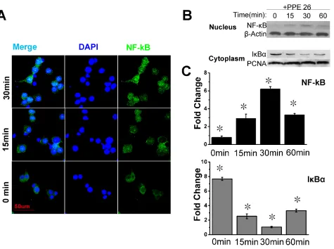
PPE26 induces the translocation of NF-κB subunits to the nucleus in mouse macrophages
NF-κB is an important transcription factor that is involved in the induction of pro-inflammatory cytokines. Our MS analysis indicated that NF-κB was up-regulated
1.55-fold in the nucleus, which suggested the activation
of the overall NF-κB-regulated signaling pathway. Thus, we examined the localization of NF-κB subunits
in RAW264.7 cells treated with or without PPE26. Confocal microscopy demonstrated the translocation of
NF-κB to the nuclei of PPE26-treated RAW264.7 cells. In unstimulated cells, NF-κB was present primarily in
the cytoplasm (Figure 5A). Western blot analysis showed that stimulation of RAW264.7 cells with PPE26 induced
the expression of nuclear NF-κB. The peak of nuclear NF-κB translocation occurred at 30 min and the nuclear expression of Iκ-Bα (the inhibitor of NF-κB) was also significantly down-regulated (Figure 5B and 5C).
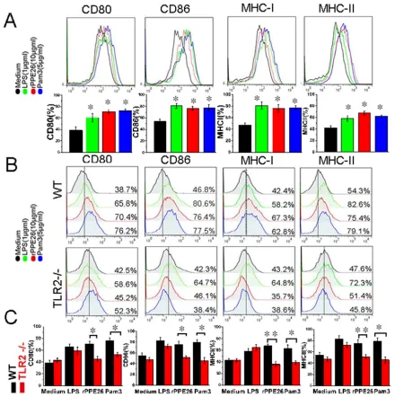
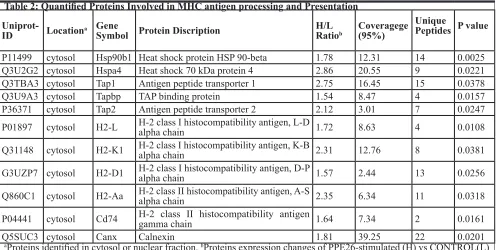
PPE26 increased the expression of co-stimulatory molecules and MHC molecules through TLR2
[image:8.612.74.549.315.669.2]Our MS data showed that H2-D1, H2-Aa, H2-L and H2-K1 were up-regulated 57%, 1.35-fold, 72% and 1.31-fold, respectively, and many proteins involved in antigen
processing and presentation were also up-regulated (Table
2), suggesting that the PPE26 can fine-tune antigens
presentation and processing (Supplementary Figure S9) , and enhance the ability of macrophages to fully activate T
cells. To clarify this finding, we examined the expression of
co-stimulatory molecules and MHC molecules in
PPE26-treated RAW264.7 cells by flow cytometry. The result demonstrated that 10μg/ml PPE26 significantly enhanced
CD80, CD86, MHC I and II expression compared with control group (Figure 6A). Then, to investigate the role of
[image:9.612.82.522.193.635.2]TLR2 in mediating the up-regulation of PPE26-induced surface markers, we measured the expression of surface molecules in PPE26-treated macrophages from WT mice and TLR2-/- mice using flow cytometry. As shown in
Figure 6B and 6C, PPE26 remained capable of enhancing the expression of co-stimulatory and MHC molecules in the WT mice macrophages, although the expression of surface markers was strongly diminished in the TLR2
-/-mouse macrophages, which suggested the enhancement of the expression of cell surface markers in response to
Figure 6: PPE26 enhances the expression of MHC molecules and co-stimulatory on macrophages via TLR2. A..
RAW264.7 cells were treated for 36 h with medium, PPE26 (10μg/ml), Pam3CSK4(5 mg/ml) or LPS (1μg/ml). The expression of cell surface markers including CD80, CD86, MHC I and MHC II was examined by FACS analysis using the respective FITC or PE-linked mAbs. B.. Macrophages derived from WT and TLR2-/- mice were treated for 36 h with medium, PPE26 (10μg/ml), Pam
3CSK4(5 mg/ml) or LPS (1μg/ml). The expression of cell surface markers including CD80, CD86, MHC I and MHC II was examined by FACS analysis using
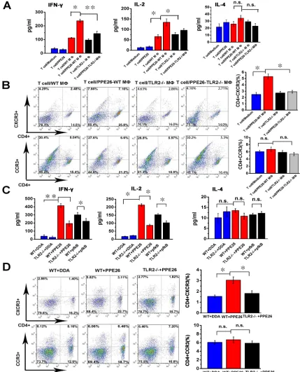
Figure 7: PPE26 induces a Th1-type immune response via TLR2-dependent macrophage activation. A.. PPE26-activated
T cells were obtained from splenocytes of C57BL/6 mice immunized with PPE26 (50 μg). Mouse peritoneal macrophages were isolated
from TLR2-/- or C57BL/6 mice. Macrophages were treated with 10μg/ml PPE26 protein for 24 h and then co-cultured with splenic T cells at a ratio of 1:10 for 3 days. The supernatant was used to measure the production of IFN-γ, IL-2 and IL-4 by ELISA. B.. T cells collected from A were stained with FITC-conjugated anti-CD4 mAbs, PE-conjugated anti-CXCR3 mAbs or PE-conjugated anti-CCR3 mAbs, and
analyzed by flow cytometry. Histograms and bar graphs show the CXCR3+ or CCR3+ T cells in the PPE26-specific CD4+ T cells. C.. TLR2-/- and C57BL/6 mice were immunized with 50 μg PPE26 mixed with DDA, 50 μg pfkB mixed with DDA or DDA alone. Splenocytes isolated from the immunized mice were stimulated with corresponding antigens (10μg/ml). The supernatants were used to measure the production of IL-2, IFN-γ and IL-4 by ELISA. D.. Lymphocytes collected from C were stained with FITC-conjugated anti-CD4 mAbs,
PE-conjugated anti-CXCR3 mAbs or PE-conjugated anti-CCR3 mAbs, and analyzed by flow cytometry. Histograms and bar graphs show
PPE26 stimulation is mediated by TLR2.
PPE26 drives a Th1-type immune response via TLR2-dependent macrophage activation
To determine the effect of PPE26 stimulation on the interaction between macrophages and T cells, we performed a mixed lymphocyte reaction (MLR) assay
using specific T cells co-cultured with
PPE26-pulsed macrophages or macrophages alone. ELISA analysis shown that T cells primed with PPE26-treated
macrophages produced significantly higher levels of IFN-γ and IL-2 compared to T cells primed with
untreated macrophages, whereas IL-4 secretion changed little (Figure 7A). Then, we investigated the expression
of chemokine receptors CXCR3 and CCR3 using flow
cytometry. As shown in Figure 7B, T cells co-cultured
with PPE26-pulsed macrophages exhibited significantly
increased CXCR3 expression compared to control group. In contrast, the expression of CCR3 in the presence of PPE26 treatment remained unaffected. Importantly, T cells co-cultured with PPE26-pulsed C57BL/6 macrophages
showed robust IFN-γ and IL-2 immune responses and
higher expression of CXCR3 when compared to those co-cultured with PPE26-pulsed TLR2-/- macrophages (Figure
7A and 7B). These findings demonstrated that
PPE26-stimulated macrophages induce the proliferation of naïve T cells towards a Th1 phenotype in vitro.
Next, to investigate whether PPE57 induced a Th1-type immune response via TLR2-mediated macrophage activation, we injected PPE26 into wild-type or TLR2
-/-mice and measured IFN-γ, IL-2 and IL-4 secretion from
T cells as well as the expression of CXCR3 and CCR3. As demonstrated in Figure 7C and 7D, PPE26 increased
IFN-γ and IL-2 production, and enhanced CXCR3 expression in T cells in WT mice; in contrast, IL-4
secretion and CCR3 expression remained unchanged. In
TLR2-deficient mice, no alteration in the IFN-γ, IL-2 and
IL-4 secretion, or CXCR3 and CCR3 expression induced by PPE26 treatment was detected. The results suggested that PPE26 appears to activate macrophages and induce Th1-type immune response.
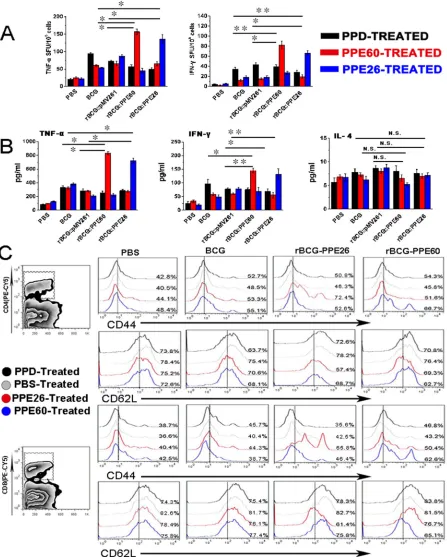
Recombinant mycobacterium bovis BCG expressing PPE26 enhances the Th1 cell-mediated response and promotes the development and maintenance of effector/memory T cells
Our data indicated that PPE26 had good potential as a vaccine for its effective induction of both cell-mediated and humoral immune responses. Thus, we constructed recombinant BCG expressing PPE26 (rBCG::PPE26) and compared the immunogenicities between rBCG::PPE26 and BCG. Figure 8A illustrated that immunization with
this strain induced stronger PPE26-specific IFN-γ and TNF-α activity than those in the control group immunized with BCG. Moreover, rBCG::PPE26 significantly increased the Th1 cytokines IFN-γ and TNF-α production
in splenocyte cultures comparable to those elicited by control group, wheras no alteration in IL-4 secretion was observed (Figure 8B).
[image:11.612.60.558.50.300.2]To assess whether the PPE26-induced macrophages
Table 2: Quantified Proteins Involved in MHC antigen processing and Presentation
Uniprot-ID Locationa GeneSymbol Protein Discription H/L Ratiob Coveragege(95%)
Unique
Peptides P value
P11499 cytosol Hsp90b1 Heat shock protein HSP 90-beta 1.78 12.31 14 0.0025
Q3U2G2 cytosol Hspa4 Heat shock 70 kDa protein 4 2.86 20.55 9 0.0221
Q3TBA3 cytosol Tap1 Antigen peptide transporter 1 2.75 16.45 15 0.0378
Q3U9A3 cytosol Tapbp TAP binding protein 1.54 8.47 4 0.0157
P36371 cytosol Tap2 Antigen peptide transporter 2 2.12 3.01 7 0.0247
P01897 cytosol H2-L H-2 class I histocompatibility antigen, L-D alpha chain 1.72 8.63 4 0.0108
Q31148 cytosol H2-K1 H-2 class I histocompatibility antigen, K-B alpha chain 2.31 12.76 8 0.0381
G3UZP7 cytosol H2-D1 H-2 class I histocompatibility antigen, D-P alpha chain 1.57 2.44 13 0.0256
Q860C1 cytosol H2-Aa H-2 class II histocompatibility antigen, A-S alpha chain 2.35 6.34 11 0.0318
P04441 cytosol Cd74 H-2 class II histocompatibility antigen gamma chain 1.64 7.34 2 0.0161
Q5SUC3 cytosol Canx Calnexin 1.81 39.25 22 0.0201
aProteins identified in cytosol or nuclear fraction. bProteins expression changes of PPE26-stimulated (H) vs CONTROL(L)
Figure 8: Recombinant BCG expressing PPE26 enhances the Th1-type immune response and induces effector/memory T cell proliferation. A.. Splenocytes were isolated from C57BL/6 mice immunized with PBS, BCG, rBCG::pMV261, rBCG::PPE60,
or rBCG::PPE26. Then, splenocytes were stimulated with PPD (10μg/ml), PPE60 (10μg/ml) or PPE26 (10μg/ml) for 36 h, and the cellular
immune response was measured by ELISPOT assay. B.. Cytokine production by mouse spleen cells was assayed by a sandwich ELISA
after stimulation of the cells with PPD (10μg/ml), PPE60 (10μg/ml), or PPE26 (10μg/ml). C.. Splenocytes were isolated from C57BL/6
activation is reflected in the ability to specifically stimulate
CD4+ and CD8+ splenic T cells isolated from rBCG::PPE26
immunized mice, we analyzed the expression of CD62L and CD44 on CD4+ and CD8+ splenic T cells using flow
cytometry. As shown in Figure 8C, PPE26 was found to induce the formation of effector/memory T cells by
displaying significantly down-regulated CD62L and
up-regulated CD44 expression on both CD4+ and CD8+
T spleen cells, demonstrating that rBCG::PPE26 could effectively promote the development of effector/memory CD4+/CD8+CD44highCD62Llow T cells. Taken together,
our findings indicated that rBCG::PPE26 enhances the
Th1 cell-mediated response, induces the development of effector/memory T cells, and may serve as a potential vaccine against M. tuberculosis.
DISCUSSION
PE/PPE family proteins of M. tuberculosis play a critical role in generating antigenic variation and evasion of host immune response. Understanding the functional characterization of mycobacterial PE/PPE proteins is essential for the comprehension of the host-pathogen interaction and the design of prospective vaccine candidates. However, little is known about the functional roles and the underlying mechanisms of PE/PPE proteins of M. tuberculosis.
In this study, to clarify the link between PPE26 and the host response, we analyzed the proteome changes during host response to PPE26 stimulation by iTRAQ subcellular quantitative proteomics. The results demonstrated that TLR2 was increased 54% in rBCG::PPE26 infected macrophages compared to BCG infected macrophages, while other TLR family members were not detected. As the key adapter protein essential for the signaling of antigens via the TLR family (except TLR3) [33, 45], Myd88 was shown to be increased by 1.88-fold in the cytosol. Moreover, MAPKs-associated
kinases and NF-κB were also significantly up-regulated.
MS data suggested that PPE26 may activate the TLR2-MyD88-mediated signaling necessary for macrophages activation. The concurrent biological validations also revealed that PPE26 could directly bind to TLR2 and
activate MAPKs and NF-κB pathway. Therefore, the
results indicate that PPE26 is a novel macrophage activation-inducting antigen through triggering a cross-talk connection between multiple pathways downstream of TLR2.
Different PE/PPE proteins appear to trigger different downstream TLR2 signaling cascades and induce the
production of either pro or anti-inflammatory cytokines
[46, 47]. PPE18 and PPE34 could activate the TLR2-MAPK pathway and induce the production of IL-10,
which is an anti-inflammatory cytokines beneficial for
the survival of M. tuberculosis [26, 27]. However, a few PE/PPE proteins are capable of triggering APC to secret
pro-inflammatory cytokines. In this study, we observed
that PPE26 could activate macrophages by binding to
TLR2 and induce higher levels of TNF-α, 6, and IL-12 p40. TNF-α is important for granuloma formation
and the clearance of mycobacterium [48]. IL-12 p40 was
previously reported to promote the production of IFN-γ
and drive the protective Th1 immune response against M. tuberculosis bacilli [49]. Furthermore, the induction of these cytokines was higher in macrophages from wild-type than those from TLR2-/- mice. These findings demonstrate
that PPE26 may be a potent TLR2 agonist and can induce
the secretion of pro-inflammatory cytokines.
T cells require two signals to become fully activated.
Antigen-specific signal is provided through the T cell
receptor which interacts with peptide-MHC molecules on the membrane of antigen presenting cells (APC) [50]. Our MS data showed that H2-D1, H2-Aa, H2-L and H2-K1 were up-regulated 57%, 1.35-fold, 72% and 1.31-fold, respectively. As the key components of the multi-protein peptide loading complex, TAP and TAPBP
were also significantly up-regulated. Moreover, PPE26 significantly increased the expression of MHC I and II in
RAW264.7, suggesting that PPE26 promotes the antigen processing and presentation, and enhances the antigen-response signal. Co-stimulatory signal is necessary for T cell activation and survival. CD80 and CD86 are the mainly co-stimulatory molecules expressed on APC, and can bind to TCR CD28 to provide co-stimulatory signaling [51, 52]. In our study, we found that PPE26 could dramatically increase the expression of CD80 and CD86 on macrophages. Taken together, our results indicate that PPE26 is capable of promoting the interaction between macrophages and T cells, and sustaining the two signals required for T cell activation.
Protection against M. tuberculosis infection depends on the rapid and continued generation of Type 1 cytokines
(especially IFN-γ and IL-2), which activate phagocytes
to constrain the intracellular mycobacterial pathogen
[53]. IFN-γ has a strong effect on the controlling/killing intracellular bacterium, the IFN-γ receptor deficiency
increased the susceptibility of patients to mycobacterial
infections [54-56]. IL-2 amplifies memory/effector T
cells functions by increasing antigenic sensitivity and improving memory capacity [57]. However, Th2 cells generate Type 2 cytokines (IL-4, IL-10) and suppress Th1-cell mediated immune response. Our results
demonstrated that PPE26 significantly increased the
production of IFN-γ and IL-2 in CD4+ T cells through
the expression of CXCR3 in CD4+ T cells, while the CCR3
level remained at the baseline. Together, our data indicate that PPE26 may regulate adaptive immunity by directing T cell immune responses towards Thl polarization.
Attempts to replace conventional BCG with
recombinant BCG to achieve stronger protective efficacy
and/or increased safety have been the focus of genetic engineering experiments [2]. In this study, we constructed the rBCG::PPE26 strain and evaluated its immunogenicity. Our results showed that immunization with this strain
induced stronger antigen-specific IFN-γ and TNF-α
activities as determined by ELISPOT assay, and higher
levels of antigen-specific CD4+ and CD8+ T-cell responses
compared to the group immunized with BCG. Likewise,
rBCG::PPE26 significantly increased the production of IFN-γ, TNF-α, and IL-2 in splenocyte cultures compared to those elicited by BCG. Our findings demonstrate
that rBCG::PPE26 enhances the Th1 cell-mediated response, and may serve as a potential vaccine against M. tuberculosis.
Immune control of M. tuberculosis depends on the rapid proliferation of effector memory T cells. These cell
subpopulations capable of producing IFN-γ are considered
to be the key components of acquired immunity and the basis for successful vaccination against TB [60]. Chief cell surface molecules include the lymph node-homing molecule CD62L and memory T cell proliferation marker molecule CD44. Effector/memory T cells express the CD44highCD62Llow surface phenotype [61, 62]. Previously,
CD4+ or CD8+ T cells with down-regulated CD62L and
up-regulated CD44 expression were reported to accumulate at
the site of infection [63]. Moreover, IFN-γ producing cells
could be described as cells expressing the CD44high and
CD62Llow phenotype. These cells enable the host to react
quickly and control a recognized pathogen if encountered again [64]. Therefore, to augment the proportion of
specific T cells with the CD44highCD62Llow phenotype is
of great importance in the context of vaccine designs and immunization strategies against TB. Our data showed that a population of CD4+ or CD8+/CD44highCD62Llow T
cells was specifically generated from splenic T cells in
rBCG::PPE26 immunized mice, suggesting that PPE26 is
a specific recall antigen to trigger Th1-mediated immune
responses. Taken together, PPE26 may link adaptive immunity and promote the formation and proliferation of effector memory T cells.
Collectively, our work indicates that PPE26 can
directly bind to TLR2 and induce pro- inflammatory
response by initiating a cross-talk of multiple pathways. PPE26 effectively modulates innate and adaptive immune responses through polarizing the development of T cells towards a Th1 phenotype. Moreover, PPE26 can augment the proportion of effector/memory T cells with a CD4+
or CD8+/ CD44highCD62Llow phenotype in rBCG::PPE26
immunized mice. These novel findings demonstrate that
PPE26 is a good antigen for the rational design of new
strategies to prevent many chronic disease caused by M. tuberculosis.
Key Messages
PPE26 triggers the cross-talk of multiple pathways involved in the host response, as revealed by an iTRAQ-based subcellular quantitative proteomics approach.
PPE26 induces cytokine production and up-regulates the function of mouse macrophages through TLR2.
PPE26 drives a Th1 immune response via TLR2-mediated macrophage functions.
Recombinant BCG over-expressing PPE26 induces
stronger antigen-specific IFN-γ activity and higher levels
of Th1 cytokines, causing Th1-polarized T-cell expansion.
MATERIALS AND METHODS
Mice and cell lines
C57BL/6 mice were purchased from the Animal Center of Slaccas (Shanghai, China). TLR2-/- and TLR4
-/-mice 6-8 weeks of age were obtained from Model Animal Research of Nanjing University (Nanjing, China). All mice
were housed under specific pathogen-free conditions in
the Animal Center of the School of Life Science of Fudan University. The experimental procedures followed the Guidelines for the Care and Use of Laboratory Animals from the National Institutes of Health and were approved by the Animal Care and Use Ethical Committee of Fudan University. The RAW264.7 cell line was purchased from the Cell Bank of the Chinese Academy of Sciences (Shanghai, China). Cells were cultured in Dulbecco’s
modified Eagle’s medium (DMEM) (Gibco, Grand Island,
NY, USA) supplemented with 10% fetal bovine serum (FBS), penicillin (100U/ml) and streptomycin (100 mg/
ml) and maintained at 37°C in a humidified incubator (5%
CO2).
Cloning and expression of recombinant PPE26
The PPE26 gene was amplified using PCR based
on the genomic NA sequence of M. tuberculosis H37Rv
with specific forward and reverse primers (Supplementary
Table 1). After treatment with the BamH1 and EcoR1 restriction enzymes, the PCR product was sub-cloned into the expression vector Pet28a and transformed into competent Escherichia coli BL21. Recombinant PPE26 was prepared by the induction of bacterial cells by
0.5 mM IPTG at 37°C for 4 h. The harvested bacteria
were suspended in 20 mM Tris-HCl (pH 8.0), 0.5 M NaCl, and 20 mM imidazole and lysed by sonication.
Nickel Affinity Gel (Sigma-Aldrich, St. Louis, MO, USA) following the manufacturer’s instructions and identified
by immunoblot using anti-His antibodies. Recombinant protein was treated with Pierce High Capacity Endotoxin Removal Resin (Pierce, USA) in accordance with the user instructions to eliminate endotoxins. The recombinant
protein was quantified with a bicinchoninic acid (BCA)
protein assay kit (Pierce, Rockford, IL, USA) and frozen
at −80 °C.
Construction of rBCG::PPE26
The PPE26 gene was amplified using PCR based
on the genomic NA sequence of M. tuberculosis H37Rv
with specific forward and reverse primers (Supplementary
Table 1). After treatment with the BamH1 and EcoR1 restriction enzymes, the PCR product was sub-cloned into the expression vector pMV261, generating pMV261::PPE26. The constructs were electroporated into
Mycobacterium bovis Bacillus Calmette-Guérin (BCG). The selected rBCG::PPE26 transformants were cultured in Middlebrook 7H9 with 10% oleic albumin dextrose
catalase (OADC) containing 50μg/ml kanamycin. The rBCG::PPE26 was identified by immunoblotting using
anti-PPE26 mouse polyclonal (Supplementary Figure S1A).
Infection of RAW264.7 macrophages
RAW264.7 cells were maintained in DMEM
supplemented with 10% FBS at 37 °C in 5% CO2. BCG
or rBCG::PPE26 was pelleted during the exponential growth phase, washed twice with PBS and resuspended. Prior to infection, a bacterial single-cell suspension was prepared by vortexing the cells with glass beads, followed by centrifugation at low speed and passage through a
5μm syringe filter to remove bacterial aggregates[29].
The RAW264.7 cells were infected with bacteria at a
multiplicity of infection (MOI) of 10 for 4 h at 37 °C in
a 5% CO2 environment, after which time the cells were
washed three times with PBS. A concentration of 200 μg/
ml gentamicin (Sigma, St. Louis, MO, USA) was added to the cells for 2h to remove the extracellular bacteria. The number of colony-forming units (CFUs) recovered from the macrophages was determined by plating the bacteria onto 7H10 agar.
Subcellular fractionation
Cytosolic and nuclear fractions were prepared using the NE-PER Nuclear and Cytoplasmic Extraction kit (Pierce, Rockford, IL, USA) following the manufacturer’s
instructions. Briefly, cells were harvested and washed
twice with PBS. The cells were suspended in buffer A
with a protease inhibitor, and incubated on ice for 10 min. The supernatant was isolated by centrifugation (10min, 6000rpm). The remaining was suspended in buffer B on ice for 30min. The soluble fractions were separated by centrifugation (15min, 14000rpm). The procedure is shown in Supplementary Figure 2. The protein concentrations were determined using the bicinchoninic acid assay (BCA) protein assay kit (Pierce, Rockford, IL, USA).
iTRAQ labeling
Protein labeling was performed as described
previously [65]. Briefly, the desalted samples were firstly mixed with 30μl of SDT buffer [4% SDS, 100 mM DTT,
and 150 mM Tris-HCl (pH 8.0)]. UA buffer [8 M urea and 150 mM Tris-HCl (pH 8.0)) was used to remove the
detergent and DTT by repeated ultrafiltration (Microcon
units, 30 kDa). After the samples were incubated with
100μl of 0.05 M iodoacetamide in UA buffer for 20 min in the dark, the protein suspensions were digested with 2μg of trypsin in 40μl of DS buffer overnight at 37°C. Finally,
the peptides were labeled using the 4-plex iTRAQ reagent according to the manufacturer’s instructions (Applied 245 Biosystems). The cytoplasmic samples were labeled as 113(control) and 115 (infection), and the nuclear samples were labeled as 116 (control) and 117 (infection).
SCX-based fractionation and LC-MS/MS analysis
SCX chromatography was performed as described previously [66]. The peptides were fractionated on a
PolySULFOETHYL A column (200 Å, 5μm, 200 X 2.1
mm) (PolyLC, Columbia, MD, USA) using an Agilent 1200 LC system (Agilent Technologies). Peptide fractions were collected using a linear gradient of solvent B (350
mM KCl in solvent A, pH 2.8) over 70 min at a flow rate
of 300 µl/min. Subsequently, the desalted peptide samples were analyzed using a Q Exactive mass spectrometer coupled to an Easy nLC (Proxeon Biosystems, now
Thermo Fisher Scientific). MS data were dynamically
acquired by choosing the most abundant precursor ions from the survey scan (300-1800 m/z) for HCD fragmentation. The dynamic exclusion duration was 60 s. Survey scans were acquired at a resolution of 70,000 at 200 m/z. The resolution for HCD spectra was set to 17,500 at 200 m/z. The normalized collision energy was 30eV.
Proteomics data analysis
Protein identification and quantification were
follows: trypsin = enzyme; missed cleavage = 1; variable modification: oxidation (M); peptide mass tolerance = 20 ppm; MS/MS tolerance = 0.1 Da; FDR≤1%; iTRAQ modification at the N-terminus of the peptide and lysine.
Relative expression pattern of proteins was determined based on the relative intensities of reporter ions of the
peptides. Criteria to select the confident list of differential proteins were set to ≥2 peptides, single peptide with multiple PSM values and p-value ≤ 0.05.
Functional clustering and network analysis
The quantified proteins were submitted to DAVID
(http://david.abcc.ncifcrf.gov/) to obtain their known biological processes and molecular functions. Proteins involved in signaling pathways were categorized by PANTHER (http://www.pantherdb.org/). A network was constructed by STRING (http://string-db.org/) according to their categorized functions. The links in the network were edited by Cytoscape (http://www.cytoscape.org/).
Measurement of cytokines
Sandwich ELISA kits were used to detect TNF-α, IL-6, and IL-12p40 levels in culture supernatants. Briefly,
RAW264.7, macrophages from WT, TLR2-/-, or TLR4 -/- mouse were cultured in 24-well plates and then were
treated with medium, PPE26(0.1-10μg/ml), Pam3CSK4
(5 mg/ml), isotype IgG (50μg/ml), proteinase K (50μg/ ml) or proteinase K (50μg/ml) + PPE26 (10μg/ml) or LPS (1μg/ml) for 24 h. Cytokine levels of TNF-α,
IL-6, and IL-12p40 in the culture media were measured as recommended by the manufacturer (BioLegend, San Diego, CA, USA). The cytokine assays were performed by measuring the absorbance at 450 nm with a microplate
reader. The modified pharmacological inhibitor experiment was designed as previously described [20]. Briefly, block
of the MAPK signaling pathway experiments involved the pretreatment of RAW264.7 cells with inhibitors
to p38 (SB203580, 10μM), ERK (U0126, 10μM) or JNK (SP600125, 10μM) for 1 h at 37 °C, followed by incubation with PPE26 for 36 h at 37°C. The IgG isotype
control A and proteinase K were purchased from Sigma-Aldrich (St. Louis, MO, USA).
RT-PCR and quantitative RT-PCR analysis
RAW246.7 cells were seeded in 24-well plates for
12 h and then treated with medium, protease K (50μg/
ml), PPE26 (10μg/ml) or Pam3CSK4 (5mg/ml) for 24 h.
The cells were harvested and rinsed twice with PBS. 1 ml/well of TRIzol (Invitrogen, Carlsbad, CA, USA) was added to each tube and cultured at room temperature for 5 min. The cells were mixed thoroughly two times
with chloroform:isoamyl alcohol (1:1), and centrifuged at 10000rpm for 15min, then rinsed with 75% ethanol (DEPC-treated water) and dissolved in DEPC-treated water. First-strand cDNA was synthesized by reverse transcription using the PrimeScript RT reagent Kit with gDNA Eraser (TaKaRa Biotechnology, Japan). The target
genes were amplified by conventional methods and
appropriate cDNA templates. RNA levels of the analyzed
genes were normalized to the amount of β-actin present in
each sample. All primers (Supplementary Table 1) were synthesized by Sangon (Shanghai, China).
Immunoprecipitation
C57BL/6 mouse macrophages were treated with
PPE26 (10μg/ml) for 6 h and lysed with RIPA lysis
buffer (Sangon, China). After pre-clearing with protein A/G sepharose beads (Santa Cruz, CA, USA) for 2 h, the mixture of cell lysates and bead was centrifuged at 10,000
x g for 5 min at 4°C. The supernatant was incubated with anti-TLR2, anti-TLR4 or anti-PPE26 overnight at 4 °C,
then the Ab-bound proteins were pull down using protein
A /G beads for 6 h at 4 °C. The beads were harvested,
washed, and boiled in 5x sample buffer for 5 min. The proteins were separated on 10% SDS-PAGE and probed with anti-TLR2, anti-TLR4 (BioLegend, CA, USA), and anti-His Abs (Santa Cruz, CA, USA) as indicated, followed by incubation with HRP-conjugated mouse anti-rat or rabbit anti-mouse secondary IgG secondary Abs. Target bands were visualized using the ECL reagent
(Thermo Fisher Scientific, MA, USA) .
Toll-like receptor binding assays
WT, TLR2-/- and TLR4-/- mouse macrophages
were incubated with PPE26-His (10μg/ml) for 1 h at 37°C. The cells were fixed in 4% PFA for 15 min, and
then permeabilized in PBST (0.1% Triton X-100) for 15 min. After blocked with 5% BSA in PBST for 2 h, the cells were incubated with TLR2 (1:200), anti-TLR4 (1:200) and anti-His Abs (1:500) overnight at 4
°C. The cells were incubated with the Alexa Fluor®568
donkey anti-mouse IgG (Santa Cruz, CA, USA) or Alexa Fluor®488 donkey anti-rabbit IgG (Santa Cruz, CA, USA)
secondary antibodies for 2 h in the dark room and then stained with 0.5 g/ml DAPI (Santa Cruz, CA, USA) for 5 min at room temperature. Between each staining step, the cells were washed three times with PBS for 5min. Finally, the cells were mounted onto slides using ProLong® Gold
Antifade Mountant (Thermo Fisher Scientific, MA, USA)
Western blot analysis
RAW264.7 cells or macrophages from C57BL/6, TLR2-/- or TLR4-/- mice were stimulated with PPE26
(10μg/ml) for indicated time, and lysed with cell lysis
buffer supplemented with a proteinase inhibitor mixture (Roche Molecular Biochemicals, Indianapolis, IN, USA). Cell pellets were processed using the NE-PER Nuclear and Cytoplasmic Extraction kit (Pierce, Rockford, IL, USA) following the manufacturer’s instructions. Equal amounts of proteins were separated on 10% SDS-PAGE and then transferred electrophoretically to PVDF membranes from Millipore (MA, USA). After treated with blocking buffer, the membranes were incubated with primary Abs
overnight at 4°C, including rabbit anti-ERK2, rabbit
anti-p38, rabbit anti-JNK, rabbit anti-phospho-ERK1/2, rabbit anti-phospho-p38, rabbit anti-phospho-JNK, rabbit
phospho-IκB-α, rabbit NF-κB p65, rabbit anti-PCNA or rabbit anti-β-actin (Santa Cruz, CA, USA). After
washed with TBST buffer, the membranes were incubated with the HRP-conjugated secondary Abs for 2 h at room temperature. Target proteins were visualized using the Pierce ECL Western Blotting Substrate (Pierce, Rockford, IL, USA).
Flow cytometric analysis
RAW264.7 or macrophages from WT or TLR2
-/-mouse were incubated with PPE26 (10μg/ml), Pam3CSK4
(5 mg/ml), or LPS (1μg/ml) for 36 h. Then, the cells were
harvested and washed with prechilled PBS, followed by
centrifugation at 1000 x g for 10 min at 4°C. The cells
were treated with Fc Block (1:100) (BD Pharmingen, CA, USA) in PBS supplemented with 1% BSA and incubated with PE-conjugated anti-mouse CD86, FITC-conjugated
anti-mouse CD80, PE-conjugated anti-mouse H-2κB for
mouse macrophages, or FITC-conjugated anti-mouse I-A/I-E (BD Pharmingen, CA, USA) on ice for 1 h in
the dark room. The cells were resuspended in 500μl PBS and analyzed using a flow cytometer (Becton Dickinson,
USA). The data were analyzed using the Cell-Quest data analysis software (10000 events per sample) and Flow4J.
Mixed lymphocyte reaction assay and Analysis of the Th1 response in vivo
TLR2-/- or C57BL/6 mice were immunised
subcutaneously three times over a 2-week
period with 50 μg of PPE26 formulated with
dimethyldioctadecylammonium (DDA) adjuvants (Sigma, Louis, MI, USA). PPE26-activated T cells were isolated from total mononuclear cells prepared from C57BL/6 mice using a MACS column. Mouse peritoneal macrophages were isolated from TLR2-/- or C57BL/6
mice as previously described[29]. Briefly, the mice were euthanized, and the peritoneal cavities were flushed
with 5 ml of ice-cold RPMI 1640 medium without FBS. Peritoneal cells were enriched by centrifugation, seeded into six-well plates in RPMI 1640 containing 10% FBS
and incubated overnight at 37 °C. Non-adherent cells were
removed, and the adherent cells were washed twice with
PBS and then treated with 10μg/ml of PPE26 for 24 h.
T cells and macrophages were co-cultured at a 1:10 ratio
for 72 h at 37 °C. Cytokines in the culture media (IFN-γ,
IL-2 and IL-4) were measured by ELISA. Harvested T cells were stained with FITC-conjugated anti-CD4 mAbs, PE-conjugated CCR3 mAbs or PE-conjugated anti-CXCR3 mAbs (BD Pharmingen, San Diego, CA, USA)
and analyzed by flow cytometry.
TLR2-/- or C57BL/6 mice were injected
subcutaneously with equal amounts (50 μg) of DDA,
DDA+PPE26 or DDA+ pfkB (Rv2029c) (an irrelevant mycobacterium antigen) to immunize the mice. After three administrations, the mice were euthanized and lymphocytes were isolated from spleen cells using Lymphocyte-M density-gradient centrifugation (Cedar Lane Lab, Burlington, NC, USA) following the manufacturer’s instructions. The cells were treated with
equal amounts (10 μg) of DDA, PPE26 or pfkB for 36
h. Lymphocytes were stained with FITC-conjugated anti-CD4 mAbs, conjugated anti-CCR3 mAbs or
PE-conjugated anti-CXCR3 mAbs, and analyzed by flow cytometry. The concentrations of IL-2, IFN-γ and IL-4 in
each supernatant sample were measured by ELISA.
Immunization of experimental animals
Groups of 12 C57BL/6 mice were immunized subcutaneously with 5×10⁶ CFUs of BCG, rBCG::PMV,
rBCG::PPE26 or rBCG::PPE60 (positive control) in 100μl
of PBS. Vaccine control mice received the Pasteur strain BCG. Lymphocytes were isolated from spleen cells 12 weeks post-vaccination using Lymphocyte-M
density-gradient centrifugation. IFN-γ and TNF-α ELISPOT
kits (U-CyTech Bio-sciences, Netherlands) were used
to determine the relative number of IFN-γ- or
TNF-α-positive cells in the single cell suspensions following the manufacturer’s instructions. The spot-forming units (SFU) were counted using a dissecting microscope. The levels
of IFN-γ, IL-4 and TNF-α were measured by ELISA.
Responder T cells were isolated from total mononuclear cells prepared from the immunized mice using a MACS column. Cells were treated with corresponding antigens for 36 h, stained with FITC-conjugated anti-CD4 mAbs, or FITC-conjugated CD8 mAbs, PE-conjugated anti-CD62L mAbs and PE-cy5-conjugated anti-CD44 mAbs
and analyzed by flow cytometry. All mAbs come from BD
Statistical analysis
Results were calculated as the mean ± SD of triplicate experiments. Statistical analysis was conducted using a one-way ANOVA followed by Tukey’s test using the origin8.0 software (Origin Lab, USA). For all tests, p
≦ 0.05 was considered statistically significant.
Abbreviations
-/- = knockout, BCG = Bacille Calmette Guerin, TLR = Toll-like receptors, CB = coomassie blue, CD62L = CD62 ligand, TB = tuberculosis, M. tuberculosis = Mycobacterium tuberculosis, PBST = PBS containing 0.1% Tween-20, TRIF = Toll/IL-1R homology
domain-containing adapter-inducing IFN-γ, PAMP =
Pathogen-associated molecular patterns, LPS = Lipopolysaccharides, CFU = colony-forming unit, IRFs = interferon regulatory factors, sulphate-polyacrylamide gel electrophoresis = SDS-PAGE, ELISPOT = enzyme-linked immune spot assay, ELISA = enzyme-linked immune sorbent assay.
MHC = histocompatibility complex IFN-γ = Interferon
gamma, CXCR3 = Chemokine receptor, CCR3 = C-C chemokine receptor type 3, CXCR5 = Chemokine receptor CXCR5, CCR4 = C-C chemokine receptor type 4, Pam3 = Pam3CSK4.
ACKNOWLEDGMENTS
This work was supported by grants from the
National Major Special Projects (2012ZX10003008); the
NSF of China (31100660) and the NSF of Shanghai Sci. Tech. Committee (11ZR1401600).
CONFLICTS OF INTEREST
On behalf of all authors of this paper, I declare that
this study will not lead to any financial or other kinds of conflicts of interest.
REFERENCES
1. O’Garra A, Redford PS, McNab FW, Bloom CI, Wilkinson RJ and Berry MP. The immune response in tuberculosis.
Annual review of immunology. 2013; 31:475-527.
2. Kaufmann SH and Gengenbacher M. Recombinant live vaccine candidates against tuberculosis. Current opinion in
biotechnology. 2012; 23:900-907.
3. Trunz BB, Fine P and Dye C. Effect of BCG vaccination on childhood tuberculous meningitis and miliary tuberculosis worldwide: a meta-analysis and assessment of
cost-effectiveness. Lancet. 2006; 367:1173-1180.
4. Orme IM, Robinson RT and Cooper AM. The balance between protective and pathogenic immune responses in
the TB-infected lung. Nature immunology. 2015; 16:57-63.
5. Kaufmann SH. Tuberculosis vaccines: time to think about
the next generation. Seminars in immunology. 2013;
25:172-181.
6. Gengenbacher M and Kaufmann SH. Mycobacterium tuberculosis: success through dormancy. FEMS
microbiology reviews. 2012; 36:514-532.
7. Schepers K, Dirix V, Mouchet F, Verscheure V, Lecher S, Locht C and Mascart F. Early cellular immune response to a new candidate mycobacterial vaccine antigen in childhood
tuberculosis. Vaccine. 2015; 33:1077-1083.
8. Majlessi L, Prados-Rosales R, Casadevall A and Brosch R. Release of mycobacterial antigens. Immunological reviews.
2015; 264:25-45.
9. Samten B, Wang X and Barnes PF. Immune regulatory activities of early secreted antigenic target of 6-kD protein of Mycobacterium tuberculosis and implications for
tuberculosis vaccine design. Tuberculosis. 2011; 91 Suppl
1:S114-118.
10. Hmama Z, Pena-Diaz S, Joseph S and Av-Gay Y. Immunoevasion and immunosuppression of the macrophage by Mycobacterium tuberculosis. Immunological reviews.
2015; 264:220-232.
11. Schafer G, Jacobs M, Wilkinson RJ and Brown GD. Non-opsonic recognition of Mycobacterium tuberculosis by
phagocytes. Journal of innate immunity. 2009; 1:231-243.
12. Cuervo AM and Macian F. Autophagy, nutrition and
immunology. Molecular aspects of medicine. 2012;
33:2-13.
13. Baena A and Porcelli SA. Evasion and subversion of antigen presentation by Mycobacterium tuberculosis. Tissue
antigens. 2009; 74:189-204.
14. Deretic V, Delgado M, Vergne I, Master S, De Haro S, Ponpuak M and Singh S. Autophagy in immunity against mycobacterium tuberculosis: a model system to dissect immunological roles of autophagy. Current topics in
microbiology and immunology. 2009; 335:169-188. 15. Bosedasgupta S and Pieters J. Inflammatory stimuli
reprogram macrophage phagocytosis to macropinocytosis for the rapid elimination of pathogens. PLoS pathogens.
2014; 10:e1003879.
16. Drage MG, Tsai HC, Pecora ND, Cheng TY, Arida AR, Shukla S, Rojas RE, Seshadri C, Moody DB, Boom WH, Sacchettini JC and Harding CV. Mycobacterium tuberculosis lipoprotein LprG (Rv1411c) binds triacylated glycolipid agonists of Toll-like receptor 2. Nature structural
& molecular biology. 2010; 17:1088-1095.
17. Noss EH, Pai RK, Sellati TJ, Radolf JD, Belisle J, Golenbock DT, Boom WH and Harding CV. Toll-like receptor 2-dependent inhibition of macrophage class II MHC expression and antigen processing by 19-kDa lipoprotein of Mycobacterium tuberculosis. J Immunol.
2001; 167:910-918.
CV. Mycobacterium tuberculosis 19-kDa lipoprotein inhibits IFN-gamma-induced chromatin remodeling of MHC2TA by TLR2 and MAPK signaling. J Immunol.
2006; 176:4323-4330.
19. Sanchez A, Espinosa P, Esparza MA, Colon M, Bernal G and Mancilla R. Mycobacterium tuberculosis 38-kDa lipoprotein is apoptogenic for human monocyte-derived
macrophages. Scandinavian journal of immunology. 2009;
69:20-28.
20. Chen ST, Li JY, Zhang Y, Gao X and Cai H. Recombinant MPT83 derived from Mycobacterium tuberculosis induces cytokine production and upregulates the function of mouse
macrophages through TLR2. J Immunol. 2012;
188:668-677.
21. Kleinnijenhuis J, Oosting M, Joosten LA, Netea MG and Van Crevel R. Innate immune recognition of Mycobacterium tuberculosis. Clinical & developmental
immunology. 2011; 2011:405310.
22. Kim TH, Park JH, Park YM, Ryu SW, Shin SJ, Park JH and Kim DJ. Synergistic effect of muramyl dipeptide with heat shock protein 70 from Mycobacterium tuberculosis on
immune activation. Immunobiology. 2015; 220:26-31.
23. Chambers MA, Whelan AO, Spallek R, Singh M, Coddeville B, Guerardel Y and Elass E. Non-acylated Mycobacterium bovis glycoprotein MPB83 binds to TLR1/2 and stimulates production of matrix metalloproteinase 9. Biochemical and biophysical research communications.
2010; 400:403-408.
24. Zumbo A, Palucci I, Cascioferro A, Sali M, Ventura M, D’Alfonso P, Iantomasi R, Di Sante G, Ria F, Sanguinetti M, Fadda G, Manganelli R and Delogu G. Functional dissection of protein domains involved in the immunomodulatory properties of PE_PGRS33 of
Mycobacterium tuberculosis. Pathogens and disease. 2013;
69:232-239.
25. Tiwari BM, Kannan N, Vemu L and Raghunand TR. The Mycobacterium tuberculosis PE proteins Rv0285 and Rv1386 modulate innate immunity and mediate bacillary
survival in macrophages. Plos One. 2012; 7:e51686.
26. Bansal K, Sinha AY, Ghorpade DS, Togarsimalemath SK, Patil SA, Kaveri SV, Balaji KN and Bayry J. Src Homology 3-interacting Domain of Rv1917c of Mycobacterium tuberculosis Induces Selective Maturation of Human Dendritic Cells by Regulating PI3K-MAPK-NF-kappa B Signaling and Drives Th2 Immune Responses. Journal Of
Biological Chemistry. 2010; 285:36511-36522.
27. Nair S, Ramaswamy PA, Ghosh S, Joshi DC, Pathak N, Siddiqui I, Sharma P, Hasnain SE, Mande SC and Mukhopadhyay S. The PPE18 of Mycobacterium tuberculosis Interacts with TLR2 and Activates IL-10
Induction in Macrophage. J Immunol. 2009;
183:6269-6281.
28. Wang H, Dong DD, Tang SW, Chen X and Gao Q. PPE38 of Mycobacterium marinum Triggers the Cross-Talk of Multiple Pathways Involved in the Host Response,
As Revealed by Subcellular Quantitative Proteomics. J
Proteome Res. 2013; 12:2055-2066.
29. Xu Y, Yang E, Huang Q, Ni W, Kong C, Liu G, Li G, Su H and Wang H. PPE57 induces activation of macrophages and drives Th1-type immune responses through TLR2. Journal
of molecular medicine. 2015; 93:645-662.
30. Fishbein S, van Wyk N, Warren RM and Sampson SL. Phylogeny to function: PE/PPE protein evolution and impact on Mycobacterium tuberculosis pathogenicity.
Molecular microbiology. 2015; 96:901-916.
31. Abou-Zeid C, Gares MP, Inwald J, Janssen R, Zhang Y, Young DB, Hetzel C, Lamb JR, Baldwin SL, Orme IM, Yeremeev V, Nikonenko BV and Apt AS. Induction of a type 1 immune response to a recombinant antigen from Mycobacterium tuberculosis expressed in Mycobacterium
vaccae. Infect Immun. 1997; 65:1856-1862.
32. Lancioni CL, Li Q, Thomas JJ, Ding X, Thiel B, Drage MG, Pecora ND, Ziady AG, Shank S, Harding CV, Boom WH and Rojas RE. Mycobacterium tuberculosis lipoproteins directly regulate human memory CD4(+) T cell activation
via Toll-like receptors 1 and 2. Infect Immun. 2011;
79:663-673.
33. Jo EK, Yang CS, Choi CH and Harding CV. Intracellular signalling cascades regulating innate immune responses to Mycobacteria: branching out from Toll-like receptors.
Cellular microbiology. 2007; 9:1087-1098.
34. Yang CS, Shin DM, Lee HM, Son JW, Lee SJ, Akira S, Gougerot-Pocidalo MA, El-Benna J, Ichijo H and Jo EK. ASK1-p38 MAPK-p47phox activation is essential for
inflammatory responses during tuberculosis via TLR2-ROS signalling. Cellular microbiology. 2008; 10:741-754.
35. Arthur JS and Ley SC. Mitogen-activated protein kinases
in innate immunity. Nature reviews Immunology. 2013;
13:679-692.
36. Kawai T and Akira S. Pathogen recognition with Toll-like
receptors. Current opinion in immunology. 2005;
17:338-344.
37. Chan J, Xing Y, Magliozzo RS and Bloom BR. Killing of virulent Mycobacterium tuberculosis by reactive nitrogen intermediates produced by activated murine macrophages.
The Journal of experimental medicine. 1992;
175:1111-1122.
38. Reba SM, Li Q, Onwuzulike S, Ding X, Karim AF, Hernandez Y, Fulton SA, Harding CV, Lancioni CL, Nagy N, Rodriguez ME, Wearsch PA and Rojas RE. TLR2 engagement on CD4(+) T cells enhances effector functions and protective responses to Mycobacterium tuberculosis.
Eur J Immunol. 2014; 44:1410-1421.
39. Cole ST, Brosch R, Parkhill J, Garnier T, Churcher C, Harris D, Gordon SV, Eiglmeier K, Gas S, Barry CE, 3rd, Tekaia F, Badcock K, Basham D, Brown D, Chillingworth T, Connor R, et al. Deciphering the biology of Mycobacterium tuberculosis from the complete genome
40. Gey van Pittius NC, Sampson SL, Lee H, Kim Y, van Helden PD and Warren RM. Evolution and expansion of the Mycobacterium tuberculosis PE and PPE multigene families and their association with the duplication of the
ESAT-6 (esx) gene cluster regions. Bmc Evol Biol. 2006;
6:95.
41. de Souza GA, Leversen NA, Malen H and Wiker HG. Bacterial proteins with cleaved or uncleaved signal peptides of the general secretory pathway. Journal of proteomics.
2011; 75:502-510.
42. Griffin JE, Gawronski JD, Dejesus MA, Ioerger TR,
Akerley BJ and Sassetti CM. High-resolution phenotypic
profiling defines genes essential for mycobacterial growth and cholesterol catabolism. PLoS pathogens. 2011;
7:e1002251.
43. Derrick SC, Yabe IM, Yang A, Kolibab K, Hollingsworth B, Kurtz SL and Morris S. Immunogenicity and protective
efficacy of novel Mycobacterium tuberculosis antigens. Vaccine. 2013; 31:4641-4646.
44. Sayes F, Sun L, Di Luca M, Simeone R, Degaiffier N,
Fiette L, Esin S, Brosch R, Bottai D, Leclerc C and Majlessi L. Strong immunogenicity and cross-reactivity of Mycobacterium tuberculosis ESX-5 type VII secretion: encoded PE-PPE proteins predicts vaccine potential. Cell
host & microbe. 2012; 11:352-363.
45. Mortaz E, Adcock IM, Tabarsi P, Masjedi MR, Mansouri D, Velayati AA, Casanova JL and Barnes PJ. Interaction of Pattern Recognition Receptors with Mycobacterium Tuberculosis. Journal of clinical immunology. 2014. 46. Etna MP, Giacomini E, Severa M and Coccia EM. Pro- and
anti-inflammatory cytokines in tuberculosis: A two-edged sword in TB pathogenesis. Seminars in immunology. 2014;
26:543-551.
47. Takeda K, Kaisho T and Akira S. Toll-like receptors.
Annual review of immunology. 2003; 21:335-376.
48. Roach DR, Bean AGD, Demangel C, France MP, Briscoe H and Britton WJ. TNF regulates chemokine induction essential for cell recruitment, granuloma formation, and
clearance of mycobacterial infection. J Immunol. 2002;
168:4620-4627.
49. Ottenhoff THM, Verreck FAW, Lichtenauer-Kaligis EGR, Hoeve MA, Sanal O and van Dissel JT. Genetics, cytokines and human infectious disease: lessons from weakly pathogenic mycobacteria and salmonellae (vol 32, pg 97,
2002). Nature genetics. 2002; 32:331-331.
50. Harding CV and Boom WH. Regulation of antigen presentation by Mycobacterium tuberculosis: a role for
Toll-like receptors. Nat Rev Microbiol. 2010; 8:296-307.
51. Cantrell D. Signaling in Lymphocyte Activation. Cold
Spring Harbor perspectives in biology. 2015; 7.
52. Kiefer F, Vogel WF and Arnold R. Signal transduction and
co-stimulatory pathways. Transplant immunology. 2002;
9:69-82.
53. Salgame P. Host innate and Th1 responses and the bacterial
factors that control Mycobacterium tuberculosis infection.
Current opinion in immunology. 2005; 17:374-380.
54. Boehm U, Klamp T, Groot M and Howard JC. Cellular responses to interferon-gamma. Annual review of
immunology. 1997; 15:749-795.
55. Sato K, Akaki T and Tomioka H. Differential potentiation of anti-mycobacterial activity and reactive nitrogen intermediate-producing ability of murine peritoneal macrophages activated by interferon-gamma (IFN-gamma) and tumour necrosis factor-alpha (TNF-alpha). Clinical and
experimental immunology. 1998; 112:63-68.
56. Ottenhoff TH, Verreck FA, Lichtenauer-Kaligis EG, Hoeve MA, Sanal O and van Dissel JT. Genetics, cytokines and human infectious disease: lessons from weakly pathogenic
mycobacteria and salmonellae. Nature genetics. 2002;
32:97-105.
57. Nunberg JH, Doyle MV, York SM and York CJ. Interleukin 2 acts as an adjuvant to increase the potency of inactivated rabies virus vaccine. Proceedings of the National Academy
of Sciences of the United States of America. 1989;
86:4240-4243.
58. Yamamoto J, Adachi Y, Onoue Y, Adachi YS, Okabe Y, Itazawa T, Toyoda M, Seki T, Morohashi M, Matsushima K and Miyawaki T. Differential expression of the chemokine receptors by the Th1- and Th2-type effector populations within circulating CD4+ T cells. Journal of leukocyte
biology. 2000; 68:568-574.
59. Khader SA, Bell GK, Pearl JE, Fountain JJ, Rangel-Moreno J, Cilley GE, Shen F, Eaton SM, Gaffen SL, Swain SL, Locksley RM, Haynes L, Randall TD and Cooper AM. IL-23 and IL-17 in the establishment of protective pulmonary CD4(+) T cell responses after vaccination and during Mycobacterium tuberculosis challenge. Nature
immunology. 2007; 8:369-377.
60. Orme IM, Andersen P and Boom WH. T cell response to Mycobacterium tuberculosis. The Journal of infectious
diseases. 1993; 167:1481-1497.
61. Hogevold HE, Moen O, Fosse E, Venge P, Braten J, Andersson C and Lyberg T. Effects of heparin coating on the expression of CD11b, CD11c and CD62L by leucocytes
in extracorporeal circulation in vitro. Perfusion. 1997;
12:9-20.
62. Graham VA, Marzo AL and Tough DF. A role for CD44 in T cell development and function during direct competition
between CD44+ and CD44- cells. Eur J Immunol. 2007;
37:925-934.
63. DeGrendele HC, Estess P and Siegelman MH. Requirement for CD44 in activated T cell extravasation into an
inflammatory site. Science. 1997; 278:672-675.
64. Gruppo V, Turner OC, Orme IM and Turner J. Reduced up-regulation of memory and adhesion/integrin molecules in susceptible mice and poor expression of immunity to
pulmonary tuberculosis. Microbiology. 2002;
65. Shadforth IP, Dunkley TP, Lilley KS and Bessant C. i-Tracker: for quantitative proteomics using iTRAQ. BMC
genomics. 2005; 6:145.
66. Lau E, Lam MP, Siu SO, Kong RP, Chan WL, Zhou Z,
Huang J, Lo C and Chu IK. Combinatorial use of offline
SCX and online RP-RP liquid chromatography for iTRAQ-based quantitative proteomics applications. Molecular