Introduction
A hallmark pathological feature of spondyloarthritis (SpA) is the development of ankylosis in axial joints. Th is process is the primary reason for the development of major disability. Its prevention therefore constitutes a key goal of management. Th ere has been a long-standing assumption that infl ammation is the process that sets in motion the chain of events that leads to ankylosis and that the two processes continue to be inextricably linked as the disease progresses over time. However, direct testing of this hypothesis has been diffi cult due to several factors, such as the lack of availability of objective clinical and laboratory measures of infl ammation, delay in diagnosis, the slow progression of disease, and the lack of access to tissue for direct histopathological analysis. Investigators have turned to indirect approaches such as animal models of SpA and the testing of various bio-markers refl ecting infl ammation. Th e signifi cant limita-tions of these approaches are not the subject of this review but highlight the increasing importance of recent advances in non-invasive assessment with imaging modalities. In particular, the advent of fat-suppressed mag netic resonance imaging (MRI) allows direct visuali-za tion of features related to infl ammation and this has, for the fi rst time, allowed the prospective non-invasive evaluation of infl ammation in axial joints. Th e reports of these studies assessing the relationship between infl
am-ma tion and structural changes have generated
con-siderable interest, culminating in numerous symposia at international meetings. Th is review sets out the reasons why this topic has generated so much interest, outlines the methodology and pitfalls surrounding the use of MRI for evaluation of the axial joints, reviews the prospective studies that focused on MRI infl ammation and its relation to radiographic changes, and suggests possible avenues of further research in SpA.
Evidence for ‘uncoupling’ between infl ammation and structural damage in spondyloarthritis
Th e detailed histopathological examination of necropsy material and tissue biopsies outlined by John Ball in his
Abstract
Demonstration of an association between infl ammation and spinal ankylosis has been challenging. Until the advent of MRI, prospective study was not possible due to inaccessibility of tissue. Recent studies using MRI have described an association between the presence of bone edema at vertebral corners on MRI and the subsequent development of syndesmophytes at the corresponding vertebral corners on radiography. Although reports have also highlighted the development of new syndesmophytes where the baseline MRI shows no infl ammation, MRI has limited sensitivity for detection of spinal infl ammation that is clearly evident on histopathology. There are also crucial methodological challenges because radiographic assessment is limited to the anterior corners of the cervical and lumbar spine while MRI lesions in the cervical spine are often small while spurious infl ammatory signal is common in the lumbar spine. Follow-up MRI evaluation in two independent studies has also shown that infl ammatory lesions that resolve after anti-TNF therapy are more prone to develop into syndesmophytes. It may be possible that very early infl ammatory lesions resolve completely without sequelae if anti-TNF therapy is introduced before new bone formation becomes largely autonomous. For an individual patient the overall development of new bone during anti-TNF therapy may therefore depend on the balance between the number of early and more mature infl ammatory lesions. Clinical trials of anti-TNF agents in early spondyloarthritis together with prospective MRI studies will allow more detailed testing of this hypothesis as a major priority for the research agenda in spondyloarthritis.
© 2010 BioMed Central Ltd
MRI and X-ray in axial spondyloarthritis:
the relationship between infl ammatory and
structural changes
Walter P Maksymowych*
R E V I E W
*Correspondence: [email protected]
562 Heritage Medical Research Building, University of Alberta, Edmonton, Alberta, T6G 2S2 Canada
1970 Heberden oration lent strong support to the concept that ankylosis was an excessive reparative res-ponse to an infl ammatory and osteo-destructive process [1]. It was expected, therefore, that anti-TNFα therapies would be benefi cial in preventing ankylosis in SpA through their profound ability to suppress infl ammation. However, the results of three studies that compared radiographic progression over the minimum required 2 year time frame in patients in anti-TNFα trials with a historical cohort of ankylosing spondylitis (AS) patients did not support a benefi cial eff ect of anti-TNFα therapy [2-4]. Several important limitations included the lack of comparability in disease severity, inadequate sample size, insuffi cient suppression of infl ammation, and possible diff erences in intake of non-steroidal anti-infl ammatory agents (NSAIDs), which have been shown to ameliorate progression [5]. Nevertheless, various sensitivity analyses aimed at demonstrating diff erences in progression within subgroups failed to identify any impact of anti-TNFα therapy. Assessment of anti-TNFα therapy in an animal model of SpA, ankylosing enthesitis, also failed to demon strate amelioration of ankylosis by anti-TNFα therapy [6]. Th is led to an alternative hypothesis whereby an as yet unknown pathogenic trigger(s) simultaneously
induces both an acute infl ammatory reaction and
activation of stromal progenitor cells, which ultimately leads to endochondral new bone [7]. But the processes of infl ammation and new bone formation proceed along essentially disconnected or ‘uncoupled’ pathways once each has been activated.
Discerning the true relationship between infl am-mation and ankylosis is pivotal to future advances in disease modifi cation because if these processes are largely uncoupled, future therapies will need to target bone formation pathways. A major challenge in understanding the link between these two processes in human SpA is the delayed recognition of disease so that most patients are assessed when both infl ammatory and reparative processes are well established. It has been proposed that initial development of infl ammation leads to erosive cartilage bone destruction followed by tissue repair and ultimately ossifi cation once infl ammation has completely resolved [8]. Consequently, this hypothesis would imply that early and eff ective suppression of
infl ammation would prevent the development of
ankylosis. MRI allows non-invasive assessment of
infl am mation but the optimal approach to the
assessment of bone is still radiography. Consequently, recent prospective studies have combined assessment of the evolution of infl ammatory lesions using MRI with the development of new bone using radiography in the spine and sacroiliac joints (SIJs) to provide further insights regarding these hypotheses.
MRI evaluation: standardization of methodology and key pitfalls
As the diversity of acute and structural lesions on MRI at diff erent stages of disease has become evident, there has been recognition for the need to develop a standardized methodology for assessment of MRI scans in SpA and to develop and validate standardized defi nitions in order to facilitate clinical research. Th is need is particularly relevant for assessment of the SIJs due to the complex anatomy of the joint. An international working group
from Canada, Denmark, and Switzerland (Th e MORPHO
Group) has developed a standardized module (available online at [9]) for evaluation of the SIJ [10]. Th e SIJs are scanned in the tilted semi-coronal plane and assessment of consecutive slices is undertaken from anterior to posterior according to anatomical landmarks and specifi c rules [11]. It is deemed essential that assessment of each slice includes corresponding T1-weighted spin echo (T1SE) and short tau inversion recovery (STIR) MRI sequences. Th e T1SE sequence detects the signal from fat and permits detailed assessment of the anatomy of the joint. Th e STIR sequence is a fat-suppression technique that allows the visualization of the free water signal associated with infl ammation and is particularly useful for identifying acute lesions in the bone marrow that may be obscured by marrow fat.
A similar standardized methodology is recommended
for assessment of the spine. Th e Canada-Denmark
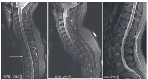
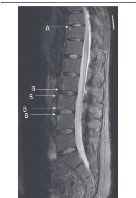
It is apparent that the appearance of a CIL may be heterogeneous and the Canada-Denmark group has defi ned two categories of CIL. In type A CILs the STIR signal is relatively homogeneous and extends to the verte-bral corner. In type B CILs, the STIR signal has receded from the vertebral corner and is typically less intense than a type A CIL and more heterogeneous (Figure 2). It is necessary to examine the corresponding T1SE sequence to determine whether there is an erosion or new bone at the vertebral corner, which then appears dark on both the T1SE and STIR sequence, or whether there is tissue metaplasia to fat, which then appears bright on the T1SE sequence. Type B CILs likely repre-sent a more advanced stage of evolution of the infl am-matory lesion. In particular, prospective analysis of MRI scans has directly shown that CILs evolve into fat lesions at the corresponding vertebral corner and that this is more likely to occur in patients receiving anti-TNFα therapy [14,15]. Reliable detection of type B CILs and assessment of resolution following anti-TNFα therapy are often more diffi cult than for type A CILs, especially in the lumbar spine due to phase encoding artifacts. Never-theless, it may be important to make this distinction because the two types of CIL may carry diff erent
prognostic implications for the development of new bone (see below).
[image:3.612.68.545.90.346.2]detection of fat infi ltration is reliably accomplished with minimal calibration, fat infi ltration is observed in the SIJs and spine of normal individuals and there is considerable heterogeneity in its morphological characteristics, particularly in the SIJs. It is presently unclear which features relate specifi cally to SpA.
Prospective studies of MRI infl ammation and radiographic progression?
Spine
Several studies, all using diff erent methodological approaches, have examined the relationship between infl ammation visible on baseline STIR MRI of the spine in patients with established AS and the development of syndesmophytes on radiographs after 2 years of follow up [20-26]. It is important to note from the outset that all radiographic assessments in each study were confi ned to the anterior cervical and lumbar spine vertebral corners because thoracic spine abnormalities cannot be reliably detected on radiography due to overlapping structures. Similarly, assessment of posterior vertebral corner
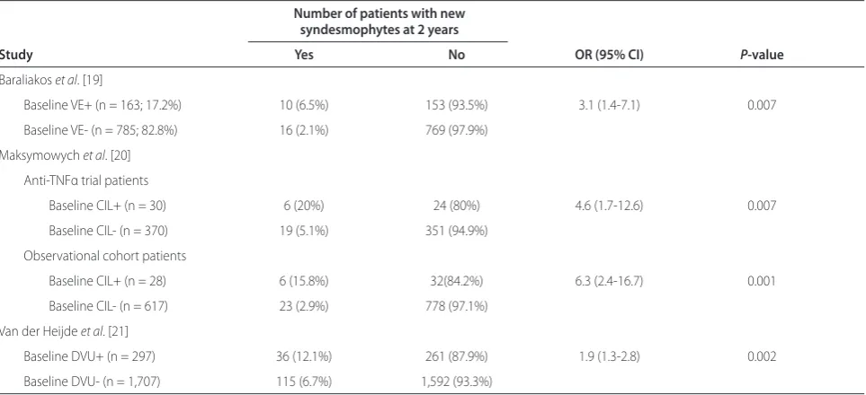
abnormalities is unreliable [27]. Consequently, MRI assessments have been confi ned to the corresponding anterior vertebral corners of the cervical and lumbar spine. As discussed above, this restricts MRI assessment to regions of the spine that may pose challenges to the reliable detection of lesions on the STIR sequence. A summary of the fi ndings from these reports is presented in Table 1 and shows that the odds ratios (ORs) for the association of baseline infl ammation and the development of new syndesmophytes after 2 years were signifi -cant for each study but varied from 1.9 to 4.6. Th e per-cen tage of individual vertebral units with infl ammation on baseline STIR MRI also varied from 7.5 to 17.2%, the percentage of vertebral regions with baseline infl amma-tion that developed new syndesmophytes varied from 6.5 to 20%, and the percentage of syndesmophytes develop-ing in the absence of baseline infl ammation varied from 61 to 76%. So while there is consistency in demonstrating a link between infl ammation and new syndesmophytes, it has also been argued that these data more strongly support a lack of coupling between infl ammation and new bone because most new syndesmophytes developed from vertebral corners that appeared normal on STIR MRI. What could account for the diff erences between studies and how does this shed light on the hypothesis that infl ammation and ankylosis are coupled and/or uncoupled?
Th e fi rst report assessed infl ammation on baseline STIR MRI in 39 patients who participated in clinical trials of anti-TNFα therapy [20]. Infl ammation was recorded at vertebral edges (VEs), and these were defi ned as positive for infl ammation ‘if the infl ammatory activity was present in the anterior half of the vertebral edge only’. Th is could refer to lesions directly at the vertebral corners as well as lesions adjacent to the endplate but not extend-ing to the vertebral corner as in a discovertebral lesion, which could have diff erent prognostic implications. Th e report does not indicate how many readers assessed either the MRI scans or the radiographs. Infl ammation was reported in 17.2% of vertebral edges at baseline, new syndesmophytes developed from 6.5% of vertebral edges with infl ammation as opposed to 2.1% without infl am-mation (OR = 3.3, 95% confi dence interval (CI) 1.5 to 7.4), and 38% of all new syndesmophytes developed from vertebral edges with baseline infl ammation while 62% had no baseline infl ammation. On follow up at 2 years, 4.4% of vertebral edges where infl ammation had resolved and 5.6% of vertebral edges that had either persistent infl ammation or had newly developed infl ammatory lesions developed a new syndesmophyte as compared to 1.3% of VEs that had no infl ammation at either baseline or follow up (P = 0.07 and 0.04, respectively).
[image:4.612.66.297.86.421.2]Th e second report assessed infl ammation on STIR MRI in 29 patients in anti-TNFα clinical trials and 41 patients Figure 2. Reference image of short tau inversion recovery MRI
followed in an observational cohort (23 on standard therapies, 18 receiving anti-TNFα therapy) [21]. Vertebral CILs were assessed as defi ned by the Canada-Denmark group [12] and the primary analysis focused on concor-dant data from three reader pairs, each reader scoring MRI scans independently. For the primary reader pair and scans from patients in anti-TNFα trials, infl ammation was reported in 7.5% of vertebral corners at baseline, new
syndesmo phytes developed from 20% of CILs at the
corresponding vertebral corner as opposed to 5.1% without CILs (OR = 4.6, 95% CI 1.7 to 12.6), and 24% of all new syndesmo phytes developed from CILs while 76% had no baseline infl ammation. Data were comparable for scans from patients in the observational cohort (Table 1). On follow up at 2 years, 25% of CILs where infl ammation had resolved had developed a new syndesmophyte compared to none of the CILs that had persistent infl ammation. Th e follow-up data on the observational cohort were extended to include 23 patients receiving anti-TNFα therapy and 27 receiving standard therapy and reported in a separate manuscript [24]. Consistent with the earlier report, new syndesmophytes developed from CILs that resolved by 2 years (42.9% in anti-TNFα and 20% in standard therapy groups, respectively) but not from a single CIL where infl ammation was persistent.
A third report assessed the relationship between infl ammation on baseline STIR MRI and development of new syndesmophytes but did not directly assess infl am-mation at the corresponding vertebral corner [22].
Infl am ma tion was recorded in MRI scans from 182
patients in a trial of infl iximab according to its presence/
absence at each cervical and lumbar discovertebral unit (DVU), which represents the region between two imagi-nary lines drawn through the middle of two adjacent vertebrae and includes the four vertebral corners, the intervertebral disc, the adjacent vertebral endplates and bone marrow [28]. Data for each of two readers scoring independently were reported but not concordant data. Infl ammation was reported in 14.8% and 20% of DVUs at baseline for readers 1 and 2, respectively. For the primary reader, new syndesmophytes developed from 12.1% of DVUs with infl ammation as opposed to 6.7% without infl ammation (OR = 1.9, 95% CI 1.3 to 2.8), and 23.8% of all new syndesmophytes developed from DVUs with base line infl ammation while 76.2% had no baseline infl am mation. At the patient level, MRI activity was not associated with change in radiography score.
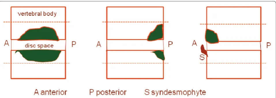
Th e studies that assessed the association between baseline infl ammation and new syndesmophytes at the corresponding vertebral corner [20,21] reported higher ORs than the study that assessed infl ammation at the level of a DVU [22]. Th is, as well as the absence of an association between MRI activity and change in radio-graphy score at the patient level [22], is not surprising. As shown in Figure 3, an infl ammatory lesion can be observed in several locations within a DVU other than the vertebral corner where a new syndesmophyte develops and this will serve to reduce the strength of the association.
[image:5.612.66.548.112.332.2]Th e consensus that most new syndesmophytes developed from vertebral corners that did not demonstrate infl ammation at baseline should not necessarily be Table 1. Summary of studies assessing the relationship between infl ammation on baseline STIR MRI of the spine and development of new syndesmophytes after 2 years of follow up
Number of patients with new syndesmophytes at 2 years
Study Yes No OR (95% CI) P-value
Baraliakos et al. [19]
Baseline VE+ (n = 163; 17.2%) 10 (6.5%) 153 (93.5%) 3.1 (1.4-7.1) 0.007
Baseline VE- (n = 785; 82.8%) 16 (2.1%) 769 (97.9%)
Maksymowych et al. [20]
Anti-TNFα trial patients
Baseline CIL+ (n = 30) 6 (20%) 24 (80%) 4.6 (1.7-12.6) 0.007
Baseline CIL- (n = 370) 19 (5.1%) 351 (94.9%)
Observational cohort patients
Baseline CIL+ (n = 28) 6 (15.8%) 32(84.2%) 6.3 (2.4-16.7) 0.001
Baseline CIL- (n = 617) 23 (2.9%) 778 (97.1%)
Van der Heijde et al. [21]
Baseline DVU+ (n = 297) 36 (12.1%) 261 (87.9%) 1.9 (1.3-2.8) 0.002
Baseline DVU- (n = 1,707) 115 (6.7%) 1,592 (93.3%)
inter preted as demonstrating uncoupling between infl amma tion and radiographic progression. First, MRI detects only about half of the infl ammatory lesions evident histopathologically in the spine of patients with AS [29]. Second, the baseline MRI represents a snapshot in time and new infl ammatory lesions can develop even in patients on anti-TNFα agents [20]. Th ird, prospective studies show that infl ammatory lesions evolve into fat lesions evident on T1SE MRI and this can be seen by 52 weeks after the start of treatment, especially in patients on anti-TNFα agents [14,15]. Th is is relevant because two recent reports have demonstrated an association between baseline fat lesions at vertebral corners on T1SE MRI and the development of new syndesmophytes at the corresponding corner [23,26]. Th e fi rst assessed 100 MRI scans from 2 cohorts of patients with AS, a clinical trial cohort of patients receiving anti-TNFα therapy (n = 38) and an observational cohort (n = 62, 26 receiving anti-TNFα and 36 receiving standard therapies), while the second report assessed 76 scans from a trial of adalimumab. Consequently, since fat lesions represent a more advanced stage in the evolution of an infl ammatory lesion and are themselves associated with radiographic progression, further studies should assess both STIR and T1SE MRI scans before concluding that a syndesmophyte has developed from a vertebral corner without current or prior infl ammation.
Another report analyzed the association between change in MRI SIJ infl ammation score after 22 weeks of anti-TNFα therapy and the development of new syn-desmophytes [25]. Patients developing new syndesmo-phytes had larger reductions in MRI SIJ infl ammation score and decreases in C-reactive protein (CRP) and IL6 so that normalization of CRP and IL6 was more
frequently observed in patients with new syndesmophytes even after adjustment for the extent of radiographic damage at baseline. Th ese patient level data demon strat-ing an association between resolution of infl ammation and new bone formation are consistent with the data reported at the individual vertebral corner level [21,24]. It is important to note, however, that the predictive capacity of biomarkers associated with infl ammation for new bone is presently unclear [30,31]. CRP has been incon-sistently associated with development of new bone [32,33], while IL6 has not been shown to be a predictor, which may refl ect its complex role in infl ammation and bone metabolism [34,35].
A major consideration in the analysis of the association between MRI infl ammatory and other lesions such as fat and the development of new bone is the necessity to adjust for within-patient variation in the extent of radiographic damage at baseline because it has been shown previously that the likelihood of fi nding a new syndesmophyte is higher in a patient that already has syndesmophytes and/ or ankylosis at baseline [36]. One study reported that the association between infl amma tion at the level of a DVU and new bone remained statistically signifi cant after adjustment for such within-patient correlation as well as after further adjustment for other confounders [22]. A second report described the use of generalized linear latent and mixed models (GLLAMMs) to show that vertebral corners that were infl ammation-positive signifi cantly predicted new syn des mophytes after adjustment for within-patient depen dence in the total number of vertebral corners with fat, infl ammation, and syndesmophytes/ankylosis at baseline [23].
[image:6.612.68.547.88.259.2]therapy is introduced before bone formation pathways become activated [37]. On the other hand, once a lesion has become more advanced as in a type B CIL and crossed a certain ‘threshold’ of maturation, introduction of anti-TNF therapy may alleviate infl ammation but bone
formation may even be enhanced through down
regu-lation of Dickkopf-1, a major inhibitor of bone formation by inhibiting signaling through Wingless proteins [38]. TNFα is a major positive regulator of Dickkopf-1. For an individual patient the overall development of new bone during anti-TNF therapy may depend, therefore, on the balance between the number of early and more mature infl ammatory lesions. Th is hypothesis could explain the lack of impact of anti-TNFα therapies on radiographic progression. A recent report supports this hypothesis, which will require further testing in early SpA cohorts [26].
Sacroiliac joint
Several small studies have assessed infl ammatory changes in the SIJ as detected by increased STIR signal refl ecting bone marrow edema in subchondral bone and the sub-sequent development of radiographic sacroiliitis. One study evaluated 17 patients with infl ammatory back pain of 3 to 14 months duration but normal pelvic X-ray who were followed for 18 to 30 months [39]. Virtually all patients had abnormalities on baseline MRI that persisted on follow-up MRI 2 to 30 months later and 11 developed plain radiographic features of sacroiliitis on follow up, suggesting that infl ammation observed on MRI is of prognostic signifi cance. In a second prospective study of 25 consecutive HLA-B27-positive patients with infl am-ma tory low back pain and ≤grade 2 unilateral sacroiliitis, the positive predictive value of ≥grade 2 sacroiliitis on baseline MRI for the development of ≥grade 2 sacroiliitis on plain radiograph after 3 years was 60% (sensitivity 85%, specifi city 47%) [40]. In contrast to the previous study, subchondral marrow edema was found in only 20 SIJs and median duration of symptoms was much longer (4 years), raising questions as to the reliability of the clinical diagnosis. In a study of 40 consecutive patients presenting with infl ammatory back pain according to the Calin criteria and followed for a mean of 7.7 years [41], the combination of severe bone marrow edema with HLA-B27 positivity was a predictor of radiographic sacroiliitis (likelihood ratio 8.0, specifi city 92%) while minor bone marrow edema, which can be found in about 25% of healthy age and sex-matched healthy controls [10], had no predictive validity. However, a detailed quantitative analysis of bone marrow edema in this cohort has recently reported that about a third of patients with substantial bone marrow edema still had minimal radiographic changes after 7.7 years of follow up [42]. Although this implies additional un identifi ed prognostic
factors, follow-up MRI examination was not available so it is unclear to what extent these infl ammatory features were persistent.
Future directions
It is likely that attention will now focus on patients with early disease to further understand the prognostic capacity of MRI. In particular, the development of the new Assessment of SpondyloArthritis international Society (ASAS) classifi cation criteria has broadened the spectrum of disease to include patients with non-radiographic disease [43]. While MRI is now accepted as a classifi cation criterion, its prognostic role is still uncertain. Preliminary evidence suggests that bone marrow edema alone may have insuffi cient prognostic capacity. Increasing attention is therefore also turning towards other features on MRI, such as fatty lesions and erosions. Although these are often termed ‘chronic’ lesions, they may be seen in a substantial proportion of patients with non-radiographic SpA [10] but little is known regarding their prognostic capacity. Th ere is keen interest in the assessment of spinal infl ammatory lesions in early disease and comprehensive follow up of suffi cient duration that will allow greater understanding of the associations between acute and structural lesions as well as between diff erent structural lesions. Th ese longitudinal data will be an essential prerequisite to the development of imaging surrogates that may supplant radiographic progression and facilitate the development of novel disease-modifying therapies.
Abbreviations
AS, ankylosing spondylitis; CI, confi dence interval; CIL, corner infl ammatory lesion; CRP, C-reactive protein; DVU, discovertebral unit; IL, interleukin; MRI, magnetic resonance imaging; OR, odds ratio; SIJ, sacroiliac joint; SpA, spondyloarthritis; STIR, short tau inversion recovery; T1SE, T1-weighted spin echo; TNF, tumor necrosis factor; VE, vertebral edge.
Competing interests
The author has received honoraria and/or unrestricted grants from Pfi zer, Merck, Abbott, Amgen, Eli-Lilly, Janssen.
Acknowledgements
Walter P Maksymowych is a Scientist of the Alberta Heritage Foundation for Medical Research.
Published: 19 April 2012
References
1. Ball J: Enthesopathy of rheumatoid and ankylosing spondylitis. Ann Rheum Dis 1971, 30:213-223.
2. van der Heijde D, Landewé R, Einstein S, Ory P, Vosse D, Ni L, Lin SL, Tsuji W, Davis JC Jr: Radiographic progression of ankylosing spondylitis after up to
two years of treatment with etanercept. Arthritis Rheum 2008,
58:1324-1331.
Williamson P, Xu W, Baker D, Goldstein N, Braun J; Ankylosing Spondylitis Study for the Evaluation of Recombinant Infl iximab Therapy Study Group: Radiographic fi ndings following two years of infl iximab therapy in
patients with ankylosing spondylitis. Arthritis Rheum 2008, 58:3063-3070.
4. van der Heijde D, Salonen D, Weissman BN, Landewé R, Maksymowych WP, Kupper H, Ballal S, Gibson E, Wong R; Canadian (M03-606) study group; ATLAS study group: Assessment of radiographic progression in the spines of patients with ankylosing spondylitis treated with adalimumab for up to
2 years. Arthritis Res Ther 2009, 11:R127.
5. Wanders A, Heijde D, Landewé R, Béhier JM, Calin A, Olivieri I, Zeidler H, Dougados M: Nonsteroidal anti-infl ammatory drugs reduce radiographic progression in patients with ankylosing spondylitis: a randomized clinical
trial. Arthritis Rheum 2005, 52:1756-1765.
6. Lories RJ, Derese I, de Bari C, Luyten FP: Evidence for uncoupling of infl ammation and joint remodeling in a mouse model of spondylarthritis. Arthritis Rheum 2007, 56:489-497.
7. Lories RJ, Luyten FP, de Vlam K: Progress in spondylarthritis. Mechanisms of
new bone formation in spondyloarthritis. Arthritis Res Ther 2009, 11:221.
8. Sieper J, Appel H, Braun J, Rudwaleit M: Critical appraisal of assessment of
structural damage in ankylosing spondylitis. Arthritis Rheum 2008,
58:649-656.
9. SPARCC MRI [http://www.arthritisimaging.ca/]
10. Weber U, Lambert RG, Ostergaard M, Hodler J, Pedersen SJ, Maksymowych WP: The diagnostic utility of MRI in spondyloarthritis: An international
multicentre evaluation of 187 subjects (The MORPHO study). Arthritis
Rheum 2010, 62:3048-3058.
11. Maksymowych WP, Dhillon SS, Chiowchanwisawakit P, Pedersen SJ, Martinez B, Østergaard M, Lambert RGW: Development and validation of web-based training modules for systematic evaluation of active infl ammatory lesions
in the spine and sacroiliac joints in spondyloarthritis. J Rheumatol 2009,
36(suppl 84):48-57.
12. Lambert RGW, Pedersen SJ, Maksymowych WP, Chiowchanwisawakit P, Østergaard M: Active infl ammatory lesions detected by magnetic resonance imaging in the spine of patients with spondyloarthritis -
defi nitions, assessment system, and reference image set. J Rheumatol 2009,
36(suppl 84):3-17.
13. Pedersen SJ, Østergaard M, Chiowchanwisawakit P, Lambert RGW,
Maksymowych WP: Validation of defi nitions for active infl ammatory lesions detected by magnetic resonance imaging in the spine of patients with
spondyloarthritis. J Rheumatol 2009, 36(suppl 84):35-38.
14. Chiowchanwisawakit P, Lambert RG, Maksymowych WP: What is the association between infl ammation and focal fat infi ltration in AS and does
treatment matter [abstract]? Ann Rheum Dis 2010, 69(suppl3):262.
15. Song IH, Hermann KG, Haibel H, Althoff CE, Poddubnyy D, Listing J, Weib A, Freundlich B, Rudwaleit M, Sieper J: Relationship between active infl ammatory lesions in the spine and sacroiliac joints and new development of chronic lesions on whole-body MRI in early axial spondyloarthritis:
results of the ESTHER trial at week 48. Ann Rheum Dis 2011, 70:1257-1263.
16. Østergaard M, Maksymowych WP, Pedersen SJ, Chiowchanwisawakit P, Lambert RGW: Structural lesions detected by magnetic resonance imaging in the spine of patients with spondyloarthritis - defi nitions, assessment
system, and reference image set. J Rheumatol 2009, 36(suppl 84):18-34.
17. Chiowchanwisawakit P, Østergaard M, Pedersen SJ, Lambert RGW, Conner-Spady B, Maksymowych WP: Validation of defi nitions for structural lesions detected by magnetic resonance imaging in the spine of patients with
spondyloarthritis. J Rheumatol 2009, 36(suppl 84):39-47.
18. WeberU, PedersenSJ, OstergaardM, Rufi bachK, LambertRG, Maksymowych WP: Erosions on MRI of the sacroiliac joints in patients with ankylosing
spondylitis: can they be reliably detected [abstract]? Ann Rheum Dis 2011,
70 (Suppl3):83.
19. Rudwaleit M, Jurik AG, Hermann KG, Landewé R, van der Heijde D, Baraliakos X, Marzo-Ortega H, Ostergaard M, Braun J, Sieper J: Defi ning active sacroiliitis on magnetic resonance imaging (MRI) for classifi cation of axial spondyloarthritis: a consensual approach by the ASAS/OMERACT MRI
group. Ann Rheum Dis 2009, 68:1520-1527.
20. Baraliakos X, Listing J, Rudwaleit M, Sieper J, Braun J: The relationship between infl ammation and new bone formation in patients with
ankylosing spondylitis. Arthritis Res Ther 2008, 10:R104.
21. Maksymowych WP, Chiowchanwisawakit P, Clare T, Pedersen SJ, Østergaard M, Lambert RG: Infl ammatory lesions of the spine on MRI predict the development of new syndesmophytes in ankylosing spondylitis: evidence
of a relationship between infl ammation and new bone formation. Arthritis
Rheum 2009, 60:93-102.
22. Van der Heijde D, Landewé R, Baraliakos , HermannK, HoubenH, HsuB, Baker D, Braun J: MRI-infl ammation of the vertebral unit (vu) only marginally contributes to new syndesmophyte formation in that unit: a multi-level
analysis. Ann Rheum Dis 2008, 67(Suppl II):130.
23. Chiowchanwisawakit P, Lambert RGW, Conner-Spady B, Maksymowych WP: Focal fat lesions at vertebral corners on magnetic resonance imaging predict the development of new syndesmophytes in ankylosing
spondylitis. Arthritis Rheum 2011, 63:2215-2225.
24. Pedersen SJ, Chiowchanwisawakit P, Lambert RG, Østergaard M, Maksymowych WP: Resolution of infl ammation following treatment of
ankylosing spondylitis is associated with new bone formation. J Rheumatol
2011, 38:1349-1354.
25. Pedersen SJ, Sørensen IJ, Lambert RG, Hermann KG, Garnero P, Johansen JS, Madsen OR, Hansen A, Hansen MS, Thamsborg G, Andersen LS, Majgaard O, Loft AG, Erlendsson J, Asmussen KH, Jurik AG, Møller J, Hasselquist M, Mikkelsen D, Østergaard M: Radiographic progression is associated with resolution of systemic infl ammation in patients with axial
spondyloarthritis treated with TNFα inhibitors. A study of radiographic progression, MRI infl ammation and circulating biomarkers of
infl ammation, angiogenesis, and cartilage and bone turnover. Arthritis
Rheum 2011, 63:3789-3800.
26. MaksymowychWP, MorencyN, Lambert RG: Evidence for autonomous new bone formation in advanced infl ammatory lesions in the spine of patients
with ankylosing spondylitis. Ann Rheum Dis 2011, 70 (Suppl3):97.
27. Wanders AJ, Landewé RB, Spoorenberg A, Dougados M, van der Linden S, Mielants H, van der Tempel H, van der Heijde DM: What is the most appropriate radiologic scoring method for ankylosing spondylitis? A comparison of the available methods based on the Outcome Measures
in Rheumatology Clinical Trials fi lter. Arthritis Rheum 2004, 50:2622-2632.
28. Braun J, Baraliakos X, Golder W, Brandt J, Rudwaleit M, Listing J, Bollow M, Sieper J, Van Der Heijde D: Magnetic resonance imaging examinations of the spine in patients with ankylosing spondylitis, before and after successful therapy with infl iximab: evaluation of a new scoring system. Arthritis Rheum 2003, 48:1126-1136.
29. Appel H, Loddenkemper C, Grozdanovic Z, Ebhardt H, Dreimann M, Hempfi ng A, Stein H, Metz-Stavenhagen P, Rudwaleit M, Sieper J: Correlation of histopathological fi ndings and magnetic resonance imaging in the
spine of patients with ankylosing spondylitis. Arthritis Res Ther 2006, 8:R143.
30. Maksymowych WP: Biomarkers in spondyloarthritis. Curr Rheumatol Rep 2010, 12:318-324.
31. Maksymowych WP: Biomarkers in spondyloarthritis: from pathophysiology
to disease assessment. Joint Bone Spine 2012, 79:4-6.
32. Van der Heijde DM, Wanders A, Mielants H, Dougados M, Landewe RBM: Prediction of progression of radiographic damage over 4 years in patients
with ankylosing spondylitis [abstract]. Ann Rheum Dis 2004, 63:OP0132.
33. Maksymowych WP, Haroon N, Morency N, Cook R, Shen H, Rahman P, Gladman DD, Inman RD: Which clinical variables predict radiographic progression in spondyloarthritis? An analysis of the Spondyloarthritis
Research Consortium of Canada (SPARCC) Cohort. Arthritis Rheum 2010,
62(10 suppl):S822.
34. BaraliakosX, LandeweR, van der HeijdeD, Listing J, Baker D, Hsu B, Braun J: A biomarker response is not associated with long-term radiographic progression in AS patients treated with infl iximab: a follow-up evaluation
of the ASSERT biomarker study. Ann Rheum Dis 2011, 70 (Suppl3):343.
35. Scheller J, Chalaris A, Schmidt-Arras D, Rose-John S: The pro- and
anti-infl ammatory properties of the cytokine interleukin-6. Biochim Biophys
Acta 2011, 1813:878-888.
36. Baraliakos X, Listing J, Rudwaleit M, Haibel H, Brandt J, Sieper J, Braun J: Progression of radiographic damage in patients with ankylosing
spondylitis: defi ning the central role of syndesmophytes. Ann Rheum Dis
2007, 66:910-915.
37. Maksymowych WP: What do biomarkers tell us about the pathogenesis of
ankylosing spondylitis? Arthritis Res Ther 2009, 11:101-102.
38. Diarra D, Stolina M, Polzer K, Zwerina J, Ominsky MS, Dwyer D, Korb A, Smolen J, Hoff mann M, Scheinecker C, van der Heide D, Landewe R, Lacey D, Richards WG, Schett G: Dickkopf-1 is a master regulator of joint remodeling. Nat Med 2007, 13:156-163.
sacroiliitis on plain radiography. A prospective, longitudinal study. J Rheumatol 1999, 26:1953-1958.
40. Blum U, Buitrago-Tellez C, Mundinger A, Krause T, Laubenberger J, Vaith P, Peter HH, Langer M: Magnetic resonance imaging (MRI) for detection of active sacroiliitis-a prospective study comparing conventional
radiography, scintigraphy, and contrast enhanced MRI. J Rheumatol 1996,
23:2107-2115.
41. Bennett AN, McGonagle D, O’Connor P, Hensor EM, Sivera F, Coates LC, Emery P, Marzo-Ortega H: Severity of baseline magnetic resonance imaging-evident sacroiliitis and HLA-B27 status in early infl ammatory back pain predict radiographically evident ankylosing spondylitis at eight years. Arthritis Rheum 2008, 58:3413-3418.
42. Aydin SZ, Maksymowych WP, Bennett AN, McGonagle D, Emery P, Marzo-Ortega H: Validation of the ASAS criteria and defi nition of a positive MRI of the sacroiliac joint in an inception cohort of axial spondyloarthritis
followed up for 8 years. Ann Rheum Dis 2012, 71:56-60.
43. Rudwaleit M, van der Heijde D, Landewe R, Listing J, Akkoc N, Brandt J, Braun J, Chou CT, Collantes-Estevez E, Dougados M, Huang F, Gu J, Khan MA, Kirazli Y, Maksymowych WP, Mielants H, Sorensen IJ, Ozgocmen S, Roussou E, Valle-Onate R, Weber U, Wei J, Sieper J: The development of Assessment of SpondyloArthritis international Society (ASAS) classifi cation criteria for
axial spondyloarthritis (Part II): Validation and fi nal selection. Ann Rheum
Dis 2009, 68:777-783.
44. Arthritis Doctor.ca [http://www.arthritisdoctor.ca/]
doi:10.1186/ar3786
Cite this article as: Maksymowych WP: MRI and X-ray in axial
spondyloarthritis: the relationship between infl ammatory and structural