leukemia cells and is an important driver of
MLL-associated leukemogenesis
Shenghao Jin, … , Anna Kalota, Alan M. Gewirtz
J Clin Invest. 2010;
120(2)
:593-606.
https://doi.org/10.1172/JCI38030
.
Mixed-lineage leukemia (MLL) is a proto-oncogene frequently involved in chromosomal
translocations associated with acute leukemia. These chromosomal translocations
commonly result in MLL fusion proteins that dysregulate transcription. Recent data suggest
that the MYB proto-oncogene, which is an important regulator of hematopoietic cell
development, has a role in leukemogenesis driven by the MLL-ENL fusion protein, but
exactly how is unclear. Here we have demonstrated that c-Myb is recruited to the MLL
histone methyl transferase complex by menin, a protein important for MLL-associated
leukemic transformation, and that it contributes substantially to MLL-mediated methylation of
histone H3 at lysine 4 (H3K4). Silencing MYB in human leukemic cell lines and primary
patient material evoked a global decrease in H3K4 methylation, an unexpected decrease in
HOXA9 and MEIS1 gene expression, and decreased MLL and menin occupancy in the
HOXA9 gene locus. This decreased occupancy was associated with a diminished ability of
an MLL-ENL fusion protein to transform normal mouse hematopoietic cells. Previous
studies have shown that MYB expression is regulated by Hoxa9 and Meis1, indicating the
existence of an autoregulatory feedback loop. The finding that c-Myb has the ability to direct
epigenetic marks, along with its participation in an autoregulatory feedback loop with genes
known to transform hematopoietic cells, lends mechanistic and translationally relevant
insight into its role in MLL-associated leukemogenesis.
Research Article
Hematology
Find the latest version:
c-Myb binds MLL through menin in human
leukemia cells and is an important
driver of MLL-associated leukemogenesis
Shenghao Jin, Huiwu Zhao, Yan Yi, Yuji Nakata, Anna Kalota, and Alan M. Gewirtz
Division of Hematology/Oncology, Department of Medicine, University of Pennsylvania School of Medicine, Philadelphia, Pennsylvania, USA.
Mixed-lineage leukemia (
MLL
) is a proto-oncogene frequently involved in chromosomal translocations
associ-ated with acute leukemia. These chromosomal translocations commonly result in MLL fusion proteins that
dysregulate transcription. Recent data suggest that the
MYB
proto-oncogene, which is an important regulator
of hematopoietic cell development, has a role in leukemogenesis driven by the MLL-ENL fusion protein, but
exactly how is unclear. Here we have demonstrated that c-Myb is recruited to the MLL histone methyl
transfer-ase complex by menin, a protein important for MLL-associated leukemic transformation, and that it
contrib-utes substantially to MLL-mediated methylation of histone H3 at lysine 4 (H3K4). Silencing
MYB
in human
leukemic cell lines and primary patient material evoked a global decrease in H3K4 methylation, an unexpected
decrease in
HOXA9
and
MEIS1
gene expression, and decreased MLL and menin occupancy in the
HOXA9
gene
locus. This decreased occupancy was associated with a diminished ability of an MLL-ENL fusion protein to
transform normal mouse hematopoietic cells. Previous studies have shown that
MYB
expression is regulated
by Hoxa9 and Meis1, indicating the existence of an autoregulatory feedback loop. The finding that c-Myb has
the ability to direct epigenetic marks, along with its participation in an autoregulatory feedback loop with
genes known to transform hematopoietic cells, lends mechanistic and translationally relevant insight into its
role in MLL-associated leukemogenesis.
Introduction
The MYB proto-oncogene was first identified as the cellular homo-log of the v-myb oncogene carried by the avian myeloblastosis viruses (AMVs) and E26 (1). MYB’s mRNA and protein (c-Myb) are highly expressed in immature hematopoietic cells and down-regulated upon terminal differentiation (2–4). The hypothesis that c-Myb might be an obligate hematopoietic transcription fac-tor was first suggested by transient gene silencing studies (5) and unequivocally demonstrated by the death in utero of Myb–/– mice
at day 15 of embryonic life secondary to disruption of definitive hematopoiesis in fetal liver (6).
The molecular and biochemical basis for MYB’s importance in normal hematopoiesis has become clearer through the efforts of many laboratories. c-Myb plays a direct role in lineage fate selec-tion, cell cycle progression, and differentiation of both myeloid and B and T lymphoid progenitor cells (5, 7–18). The mechanistic basis for c-Myb’s myriad effects on blood cell development remains to be fully elucidated, but it participates, critically, in all of these activities through modulation of its effective intracellular levels (12, 19) and through effects, both activating and suppressive, on gene expression. These in turn are mediated either by direct bind-ing to gene targets or as a result of interaction with various protein binding partners, such as p300 (20, 21), CREB-binding protein (CBP) (22), and FLASH (23). For example, c-Myb and p300 inter-actions have been reported to play a vital role in regulating normal thrombopoiesis (24) and more globally to control the proliferation and differentiation of hematopoietic stem and progenitor cells (21). Disruption of normal c-Myb protein interactions might also
contribute to its ability to transform cells by removing control of a so-called pacifying protein (25, 26).
The MLL (mixed lineage leukemia) gene, a human homolog of Drosophila melanogaster trithorax (trxG), was the first trxG gene identified as a proto-oncogene (27–30). MLL is a very large protein (~430 kDa) with a myriad of functions. It has been known to be required for maintenance of Hox gene expression during embry-onic life (31), an attribute that may derive, at least in part, from its intrinsic histone methyltransferase (HMT) activity (32, 33). It is also known to be cleaved by the threonine aspartase taspase 1 into 2 fragments, MLLN and MLLC, which have opposing effects
on transcription. MLLN silences transcription when it partners
with corepressor proteins, while MLLC is a strong activator when
partnered with CBP (34). The MLL gene is frequently involved by chromosomal translocations in acute leukemia, and at least 50 different chimeric MLL proteins have been reported to result from these translocations (35). These chimeric proteins appear to be functional, resulting in dysregulated transcription.
Recent progress in purifying MLL-containing protein com-plexes from cell lines has shown that the wild-type protein has great propensity to interact with other proteins. These interac-tions lead to a plethora of functionally diverse roles for MLL in cell development and function as a result of the ability to also affect chromatin remodeling (36–39) and RNA processing (40). Consistent core components of these complexes are the SET1 domain–associated proteins WDR5, Ash2L, and RbBP5, which are required for the assembly and targeting of the native MLL complex (41, 42). Specifically, they are thought to orient the C-terminal SET domain adjacent to the PHD domain (43, 44) so that methylation of histone H3 at lysine 4 (H3K4) can pro-ceed efficiently (32, 45, 46).
Conflict of interest: The authors have declared that no conflict of interest exists.
Menin, the product of the MEN1 gene mutated in familial mul- tiple endocrine neoplasia type 1, has also been found in MLL fam- ily HMT complexes (39, 47). Menin binds MLL through the con-sensus RXRFP sequence within the first 10 amino acids of MLL. Menin and MLL both associate with the HOXA9 promoter, and in the absence of menin, MLL and its fusions fail to regulate HOXA9 expression, which is believed to be critical for transformation by MLL fusion proteins (39, 48). Very recently, it has been suggested that the sole function of menin is to recruit proteins into the MLL complex, and one of these, LEDGF, has been shown to be criti-cal for leukemic transformation (49, 50). It was speculated that other, as-yet-unidentified proteins, might also be recruited to the MLL complex and that such proteins might also be important for inducing the leukemic phenotype. MYB has recently been shown to be essential for MLL-ENL–mediated transformation (51), suggest-ing that it too might interact in some manner with MLL. Herein we provide data, in both cell lines and primary patient material, that strongly suggest that menin also recruits c-Myb to the MLL complex and that this interaction has important functional signif-icance with respect to expression of downstream MLL gene targets on MLL HMT activity, on MLL fusion–mediated transformation, and on global methylation of H3K4.
Results
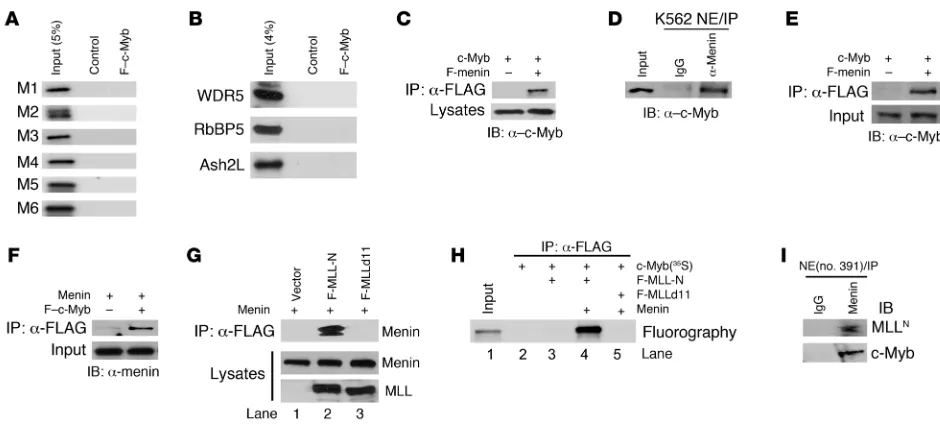
c-Myb associates with the MLL-menin complex. c-Myb has been reported to be an important downstream element of the MLL/HoxA/Meis1 leukemic transformation pathway (51). To better understand the relationship among these proteins, we first examined the possibil-ity that c-Myb might physically interact with them. As described in Figure 1A, we began by transfecting HEK 293T cells with a FLAG-tagged c-Myb (F–c-Myb) expression vector and then purifying the protein from cell lysates by immunoprecipitation with anti-FLAG affinity M2 beads. 293T cells transfected with empty FLAG-tagged vector served as a control. We then mixed the M2 agarose beads, pre-pared from either F–c-Myb– or control vector–transfected cells, with nuclear extracts derived from untransfected K562 cells. The beads were washed, and bound proteins were eluted. As shown in Figure 1B, c-Myb was detected in the immunoprecipitate derived from F–c-Myb–expressing cells, but not in control vector–transfected cells. Immunoblot assays (Figure 1C) revealed that endogenous
MLL as well as other core components of the MLL complex (WDR5, RbBP5, and Ash2L) were coprecipitated with purified F–c-Myb. Menin was also detected in the F–c-Myb immunoprecipitates, indi-cating that this protein too was associated with the MLL complex. c-Myb interacts with the MLL complex through menin. The above experiments suggested that c-Myb could specifically interact with the MLL complex proteins. We next sought to determine how this association was constructed. We first tested the hypoth-esis that c-Myb might directly interact with MLL and sought to determine which domains might be involved. To this end, we generated 6 MLL fragments derived from both the MLLN region
(M1–M4), and the MLLC
region (M5, M6) by in vitro transcrip- tion/translation reactions and then utilized them for direct bind-ing assays. Under the conditions used for these studies, none of the 6 fragments was demonstrated to interact with c-Myb (Figure 2A). Additional experiments of this type carried out with in vitro translated WDR5, RbBP5, and Ash2L proteins suggested that c-Myb does not directly bind to any of these SET domain super-family proteins either (Figure 2B). These results strongly suggest that c-Myb does not directly interact with either MLL or the 3 core SET domain–associated proteins.
Since c-Myb and MLL did not appear to interact directly, we next explored the possibility that c-Myb might interact with the complex through menin. This seemed a tenable alterative, since the MLL-menin interaction is known to be important for the induction of leukemia (48), and menin is reported to inter-act with a myriad of protein partners (52). A similar strategy was employed. HEK 293T cells were cotransfected with vectors expressing c-Myb and FLAG-tagged menin (F-menin). Analysis of anti-FLAG affinity M2 immunoprecipitates from transfected cell lysates revealed the presence of c-Myb along with F-menin when the 2 proteins were coexpressed, but not when c-Myb was expressed along with an empty control vector (Figure 2C). Immunoprecipitation of K562 cell nuclear extracts with anti-menin antibody also showed the presence of endogenous c-Myb protein in the menin immunoprecipitate (Figure 2D). Together, these results provide strong evidence of an interaction between c-Myb and menin in living hematopoietic cells.
[image:3.585.46.309.84.283.2]As the association between c-Myb and menin was demonstrated in cell lysates, it was not certain whether this interaction was direct
Figure 1
c-Myb coprecipitates with the menin and MLL complex. (A) Schematic depiction of experimental scheme. HEK 293T cells were transiently transfected with expression vector encoding FLAG-tagged c-Myb. FLAG-tagged empty vector was used as a control. Cell lysates prepared from transfectants were mixed with α-FLAG antibody affinity M2 agarose to purify FLAG– c-Myb protein. After washing with lysis buffer (1% NP-40), FLAG– c-Myb–containing M2 beads were incubated with nuclear extracts (NE) from K562 cells to isolate the c-Myb–containing complex. Beads were washed twice with nuclear extract buffer (0.05% NP-40). (B and C) Isolated samples were probed by immunoblotting with α–c-Myb (B), α-menin, α-MLLC, α-WDR5,
or required an adapter protein. We addressed this question by expressing both proteins, with or without a FLAG tag, using an in vitro transcription/translation system. The expressed proteins were then employed in in vitro binding assay. We found that c-Myb was coprecipitated when it was mixed with F-menin, but not when mixed with sample generated from empty vector (Figure 2E). Simi-lar reciprocal binding assays showed that menin was only detected when it was mixed with F–c-Myb (Figure 2F), indicating clearly that c-Myb directly interacts with menin.
To further define the various protein interactions within the c-Myb–MLL complex, 2 FLAG-tagged fragments of MLL (MLL-N: aa 1–400; MLLd11: aa 12–400) were transiently expressed in 293T cells that were also engineered to express menin. The ability of these MLL fragments to interact with menin was assessed by immunoprecipitation and Western blotting. As shown in Figure 2G, lane 2, menin was easily detected in the anti-FLAG immuno-precipitate prepared from cells expressing F–MLL-N. In distinct contrast, menin was not detected in the immunoprecipitate prepared from cells expressing F–MLLd11, a mutant that lacks aa 1–11 at the N terminus (Figure 2G, lane 3). This site has previ-ously been defined as a high-affinity menin binding site (48). The previously described in vitro translated protein binding assay also demonstrated that MLL-N directly interacts with menin (Supple-mental Figure 1A; supplemental material available online with this article; doi:10.1172/JCI38030DS1). A further binding assay using
in vitro translated proteins indicated that c-Myb only coprecipi-tates with MLL-N in the presence of menin (Figure 2H, lane 4). In the absence of menin, c-Myb failed to immunoprecipitate with MLL-N (Figure 2H, lane3). As expected, even in the presence of menin, c-Myb did not immunoprecipitate with MLLd11 (Figure 2H, lane 5). These results demonstrate that the N-terminal menin binding site is required for the association of MLL with c-Myb, and in aggregate with the rest of these studies, they strongly sup-port the suggestion that c-Myb associates with MLL complex via a direct interaction with menin.
[image:4.585.55.525.79.294.2]Finally, to ascertain the potential physiologic relevance of this observation, we investigated whether a c-Myb–menin–MLL com-plex could be detected in human leukemia cells isolated from a patient with a t(6;11) chromosomal translocation of MLL gene. To this end, we carried out an immunoprecipitation with anti-menin antibody on nuclear extract prepared from this patient’s cells and clearly detected both c-Myb and MLL, which strongly suggested the existence of such a complex in vivo (Figure 2I). Furthermore, the c-Myb–menin–MLL complex was also identified in human primary T lymphocytes stimulated in culture with phytohemag-glutinin (PHA) and IL-2 (Supplemental Figure 1C), as well as in SEM-K2 leukemia cells, which express the MLL-AF4 fusion as a consequence of t(4;11) chromosomal translocation (Supplemental Figure 1B). Taken together, these results indicate that c-Myb forms a multiprotein complex with MLL through menin both in vitro and in vivo, in human normal primary cells and human leukemia cell lines with or without the MLL chromosomal translocation. Figure 2
c-Myb associates with MLL through menin. (A) In vitro translated, 35S-labeled MLL fragments (indicated on the left) and F–c-Myb (indicated at the top) were mixed, followed by IP with anti-FLAG M2 agarose. Samples were resolved on SDS-PAGE, then amplified, dried, and fluorographed. (B) In vitro protein-binding assay of WDR5, RbBP5, and Ash2L with F–c-Myb as described in A. (C) Cell lysates prepared from 293T cells cotransfected with c-Myb and FLAG-menin were subjected to IP with anti-FLAG M2 agarose, followed by immunoblotting with α–c-Myb antibody. (D) Nuclear extracts prepared from K562 cells were subjected to IP with α-menin antibody or control IgG. Immunoprecipitates were analyzed by immunoblot with α–c-Myb antibody. (E and F) c-Myb and menin proteins were in vitro translated with or without a FLAG tag separately. The various proteins were mixed as indicated in the blots of E and F and then subjected to IP with anti-FLAG M2 agarose. Immunoprecipitates were analyzed by immunoblotting with α–c-Myb (E) or α-menin (F) antibody. (G) FLAG-tagged MLL deletion mutants (F-MLL-N: aa 1–400; F-MLLd11: aa 12–400) and menin were transiently expressed in 293T cells. Cell lysates were subjected to IP with α–FLAG M2 agarose. Immunoprecipitates were analyzed by immunoblotting with α-menin antibody. (H) In vitro translated proteins were mixed as indicated on the right, followed by IP with
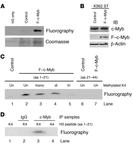
c-Myb–containing MLL complexes have HMTase activity specific to histone H3K4. We next explored the functional significance of c-Myb’s association with the MLL by determining c-Myb’s effect on the HMT activity of the MLL complex (Figure 3). To do this, we first performed the immunoprecipitations using anti-FLAG affinity M2 beads from vector control or F–c-Myb–transfected 293T cell lysates and then mixed the beads with nuclear extracts of K562 cells as described in Figure 1A. Immunoprecipitates were incubated with recombinant human histone H3, together with the radioactive methyl donor S-adenosyl-l-[methyl-3H] methionine
([3H]SAM). We observed that the immunoprecipitate derived from
lysates made from F–c-Myb–transfected cells methylated histone H3 robustly, while the immunoprecipitate derived from control vector–transfected cells was completely inactive (Figure 3A).
[image:5.585.51.277.78.324.2]To delineate which lysine residues in histone H3 are specific sub-strates for the c-Myb–MLL complex, we employed K562 stable cell lines (K562ST) transfected with F–c-Myb or an empty vector that served as a control. Immunoblots indicated that F–c-Myb was only present in the F–c-Myb stable cells and that the total amount of c-Myb protein in F–c-Myb stable cells (F–c-Myb plus endogenous c-Myb) was approximately the same as in the K562 vector control cells (Figure 3B). This result alleviated the potential concern that gross overexpression of F–c-Myb in the K562 lysate might result in activity artifacts. Accordingly, we then incubated anti-FLAG immunoprecipitates prepared from nuclear extracts of control or F–c-Myb K562 stable cells with histone H3 peptides (aa 1–21) that were either non-, mono-, di-, or trimethylated on K4 (Figure 3C, lanes 1–5) or nonmethylated H3 peptide (aa 21–44) (Figure 3C, lanes 6 and 7). The F–c-Myb immunoprecipitates showed Figure 3
The c-Myb–containing complex methylates histone H3 on lysine 4. (A) F–c-Myb or control immunoprecipitate (described in Figure 1A) was incubated with recombinant human histone H3 and the methyl donor [3H]SAM. Samples were resolved on 15% SDS-PAGE, stained with Coomassie blue (bottom panel), amplified, dried, and flourographed (top panel). (B) Cell lysates (top panel), α-FLAG immunoprecipitates (middle panel) prepared from F–c-Myb stably transfected K562 cells, or empty vector control K562 stable cells were immunoblotted with
[image:5.585.307.542.481.741.2]α–c-Myb and α-FLAG antibodies. (C) Methylation of histone H3 pep-tides with different levels of K4 premethylation (aa 1–21) or without methylation (aa 21–44) by F–c-Myb–containing complexes prepared from K562 stable cells. (D) Mutation of H3 peptide from K4 to A4 abro-gated the methylation activity of the c-Myb complex.
Figure 4
Loss of c-Myb affects global H3K4 methylation level in K562 cells but not in human SEM-K2 leukemic cells. (A and B) K562 cells were nucleofected with control siRNA or MYB siRNA. Whole-cell extracts were prepared 3 days after the initial nucleofection according to the cell number counts and probed by immunoblotting with α–c-Myb, α-menin,
α-WDR5, α-MLLC, α-RbBP5, α–β-actin (A), α-H3K4(Me)1-3, and α-H3 (B) antibodies. Mock indicates K562 cells nucleofected with the same amount of nuclease-free water without siRNA. (C) ChIP assay was performed to assess the occupancy of c-Myb on the HOXA9 gene locus of HL-60 cells. Chromatin was immunoprecipitated with α–c-Myb antibody. The presence of the HOXA9 locus DNA in the chromatin precipitates was assessed by standard PCR. Negative and positive controls consisted of IgG and α–histone H3 antibody, respectively. (D) ChIP analysis of HL-60 cells nucleofected with control siRNA or MYB
siRNA was performed using antibodies specific for MLLC and menin. The presence of the HOXA9 locus DNA in the chromatin precipitates was assessed by standard PCR. ChIP using α–histone H3 antibody served as a positive control. (E) SEM-K2 cells were nucleofected with control siRNA or MYB siRNA. Whole-cell extracts were prepared 3 days after the initial nucleofection according to the cell number counts and probed by immunoblotting with α–c-Myb, α-H3K4(Me)1-3,
unambiguous HMT activities on non-, mono-, and dimethylated H3 peptides (aa 1–21) (Figure 3C, lanes 2–4). In contrast, con- trol cell immunoprecipitates showed no activity on these 2 non-methylated peptides (Figure 3C, lanes 1 and 6). The inability of the trimethylated H3K4 peptide and nonmethylated H3 peptide (aa 21–44) to be further methylated by the F–c-Myb–containing immunoprecipitates (Figure 3C, lanes 5 and 7), despite the fact that these peptides have unmethylated K9, K27, and K36 resi-dues, demonstrates convincingly that the c-Myb–containing MLL complex has a preference for methylating K4. Finally, there was no detectable H3 methylation when a mutated (K4A) H3 peptide (1–21 aa) (Figure 3D, lane 4) as employed in the same assay. Hence, the c-Myb–containing HMT activity is specific for H3 lysine (K4).
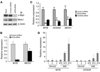
Deletion of c-Myb decreases global H3K4 methylation levels in K562 cells but not in SEM-K2 cells. To provide additional proof of c-Myb’s role in facilitating MLL complex–mediated methylation of H3K4, we nucleofected K562 cells with MYB-targeted siRNA. Mock-nucleo-fected or control siRNA–nucleofected K562 cells served as controls for nonspecific effects of the transfection procedure itself or the siRNA, respectively. Whole-cell lysates were prepared for immu-noblotting from all of these cells 72 hours after nucleofection.
The MYB siRNA resulted in an efficient knockdown of c-Myb pro-tein, with an at least 80% reduction in protein expression level relative to that observed with mock- or con-trol siRNA–treated cells (Figure 4A, top panel). The expression levels of menin and other components of MLL complex (MLL, WDR5, and RbBP5) were unaffected by either the MYB-targeted or control siRNA (Fig-ure 4A). Remarkably, in K562 cells in which c-Myb levels were downregu-lated with siRNA, levels of H3K4 methylation were also reduced. The changes in H3K4 methylation and histone H3 protein were quantitated (Figure 4B). Trimethylation of H3K4 decreased by 30%–50%, while a more modest effect on monomethylation was observed (~15%). There was no apparent effect on the dimethylated form of H3K4 (Figure 4B). A similar pattern of effects on H3K4 methyla-tion was reported after knockdown of WDR5 (38), suggesting that c-Myb might interact with WDR5 in some as-yet-unspecified manner. The expression level of histone H3 was unchanged.
It has been reported that MLL binding in the HOXA9 locus is asso-ciated with the ability to methylate H3K4, and in particular to trimeth-ylate the dimethylated form of H3K4 (36, 38). One might then anticipate that if c-Myb was responsible for targeting the MLL complex to this locus, then a MYB knockdown ought to be associated with a decrease in MLL and menin at this site. To test this hypothesis, we carried out a series of ChIP assays in HL-60 cells using DNA primers specific for the first exon of HOXA9, a region previously reported by Dou et al. (36) and Wysocka et al. (38) to be occupied by MLL. We first carried out a ChIP assay to confirm the presence of c-Myb in the HOXA9 locus in untreated HL-60 cells (Figure 4C). We then investigated whether MYB silenc-ing would affect the occupancy of MLL and menin on the HOXA9 locus. For this experiment, we nucleofected HL-60 cells with control or MYB-targeted siRNA. ChIP assays were performed 72 hours after nucleofection using anti-MLLC
[image:6.585.41.393.80.333.2]or anti-menin antibod-ies. The occupancy of MLL and menin on the HOXA9 locus was substantially decreased or undetectable, depending on which pri- mary antibody was employed for the immunoprecipitation (Fig-ure 4D). To demonstrate that these results were not dependent on the use of a particular cell line, we carried out identical silencing experiments and ChIP assays using KCL-22 cells (human chronic myeloid leukemia). As showed in Supplemental Figure 2, B–E, the results obtained with these cells were entirely consistent with those obtained with HL-60 cells. Of note, there are several canonical c-Myb–binding sequences in 2 HOXA9 exons (2 in exon 1 and 6 in Figure 5
Loss of c-Myb results in a decrease in HOXA9 and MEIS1 gene expression. (A) K562 cells were nucleofected with control siRNA or MYB siRNA. Whole-cell extracts were prepared 3 days after the initial nucleofection. Proteins were subjected to immunoblot analysis with α–c-Myb, α-Meis1, and
α–β-actin antibodies. Mock indicates K562 cells nucleofected with the same amount of nuclease-free water without siRNA. (B) K562 cells were nucleofected with control siRNA or MYB siRNA. RNA samples, prepared 2 days after the initial nucleofection, were reverse transcribed and used for qRT-PCR analysis for MYB and MEIS1 expression, determined in triplicate. Relative expression of
exon 2) (Supplemental Figure 3A). A ChIP analysis conducted on KCL-22 cells with primer sets specific for the c-Myb–binding sites revealed that c-Myb, menin, and MLL are localized to these exons (Supplemental Figure 3, B–E). These results strongly suggest that c-Myb recruits MLL and menin to the HOXA9 locus and that it does so by binding to its canonical recognition sites. In support of this statement, we also found that ChIP experiments conducted in cells expressing a c-Myb construct with deletion of the DNA-bind-ing domain (c-Myb 194–636) led to the decrease in binding of MLL and menin on the HOXA9 locus (see below).
Finally, to further investigate c-Myb’s role in regulating overall H3K4 methylation, and specifically whether c-Myb might aug-ment the activity of other cellular HMTs, we determined the effect of a MYB knockdown on H3K4 methylation in human SEM-K2 leukemia cells. SEM-K2 cells were derived from a child with ALL, and though they retain a copy of the wild-type MLL gene, they express an MLL-AF4 fusion protein as a consequence of a t(4;11) chromosomal translocation (53). Accordingly, we postulated that a decrease in H3K4 methylation might result from the loss of non-MLL c-Myb–dependent HMT activity. The SEM-K2 cells were nucleofected with identical control or MYB-targeted siRNA.
Whole-cell lysates were prepared 72 hours after nucleofection. Immunoblotting with anti–c-Myb antibody showed that c-Myb protein expression levels in the MYB siRNA–treated SEM-K2 cells was approximately the same as in K562 cells (~20%) relative to that observed with control siRNA–treated cells (Figure 4E). However, in contrast to the results obtained with the myeloid K562 cells, no detectable change in the levels of mono- or dimethylated H3K4 was observed, while the trimethylated form was reduced only approxi-mately 6% (Figure 4E). Again, the total protein level of histone H3 was unchanged. These results suggest that c-Myb has limited abil-ity to promote non–MLL-dependent HMT activity, at least in this particular lymphoid leukemia cell line.
[image:7.585.44.364.81.495.2]MYB silencing is associated with decreased HOXA9 and MEIS1 gene expression. The results described above suggested that c-Myb might well influence the biological activity of the MLL complex. To inves-tigate this possibility, we investigated the effect of MYB silencing on the ability of the MLL complex to regulate HOXA9 and MEIS1 expression in different types of leukemia cells. In K562 cells, c-Myb protein and mRNA expression were effectively eliminated using siRNA (Figure 5, A and B), and specificity was suggested by lack of effect on β-actin (Figure 5A). The MYB knockdown resulted in
Figure 6
Decrease in trimethylation of H3K4 and HOXA9
gene expression is specific for MYB silencing but not secondary to the antiproliferation effect of MYB silencing. (A and B) KCL-22 cells were nucleofected with control siRNA or B-MYB
siRNA. Whole-cell extracts were prepared 3 days after the initial nucleofection accord-ing to the cell number counts and probed by immunoblotting with α–c-Myb, α–B-Myb, and
α–β-actin (A), α-H3K4(Me)1-3, and α-H3 (B) antibodies. (C) Cell proliferation assays were carried out with KCL-22 cells nucleofected with mock, control, MYB, and B-MYB siRNAs at indicated time points. Silencing MYB or
B-MYB inhibited cell proliferation. (D–G) KCL-22 cells were nucleofected with control, MYB, or B-MYB siRNA. RNA samples, prepared 2 days after the initial nucleofection, were reverse transcribed and used for qRT-PCR analysis for
MYB (D), B-MYB (E), HOXA9 (F), and MEIS1
(G) gene expression determined in triplicate as described in Figure 5B. (H and I) KCL-22 cells were nucleofected with control, MYB, or HOXA9
a considerable reduction in Meis1 protein relative to the decrease observed with mock- or control siRNA–treated cells (Figure 5A). Quantitative real-time RT-PCR (qRT-PCR) analysis also showed that knockdown of MYB expression resulted in a moderate down-regulation (~40% decrease) in MEIS1 mRNA level relative to that of GAPDH (Figure 5B). The effect of a MYB knockdown on HOXA9 mRNA could not be determined in these experiments because
[image:8.585.96.490.80.500.2]HOXA9 was undetectable in control (untreated) K562 cells. There-fore, to further establish whether c-Myb is required for expression of HOXA9 as well as MEIS1, we also performed a MYB-directed siRNA silencing experiment in human ML-2 leukemia cells (Figure 5C). These cells were of particular interest because they bear a t(6;11) chromosomal translocation and thus express an MLL-AF6 fusion protein exclusively. After silencing MYB, qRT-PCR analysis showed that HOXA9 expression was moderately downregulated (50% reduc-tion) in the MYB-knockdown cells compared with mock- or control Figure 7
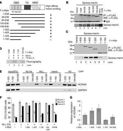
The interaction between c-Myb and the menin-MLL complex is required for localization of MLL and menin on the HOXA9 gene, transformation of myeloid progenitors, and HOXA9 gene expression. (A) Schematic representation of the constructions used. DBD, DNA binding domain; TA, transactivation domain; NRD, negative regulation domain. Various c-Myb mutants containing FLAG tag at their N termini and Xpress-tagged menin were transiently transfected in 293T cells. Cell lysates prepared from transfectants were subjected to IP with anti-FLAG M2 agarose. High-affinity menin-binding capacities are indicated on the right. (B and C) Immunoprecipitated proteins were separated by 10% SDS-PAGE and immunoblotted with anti-FLAG antibody (upper panel) or anti-Xpress antibody (bottom panel) antibody. (D) F–c-Myb mutants, menin, and 35S-labeled F–MLL-N were generated by in vitro transcription/translation. c-Myb was incubated with protein reaction mixtures as indicated on the right in the presence of α-FLAG M2 agarose. Samples were resolved on 10% SDS-PAGE, amplified, dried, and fluorographed. (E) ChIP assay of HL-60 cells transfected with c-Myb mutants was performed using the antibodies indicated at the top. Amplicons upstream of the HOXA9 and
siRNA–treated cells (Figure 5C). Interestingly, the converse was also found to be true. That is, siRNA-mediated knockdown of HOXA9 caused a large downregulation of MYB expression (Figure 5C), a result consistent with the recent observation of the Hess group (51). Unexpectedly however, silencing of MYB was also associated with decreased expression of MEIS1, as was a siRNA-mediated knockdown of HOXA9. In accordance with these results, in AML patient samples (nos. 70, 972, 973; Supplemental Table 1) in which MYB expression was found to be elevated compared with that in normal bone marrow cells (sample nos. 1, 2, and 3), qRT-PCR analy-sis revealed that HOXA9 mRNA expression was elevated as well, and in proportion to the elevation of MYB (Figure 5D).
It has been reported that c-Myb plays a direct role in cell prolif- eration (8–12). Therefore, one could question whether the observa-tions reported above were the specific result of MYB silencing or merely secondary phenomena resulting from decreased cell prolif-eration. To address this issue, we examined the effects of silencing B-MYB, a Myb family member also involved in regulating cell pro-liferation (54, 55), on H3K4 methylation and expression of HOXA9 and MEIS1. For these experiments, all assays were carried out with MYB and B-MYB siRNAs in KCL-22 human leukemia cells. Silenc-ing B-MYB had no effect on c-Myb protein levels (Figure 6A) or on H3K4 methylation (Figure 6B). As expected, however, silenc-ing MYB or B-MYB inhibited cell proliferation to a similar extent (Figure 6C). Specificity of silencing was confirmed by qRT-PCR (Figure 6, D and E). Consistent with results obtained in other cell lines, silencing MYB in KCL-22 cells decreased the trimethylation of H3K4 (Supplemental Figure 2C). Silencing MYB also led to the very large decrease in HOXA9 gene expression (≥90%) in KCL-22 cells and a more modest reduction in MEIS1 gene expression (~25%). Silencing B-MYB did not affect the expression of either gene (Figure 6, F and G). Taken together, these results indicate that the observed decrease in trimethylation of H3K4 and HOXA9 gene expression is not a secondary phenomenon related to decreased cell proliferation but is in fact specific to the loss of c-Myb. Finally, we noted that as expected, silencing HOXA9 reduced MYB gene
expression (Figure 6H), but quite unexpectedly, silencing MYB led to a dramatic decrease in HOXA9 gene expression (Figure 6I). These results suggest the existence of an autoregulatory feedback loop between MYB and HOXA9 that has not previously been described and whose exact control remains to be defined.
Delineation of the c-Myb–menin interaction domain. To develop a more mechanistic understanding of how c-Myb and menin interact, we sought to localize more precisely the c-Myb–menin interaction domains. This was accomplished by construction of a series of c-Myb FLAG-tagged deletion mutants (Figure 7A) in pcDNA3 expression vectors. These were cotransfected with Xpress-tagged menin (Xpress-menin) in 293T cells (Figure 7, B and C). Immunoprecipitation of the various FLAG-tagged c-Myb constructs from transiently trans- fected 293T cells revealed that deletion of the c-Myb DNA–bind-ing repeats R1, R2, and R3 had no effect on the ability of c-Myb to bind to menin (Figure 7B). Further, the c-Myb DNA–binding domain (aa 39–193), which is contained within the aa 1–193 expres-sion fragment, was not only dispensable for a stable c-Myb–menin interaction; by itself, it had no ability to bind menin at all, at least under these assay conditions (Figure 7C, lanes 6 and 7). In contrast, c-Myb fragment 1–325 strongly interacted with menin (Figure 7C, lane 5). Reciprocal experiments conducted in vitro with recombi-nantly expressed proteins indicated that full-length c-Myb and c-Myb 194–636 were both able to form a complex with MLL in the presence of menin (Figure 7D, lanes 2 and 4, respectively). As expected from the experiments carried out with 293T lysates, c-Myb DNA– binding domain fragment 1–193 was unable to form a complex with MLL even in the presence of menin (Figure 7D, lane 3). Thus, the c-Myb–menin interaction domain was localized to a region within c-Myb spanning residues 194–325.
[image:9.585.82.506.82.247.2]c-Myb binding to its specific DNA recognition site is required for localiza-tion of MLL and menin to the HOXA9 promoter locus, for HOXA9 gene expression, and for transformation of normal myeloid progenitor cells. To ascertain whether the association of c-Myb with menin-MLL is required for localization of the complex to the HOXA9 gene promoter region in vivo, we transfected HL-60 cells with 3 of the Figure 8
c-Myb mutants (1–325, 1–193, and 194–636, respectively) described immediately above. We then carried out ChIP assays employing antibodies specific for either menin or MLL protein and the pre-viously described cDNA primers spanning the c-Myb–binding sites in the HOXA9 promoter locus. We found that transfection of HL-60 cells with c-Myb 1–193 decreased the localization of both MLL and menin on the HOXA9 promoter as compared with that observed with c-Myb 1–325, which contains the menin-binding domain (Figure 7E). These results strongly suggest that c-Myb’s ability to bind to its DNA recognition region is necessary for recruitment of the menin-MLL complex to the HOXA9 promoter. In support of this conclusion, transfection of HL-60 cells with c-Myb fragment 194–636, which is competent to interact with menin-MLL but is unable to bind DNA, recruits considerably less menin-MLL to the HOXA9 locus (Figure 7E). We speculate that the menin-MLL protein detected in the HOXA9 promoter in the presence of 194–636 is bound there by either endogenous c-Myb or perhaps LEDGF, which has previously been reported to recruit menin-MLL to this locus.
Finally, to investigate the potential role of c-Myb in MLL-medi-ated leukemogenesis, we carried out in vitro transformation assays using normal mouse bone marrow cells obtained from 5-fluoro- uracil–treated (5-FU–treated) donors transfected with an MLL-ENL fusion construct and c-Myb deletion mutant 1–325, 1–193, or 194–636. We then assessed the cells for growth characteristics consistent with transformation (51, 56) in serial methylcellulose replating assays (Figure 7F). A dominant-negative c-Myb expres-sion construct consisting of a C-terminal fusion of the c-Myb DNA–binding domain (1–200 aa) to the KRAB repressor domain (1–74 aa) (DNMyb-KRAB) was used as a negative control for these experiments. After the first round of plating, cells transfected with the various constructs yielded a similar number of colonies. When the colonies present in the dishes were disaggregated and replated in a second round, colony formation was, as expected, diminished under all conditions. However, in the third replating round, cells expressing the full-length c-Myb expression fragment (aa 1–636) or the c-Myb deletion mutants capable of interacting with menin (aa 1–325 and 194–636, respectively) recovered their original clon-Figure 9
[image:10.585.51.541.82.419.2]ing efficiency, a hallmark of transformation (56), in contrast to those expressing the 1–193 mutant, which is incapable of binding menin, or the DNMyb-KRAB. We believe that the transformation observed in cells transfected with MLL-ENL alone occurs because of the presence of endogenous c-Myb protein in the murine mar-row cells. This hypothesis is supported by the work of Hess et al. (51) and is explained mechanistically by results shown in Figure 7G. Here we explored the effect of c-Myb mutations on HOXA9 gene expression in SEM-K2 cells. When cells were transfected with c-Myb 1–636 (full-length c-Myb) or c-Myb 1–325, HOXA9 expression lev- els increased 50% and 70%, respectively, compared with cells trans- duced with an empty vector control. In contrast, HOXA9 expres-sion fell by 50% compared with control in cells transduced with the c-Myb 1–193 construct. Therefore, if c-Myb cannot bind to its DNA recognition sequence, menin-MLL is not effectively recruited to the HOXA9 locus, HOXA9 expression falls, and transformation diminishes accordingly. The functional relationship between these proteins holds regardless of whether MLL is wild-type or part of a fusion protein as long as the menin interaction domains persist. A molecular basis for c-Myb’s role in oncogenic MLL-fusion pro-tein transformation is therefore provided by these studies.
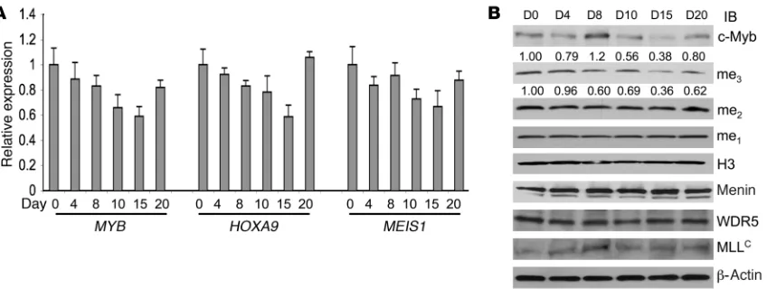
c-Myb inhibition is associated with decreased HOXA9 and MEIS1 mRNA expression in AML patients being treated with a MYB–targeted antisense oligodeoxynucleotide. To further explore the relationship among MYB, HOXA9, and MEIS1 in primary leukemia cells in vivo, we examined the expression of all 3 genes in patients participating in a phase I clinical trial evaluating the toxicity of a MYB-targeted phosphorothioate-modified antisense oligodeoxynucleotide (ASODN; http://clinicaltrials.gov/ct2/show/NCT00780052?term =UPCC+04701&rank=1). The ASODN was delivered by con-tinuous intravenous infusion for 7 days at a dose of 3 mg/kg/d. Peripheral blood mononuclear cells (95%–100% blasts) were iso-lated from 3 patients (nos. 866, 995, 1207; Supplemental Table 1) on days 0, 4, 8, 10, 15, and 20 after the start of the infusion and analyzed by qRT-PCR for MYB and HOXA9 expression (Figure 8A, Supplemental Figure 4). In the case of patient no. 866, additional analysis for MEIS1 mRNA expression as well as Western blotting for c-Myb, menin, WDR5, and MLL protein expression and H3K4 methylation was also carried out (Figure 8B). In patient 866, MYB mRNA levels were reduced 40% from day 0 to day 15 and returned to baseline by day 20 (Figure 8A). HOXA9 and MEIS1 mRNA levels in this patient’s samples paralleled those of MYB. Also in agree-ment with our in vitro data, trimethylation of H3K4 correlated directly with relative c-Myb expression, whereas mono- and di-K4 methylation, also as expected, was unchanged (Figure 8B). Of note, protein expression levels of menin, WDR5, MLLC, and
histone H3 were not effected by the c-myb–targeted ASODN, sug-gesting specificity of the effect (Figure 8B).
Discussion
c-Myb’s ability to transactivate genes required for lineage com-mitment, cell proliferation, and differentiation underlie its importance for normal hematopoietic cell development (5, 6). It is intuitive that c-Myb’s participation in such critical functions might also suggest mechanisms whereby c-Myb could contribute to hematopoietic cell transformation (57). Simple overexpression is one such mechanism, but overexpression of MYB is not found in the majority of leukemia patients (58). Indeed, direct evidence implicating MYB overexpression in the pathogenesis of leukemia in primary patient material has only recently been provided in a
small subset of acute lymphoblastic leukemia patients (<10%) in whom a translocation that juxtaposed the TCRB and MYB loci [i.e., t(6;7)(q23;q34)] or a short, somatic gene duplication increased MYB expression (59, 60). Therefore, how MYB might facilitate leu-kemic transformation remains incompletely understood. What is known is that failure to downregulate MYB expression is associated with a block in differentiation (61, 62) and a failure of proliferation arrest (57). Yet transgenic mice that overexpress c-Myb protein do not develop leukemia (63), suggesting that c-Myb protein requires additional cell-intrinsic factors in order to manifest its transfor-mation ability. Cells capable of supplying such factors have been hypothesized to be both rare and immature (64), and the identity of factors supplied remains uncertain. Cooperation of oncogenes is one possibility (65). A recent example of this has been described in chronic myeloid leukemia, where c-Myb cooperates with Bcr/ Abl (66) to drive leukemic transformation, perhaps by blocking differentiation as a result of c-Myb–mediated downregulation of C/EBPα (67). Aberrant gene activation by alternately spliced but transcriptionally active c-Myb proteins, translated from alternately spliced MYB mRNAs, is another way that c-Myb may contribute to leukemogenesis (68). We now extend these findings, presenting a series of observations that not only provide mechanistic insight into how c-Myb may facilitate cellular transformation, even in the absence of overexpression, but may also explain how c-Myb exerts such a myriad of effects on cell proliferation and differentiation.
c-Myb forms a complex with MLL through menin both in vitro and in vivo. The results presented here clearly show that c-Myb associates with the MLL complex in human hematopoietic cells and that this association occurs through menin, a protein first identified as the tumor suppressor of the MEN1 gene. The existence of a c-Myb–menin–MLL complex was confirmed in vitro, in vivo, and in a variety of normal and neoplastic cell types using a host of complementary methods including: (a) coimmunoprecipitation of F–c-Myb with menin, MLL, and its core components (includ-ing WDR5, RbBP5, and Ash2L) from K562 nuclear extracts using purified F–c-Myb protein; (b) coimmunoprecipitation of MLL and c-Myb with menin in K562 cells, KCL-22 cells, SEM-K2 cells, human leukemia cells from a patient with MLL gene transloca-tion, and normal human primary T lymphocytes using anti-menin antibody; (c) studying direct in vitro interactions among c-Myb, menin, and MLL using purified proteins; and (c) demonstrating clearly that menin is required in order for c-Myb and MLL to form a complex. Copurification of these proteins was also obtained by 2-step ion exchange chromatography (data not shown). The possibility that c-Myb might be directly associated with WDR5, Ash2L, and RbBP5 was also ruled out by the failure of F–c-Myb to coprecipitate 35
S-labeled forms of these proteins in direct bind-ing assays. These results strongly suggest that c-Myb interacts with the MLL complex exclusively through menin. Nonetheless, once incorporated into the complex, other indirect effects on pro-tein members of the complex are possible, as suggested by the loss of H3K4 trimethylation, a WDR5 function (38), when c-Myb is silenced with siRNA (Figure 4B and Figure 8). Therefore, by associating with the menin-MLL complex, c-Myb may direct the complex to its cognate binding sites, where effects on expression of c-Myb target genes may be induced.
4 core histones (H3, H4, H2A, H2B) around which 147 base pairs of DNA are wrapped. Nucleosomal histones, and their unstructured N-terminal tails, are the target of a variety of covalent modifications. It is now well established that posttranslational acetylation, meth-ylation, phosphorylation, and ubiquitination of these histones play an intrinsic role in transcriptional regulation. It has also been sug- gested that histone modification may be involved in the propaga-tion of the transcriptional state through cell division (69, 70).
Methylation of histone H3 on lysine 4 was first identified in trout testes (71). It has been one of the characteristic of active chromatin in various organisms (70, 72). The existence of distinct patterns of mono-, di-, and trimethylation at H3K4 suggests that the transition between methylation states is a regulated event and that different methylation states are associated with distinct regu-latory outcomes. MLL family proteins have already been reported to display H3K4 methyltransferase activity (32, 73, 74).
The c-Myb–menin–MLL complex appears to be functionally simi-lar to the previously described yeast Set1 complex, which promotes transcriptional activation by methylating H3K4. Like the Set1 complex, the isolated c-Myb–MLL complex shows robust methyl-transferase activity on both recombinant human histone H3 and synthetic histone H3 peptides. More importantly, the c-Myb–MLL complex methylates H3K4 non-, mono-, and dimethylated peptides efficiently and specifically (Figure 3, C and D). Subunits of the c-Myb–associated HMTase complex are homologous to members of the yeast Set1 assembly. These results are consistent with the reported ability of the MLL complex, and specifically the MLL SET domain, to effect H3K4 methylation. In fact, the observed reduc-tion of global H3K4 trimethylation in MYB-knockdown human leukemia cells suggests that the specificity of MLL methyltransfer-ase might be influenced by c-Myb, as proteins that associate with the enzyme may affect its selection of residues to modify (75), or the degree of methylation at a specific site (42). In this specific regard, our data appear to indicate that c-Myb may perturb WDR5 func-tion, though with respect to effects on global methylation, effects on other HMTs are not ruled out. Regardless, since methylation of H3K4 is associated with open chromatin and active transcrip-tion (32, 45, 46), this work strongly suggests that in addition to its direct effects on transcription, c-Myb may regulate hematopoietic cell gene expression by leading complexes such as menin-MLL to c-Myb–bindings sites throughout the genome where the MLL set domain may exert effects on H3K4 methylation.
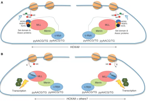
MLL and menin both associate with the HOXA9 promoter. In the absence of menin, MLL and its fusions fail to maintain HOXA9 gene expression, an event that has been reported to be critical for MLL fusion protein–mediated transformation (39, 48). Recently, lens epi-thelial-derived growth factor (LEDGF) has been shown to recruit the menin-MLL complex to the Hox locus and, because of this activity, to play a critical role in leukemic transformation by MLL fusion pro-teins (49). c-Myb has also been shown to be essential for MLL-ENL– mediated transformation (51) and a critical component of the MLL myeloid leukemia stem cell maintenance signature (76), suggesting that it too might interact in some manner with MLL. Our observa-tions that c-Myb recruits MLL to the HOXA9 gene locus by binding to its canonical DNA recognition sites, that this recruitment is criti-cally dependent on c-Myb’s interaction with menin, and finally that HOXA9 gene expression falls in the absence of c-Myb, in concert with its ability to transform normal myeloid progenitor cells, all provide mechanistic insight into c-Myb’s potential role in leukemic trans-formation in those leukemias associated with MLL fusion proteins.
Whether c-Myb plays a similar role in leukemic transformation in cells expressing wild-type MLL remains to be determined. Potential interactions between c-Myb and LEDGF and whether either makes the other dispensable with respect to transformation in vivo also remain to be determined.
MYB, HOXA9, and MEIS1 participate in an autoregulatory feedback loop. As part of our evaluation of the biological implications of c-Myb’s incorporation into the MLL complex, we evaluated the expression levels of HOXA9 and its cofactor MEIS1, 2 well-estab-lished MLL target genes, in human leukemia cell lines nucleofected with MYB-targeted siRNA and, perhaps of greater relevance, in cells isolated from AML patients undergoing treatment with a MYB-targeted ASODN. We noted with interest that in cells with (ML-2) or without (K562) an MLL translocation expression of both HOXA9 and MEIS1 was specifically downregulated by the MYB-targeted siRNA. These results suggest that binding of c-Myb in the HOXA9 locus, independent of effects on H3K4 methylation, is sufficient for upregulation of HOXA9 expression. Importantly, expression of HOXA9 and MEIS1 paralleled MYB expression levels in leukemia patients undergoing treatment with a MYB-targeted ASODN. These results support the physiologic relevance of the observations in cell lines just mentioned. Previous work demon-strating a connection between MYB expression and HOX cluster genes placed c-Myb downstream of Hoxa9 and Meis1 (51, 77). The results reported in this study (Figures 5–8) clearly indicate that c-Myb plays a role in regulating expression of HOXA9 and MEIS1. In aggregate, these studies strongly suggest that c-Myb and these Hox cluster proteins are all in linked in an autoregulatory loop, the regulation and functional significance of which is presently under investigation in our laboratory.