IJPSR (2016), Vol. 7, Issue 11 (Research Article)
Received on 26 May, 2016; received in revised form, 28 July, 2016; accepted, 04 August, 2016; published 01 November, 2016
QUANTUM AND HISTOLOGICAL STUDIES OF PARACETAMOL ALTERNATIVES TO AVOID CANCER EFFECT
Anwar El-Shahawy *1, Nesreen G. Abdelhaliem 2, Hana Gashlan 3 and Safaa Qusti 3
Chemistry Department 1, Faculty of Science, Assiut University, Assiut, Egypt Histology Department 2, Faculty of Medicine, Sohag University, Sohag, Egypt
Biochemistry Department 3, Faculty of Science, King Abdulaziz University. Jeddah, KSA
ABSTRACT: Paracetamol (acetaminophen) (PA) is a widely used as an analgesic and antipyretic drug that is commonly available in without a prescription. Paracetamol has been used to treat many diseases such as headache, muscle aches, arthritis, backache, toothaches, colds, and fevers. It cures pain in mild arthritis but has no effect on the underlying inflammation and swelling of the joints. It has been reported as the common cause of drug toxic ingestion. These studies aim to compare the effect of some Paracetamol derivatives such Phenacetin, p-nitroacetanilide, p-bromoacetanilide and n-acetylanthranilic acid on liver structure to avoid Paracetamol cancer effect during its metabolism. From the histological point of view, it has been found that p-nitroacetanilide is the best alternative due to its metabolite products.
INTRODUCTIOIN: Paracetamol (acetamino
phen) (PA) is a widely used as an analgesic and antipyretic drug that is commonly available without a prescription. Paracetamol has been used to treat many diseases such as headache, muscle aches, arthritis, backache, toothaches, colds, and fevers. It cures pain in mild arthritis but has no effect on the underlying inflammation and swelling of the joints. It has been reported as the common cause of drug toxic ingestion. Paracetamol or acetaminophen (N-acetyl-p-aminophenol; PA) is a commonly used analgesic antipyretic drug and is used for wide range in different disease.
QUICK RESPONSE CODE
DOI:
10.13040/IJPSR.0975-8232.7(11).4387-99
Article can be accessed online on: www.ijpsr.com
DOI link: http://dx.doi.org/10.13040/IJPSR.0975-8232.7 (11).4387-99
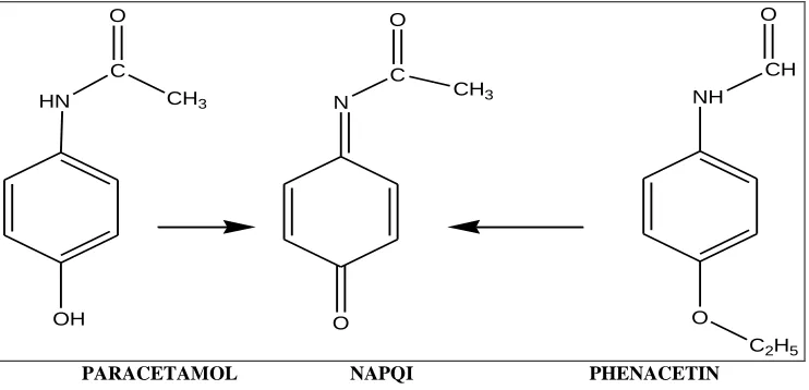
It was found that cumulative overdose can cause hepatic toxicity in both humans and experimental animals [Khandelwal et al.1. It was reported that, in certain circumstances, individuals died after taking less than the known minimum threshold toxic dose because of their higher sensitivity to its toxic effects; so, personals risk of toxicity following the overdose was difficult to be estimated by Ucheya and Igweh 2. Paracetamol at therapeutic doses is rapidly metabolized in the liver and then it is oxidized by cytochrome P450. The exact mechanisms of Paracetamol-induced toxicity still not clear. The general concept is that drug oxidation by various cytochromes P450 generates a highly cytotoxic N-acetyl-p-benzoquinone imine (NAPQI) that conjugates with glutathione (GSH), leading to the depletion of cellular GSH pools and an increasing the oxidative stress. The induced oxidative stress in the cell may ultimately cause cell death by Al-Belooshia et al. 3 and Saito et al.13.
Keywords:
DFT, histology studies, Phenacetin, p-nitroacetanilide, p-bromoacetanilde
Correspondence to Author: Anwar El-Shahawy
Chemistry Department,
Faculty of Science, Assiut University, Assiut, Egypt.
Caspase 3 (cysteinyl aspartate proteinase) is a crucial molecule in regulating both mitochondrial and death receptor apoptotic pathways by Lavrik
4
. A number of genetic and biochemical studies suggest that caspase activation is essential for the occurrence of the apoptotic phenotype of cell death by Janicke et al.5. Caspase-3 substrate cleavage has been observed under oxidative stress in different pathological conditions by Meki et al. 6. Heat shock proteins are molecular chaperones that play a vital physiological role enabling correct folding of freshly synthesized proteins.
It is believed that they have various cytoprotective functions through mechanisms that include refolding of proteins impacted by stress-induced denaturation and inhibition of apoptotic molecules by Turturici. et al.7. In normal cells, HSP70 is expressed at low levels, but the expression was enhanced by stressful situation as oxidative stress by Tas et al. 8. It has been speculated that HSP70-mediated inhibition of cell death may involve two distinct mechanisms: in the case of stimuli that cause stress-induced necrosis associated with protein damage, the key role appears to reflect a chaperone function in repairing the protein machinery, whereas separate and distinct mechanisms appear to be responsible for inhibition of apoptosis when protein damage is not a main feature by Steel et al.9.
El-Shahawy 10 studied the metabolism of Paracetamol using DFT method of electron transfer between nucleic acid bases and the carcinogenic metabolite of Paracetamol, NAPQI. It has been concluded that the electron transfer between NAPQI and guanine equals to 0.4 eV; this means that once NAPQI contact the liver cell in absence of glutathione, a spontaneous electron transfer takes place from guanine to NAPQI forming cationic nucleus i.e positive cancer.
Quantum Calculations:
DFT-quantum calculations have been carried out using Gaussian 03 and Chemoffice 2005 as has been described before by El-Shahawy 10. Charge transfer studies between Paracetamol and its analogues such as Phenacetin, nitroacetanilide, p-bromoacetanilide and n-acetylanthranilic acid with nucleic acid bases were performed using DFT
method. It has been found that p-bromoacetanilide has the highest cancer energy barrier and it was concluded that this product can be used as an a Paracetamol alternative.
Metabolism of Paracetamol:
To understand well the denotation of cancer, it is mutual electron transfer from/to the nucleic acid bases and electron donor or electron acceptor, i.e. free radicals, some drugs even food like grills and fries. Losing an electron from the nucleic acid bases inside the nucleus of liver cell produces carcinogenic cell in which the nucleus acts as electron donor to any electron acceptor such as in Paracetamol metabolite in the liver. PA is metabolized primarily in the liver by El-Shahawy 10 into toxic and non-toxic products. Three metabolic pathways are notable, Fig. 1.
[image:2.612.341.535.315.509.2]
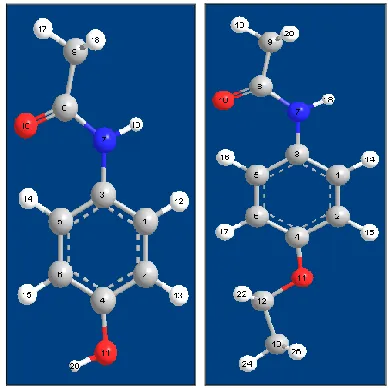
PARACETAMOL(PA) PHENACETIN (PH)
FIG. 1: MINIMUM ENERGY STRUCTURES OF PA AND PH.
radical which can behave as a positive carcinogenic cell. The positive cancer means that the nucleus lacks an electron due to the electron transfer;
therefore it behaves abnormally i.e. carcinogenic behavior.
O H
NH
O
O
NH
O
GlcA
O
N
O
O
NH
O S
O H
O O
O H
NH
O
GSH
Toxic reactions with proteins and nucleic acids
Glucuronidation
Sulfa tion
Hepatic enzymes
GSH Conjugation NAPQI
[image:3.612.122.493.553.732.2]Paracetamol
FIG. 2: METABOLISM PATHWAYS OF PARACETAMOL
To deepen the denotation of cancer, it is mutual electron transfer between the nucleic acid bases and electron donor or electron acceptor, i.e. free radicals, drugs even some food like grills and fries. Losing an electron from the nucleic acid bases inside the nucleus produces carcinogenic cell in which the nucleus acts as electron donor to any electron acceptor such as in case of Paracetamol
metabolite in the liver , NABQI, which has high electron affinity being sufficient to withdraw an electron from guanine in the nucleus of liver cell in absence of glutathione. Therefore the nucleus has electron deficiency producing cationic nucleus as a free radical which behaves as positive carcinogenic cell.
HN C O
CH3
OH
N
O C O
CH3 NH
O
CH O
C2H5
PARACETAMOL NAPQI PHENACETIN
DFT-Quantum Studies:
After administration of PA and after its arrival to the blood of pH 7.4 from the gastrointestinal tract, PA has some anionic forms. The charge density anionic form of PA tends to be close to the NAPQI molecules and the anionic form enthalpy change to NAPQI is less than that of PA molecule, Table 1.
TABLE 1: DFT-ENTHALPY CHANGE OF PA AND PA ANION TO NAPQI AT 37 OC.
Compounds Total Energy eV ΔH k cal mol-1
NAPQI -13989.07243 ---
PA anion -14006.3960 399.270
PA molecule -14023.51472 794.428
From the previous Table 1, it can be concluded that the enthalpy change for the anionic form of PA is lower than that of the PA molecule. Hence the alteration of the anionic form to NAPQI is easier than that of PA molecule. Paracetamol has ionization constant pKa being equal to 9.5 in the
human blood medium of pH=7.4, therefore the ratio of the PA anions with respect to PA molecules equals to 0.8%. This means that there are some PA anions in the human blood with the major PA molecules before the drug arrival to the human liver.
MATERIAL AND METHODS: Animals and treatments:
Thirty adult male albino rats (3 months old; 200– 250 g body weight) were obtained and maintained in the Animal Nutrition and Care House, Faculty of Medicine, Sohag University. The animals were treated in accordance with the published guidelines established by Sohag Council on laboratory Animal Care, and the experimental protocol was approved by the Institutional Animal Use Committee of Faculty of Medicine, Sohag University (Egypt). Animals were housed in properly ventilated cages with controlled temperature (25°C), humidity, and 12hs light/dark cycles and were allowed free access to rodent laboratory food and water throughout the experiment. After 1 week of acclimatization, the rats were divided randomly into six groups of 5 animals each:
(1) Control Group I: This group served as the control group and received the corresponding volume of PA vehicle (DMSO).
(2) Group II: This group was treated with 8 mg/kg/day Paracetamol compound (A) orally through a gastric tube for 14 consecutive days.
(3) Group III This group was treated with 8 mg/kg/day Phenacetin (B) orally through a gastric tube for 14 consecutive days.
(4) Group IV This group was treated with 8 mg/kg/day P-nitroacetanilide (C) orally through a gastric tube for 14 consecutive days.
(5) Group V This group was treated with 8 mg/kg/day P-bromoacetanilide (D) orally through a gastric tube for 14 consecutive days.
(6) Group VI This group was treated with 8 mg/kg/day N-acetylanthranilic acid (E) orally through a gastric tube for 14 consecutive days.
The rats were administered the treatments in the morning after food supplementation to be sure that the stomach of the animals was full.
METHOD:
Twenty-four hours after the last dose of the experiment, liver specimens were obtained from each animal for histological studies.
Histological study:
using the primary antibody. Brown color in the cytoplasm was considered a positive reaction. The number of positive cells was counted by means of image analysis in five randomly selected, separate, ×400 magnified fields from each slide. All images were semi quantitatively analyzed using the image analyzer (Leica Q 500 MC program, Wetzlar, Germany) in the Histology Department, Faculty of Medicine, Sohag University.
Statistical analysisfor histological Samples:
Data, expressed as means±SEs, were subjected to analysis using the paired t-test: by SPSS software version 16 for Windows (SPSS, IBM, Chicago, IL, USA) with P values < 0.05 regarded as statistically significant.
RESULTS:
Light microscopic study:
H & E stain: Fig. 4 in (a, b) control group I
sections showed a normal histological pattern with the hepatic lobules and portal tract in between them. The lobules consisted of branching and anatomizing cords of hepatocytes radiating from the central vein and extending to the periphery. The hepatocytes were polygonal in shape and had acidophilic granular cytoplasm and vesicular nuclei. The cords were separated by blood sinusoids, which were narrow spaces and lined by flat endothelial cells. The portal tracts were located at the periphery of the lobules. The cell cords were separated by narrow blood sinusoids. The portal tract contained branches of portal vein, hepatic artery, and bile duct.
In (c, d) group II sections revealed marked degenerative changes in most of the hepatic lobules. Wide areas of centrilobular necrosis were
also observed. The necrosis was characterized by loss of cellular architecture of hepatic parenchyma. The hepatocytes exhibited deeply stained acidophilic cytoplasm with dark nuclei. Some hepatocytes showed apoptotic changes with appearance of apoptotic bodies and pyknotic nuclei. Inflammatory cellular infiltration of lymphocytes, neutrophils and eosinophils appeared in the portal areas.
In (e, f) group III sections observed that the histological structure of some hepatic lobules was more or less similar to the control group. Some lobules showed dilated central veins and sinusoids. Some hepatocytes exhibited necrotic changes while others had pyknotic nuclei and vacuolated cytoplasm.
In (g, h) group IV sections demonstrated less degree of affection in comparison to paracetamol group I. Most of the hepatic lobules appeared similar to the control. Few lymphocytic infiltrations in both portal areas and in between the hepatocytes were noticed.
In (i, j) group V sections revealed the histological structure of most of the hepatic lobules was more or less similar to the control group apart from focal areas of priportal necrosis. Some cells showed dense nuclei and vacuolated cytoplasm.
FIG. 4: PHOTOMICROGRAPHS OF A SECTIONS OF THE LIVER OF (a, b) GROUP I SHOWING CLASSICAL HEPATIC LOBULE CONTAINING CENTRAL VEIN (*) SURROUNDED BY RADIATING HEPATIC CORDS (THIN ARROW) FORMED FROM HEPATOCYTES WITH VACUOLAR CYTOPLASM AND LARGE, ROUNDED CENTRAL VESICULAR NUCLEI (THICK ARROW) AND SEPARATED FROM EACH OTHER BY HEPATIC SINUSOIDS (ARROW HEAD). NOTE THE PORTAL TRACT WITH BRANCHES OF PORTAL VEIN (V), HEPATIC ARTERY (A), AND BILE DUCT (D). (c, d) GROUP II SHOWING DILATED CENTRAL VEIN (*) AND INTENSE CENTRILOBULAR NECROTIC FOCI. THE HEPATOCYTES APPEAR HIGHLY ACIDOPHILIC WITH DENSE NUCLEI (THICK ARROW). SOME CELLS ARE BALLOONED WITH CYTOPLASMIC VACUOLATION AND DISINTEGRATED NUCLEI (THIN ARROW). OTHERS CONTAIN PYKNOTIC NUCLEI (RED ARROW). NOTE: APOPTOTIC BODIES (ARROW HEAD) AND LOSS OF HEPATIC ARCHITECTURE (ARROW HEAD). INFLAMMATORY CELLULAR INFILTRATION OF LYMPHOCYTES (L), EOSINOPHILS (E) AND MACROPHAGE (M) APPEAR AT PORTAL AREA. (e, f) GROUP III SHOWING MOST OF THE HEPATOCYTES MORE OR LESS SIMILAR TO CONTROL GROUP (ARROW HEAD). SOME HEPATOCYTES SHOW NECROTIC CHANGES CYTOPLASM WITH HIGH ACIDOPHILIC AT PORTAL AREA (ARROW). CENTRAL VEIN AND BLOOD SINUSOID SHOW MILD DILATATION (*). NOTE: PORTAL TRACTS (PT). (g, h) GROUP IV SHOWING MOST OF THE HEPATOCYTES ARE MORE OR LESS SIMILAR TO CONTROL GROUP (ARROW HEAD). LYMPHOCYTIC INFILTRATION AT PORTAL AREA AND IN BETWEEN THE HEPATOCYTES (ARROW) IS SEEN. (i, j) GROUP V SHOWING FOCI OF NECROSIS WITH DENSE NUCLEI AND HIGHLY ACIDOPHILIC CYTOPLASM (ARROW). SOME HEPATOCYTES HAVE VACUOLATED CYTOPLASM (ARROW HEAD). NOTE: PYKNOTIC NUCLEI (RED ARROW). (k, l) GROUP VI SHOWING DILATED CENTRAL VEIN AND BLOOD SINUSOIDS (*) WITH NEW VASULARIZAION (CURVED ARROW) BETWEEN HEPATIC LOBULES. SOME HEPATOCYTES SHOW NECROSIS (ARROW) AND OTHERS SHOW BALLOONED DEGENERATION (ARROW HEAD). NOTE: CENTRAL VEIN (*). SCALE BAR (a, e, g, i, k) 1µm, SCALE BAR (b, c, d, f, h, j, l) 0.7µm.
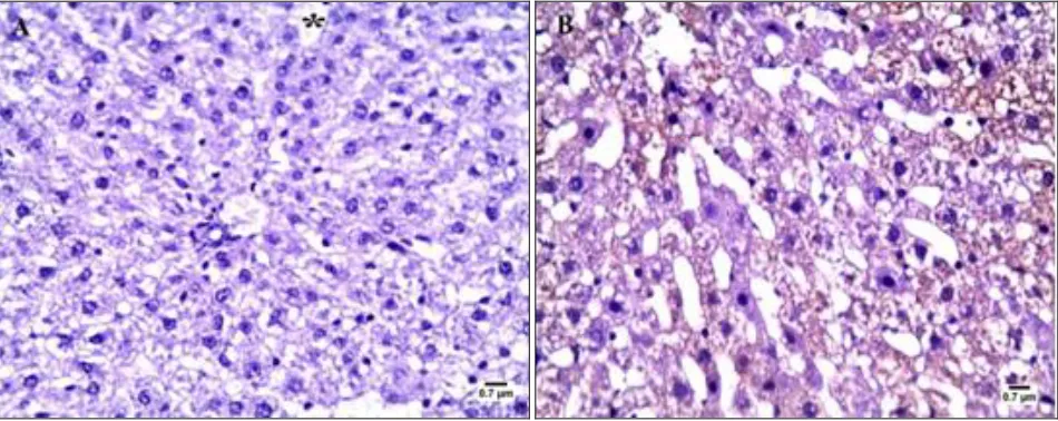
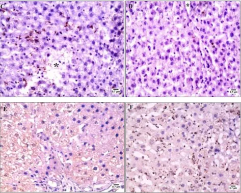
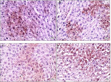
Quantitative immunohistochemical assessment and statistical analysis (Fig. 5, 6 and Histogram 1 and
Table 2) The expression of caspase-3 as well as HSP70 positive cells was significantly increased in groups II, III, V and VI as compared with control
[image:7.612.103.512.54.220.2] [image:7.612.69.544.551.740.2]
FIG. 5: PHOTOMICROGRAPHS OF IMMUNOHISTOCHEMICAL STAINING OF CASPASE-3 IN THE LIVER SHOWING; A- CONTROL GROUP I NEGATIVE FOR CASPASE-3 IMMUNOREACTION. B- HIGH EXPRESSION OF BROWN CYTPLASMIC GRANULES IN CASPASE-3 IMMUNOSTAINED HEPATOCYTES IN GROUP II. C, E & F- MODERATE EXPRESSION OF CASPASE-3 APPEARS IN GROUP III, V AND VI RESPECTIVELY. D- MILD
[image:8.612.71.544.53.432.2]
[image:9.612.75.551.54.405.2]
FIG. 6: PHOTOMICROGRAPHS OF IMMUNOHISTOCHEMICAL STAINING OF (HSP) 70 IN THE LIVER A- LIVER OF CONTROL GROUP I SHOWING VERY FEW POSITIVE HSP70 IMMUNOSTAINED HEPATOCYTES. B & C- LIVER OF GROUP II &III SHOWING HIGH EXPRESSION OF IMMUNOSTAINED HSP70 ANTIBODY D& F- LIVER OF GROUP IV AND VI SHOWING MODERATE EXPRESSION OF HSP70. E- LIVER OF GROUP V SHOWING MILD EXPRESSION OF HSP70. SCALE BAR 0.7µm.
TABLE 2: MEAN ± SE FOR EACH OF BOTH CASPASE-3 AND HSP70 POSITIVE CELLS IN THE DIFFERENT GROUPS
Group I Group II Group III Group IV Group V Group VI
Caspase 3 antibody positive cells
13.86±1.9 47.73±4.9++ 25.13±2.5++* 13.26±2.2* 24.73±2.8++* 31.20±2.7++#
HSP 70 antibody positive cells
6.8± 1.4 39. 7±1.9++ 32.10±2.3++ 15.20±3.6* 17.24±3.2++# 27.9±3.3++
++ P < 0.01 in comparison with group I (control).
+P < 0.05 in comparison with group I (control).
* P< 0.01 in comparison with group II (paracetamol).
# P < 0.05 in comparison with group II (paracetamol).
[image:9.612.136.486.590.751.2]DISCUSSION: The liver is the primary organ involved in xenobiotics metabolism and is a major target organ for chemicals and drugs. Hepatotoxicity is therefore an important endpoint in the evaluation of the effect of particular xenobiotics. Administration of paracetamol causes oxidative stress and generation of ROS, especially in the liver by Saito et al.13. Its side effects are worldwide and damage to the liver is a major complication. It could cause a significant decrease in the activities of antioxidant enzymes and increase in the amount of lipid peroxidation product in the treated rats by Madkour, and Abdel-Daim, 14.
This study was designed to compare between some Paracetamol derivatives to reach the maximum safety in its use. The low therapeutic dose used in this work was calculated according to Paget’s formula by Paget et al.15. This choice was supported with the results of some researchers who found that similar low dose caused spermatogenic cell alterations in testis by Wafaa et al.16. On the other hand other studies cited that there were no significant histopathological changes with low dose and they attributed the negative toxic effect to the different route of administration by Payasi et al.17.
In the present study the histological examination revealed that Paracetamol induced variable degrees of hepatocellular degeneration. In the present study centrilobular necrosis was observed as loss of architecture and hepatocytes exhibited highly acidophilic cytoplasm, pyknotic nuclei and irregular outlines. The expression of caspase-3 (a key executioner of apoptosis) positive cells was significantly increased in liver tissues compared to the control. These changes could be due to increased NAPQI production and lipid peroxidation as a result of mitochondrial GSH depletion by NAPQI conjugation which lead to cytochrom P450 bioactivation especially in the centrilobular cells by Oz et al. 18 and Galal et al. 19. Moreover the increased oxidative stress leads to release of more proinflammatory cytokines particularly TNF-alpha that leads o apoptotic damage by Murat et al. 20.
It is well known that liver tissue contains a relatively high content of polyunsaturated fatty acids, which are sensitive to peroxidative damage by Catala 21. In agreement with our results some
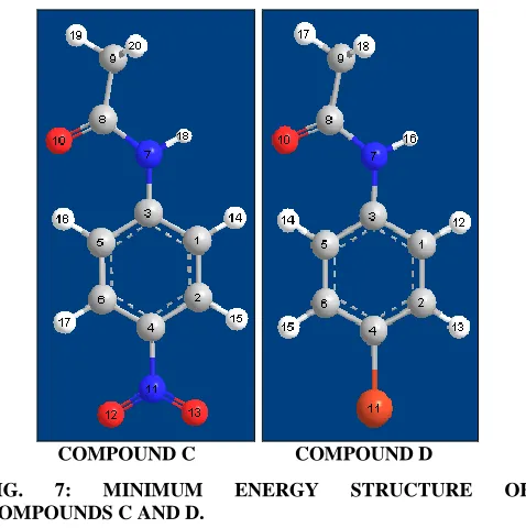
authors found that paracetamol induced necrotic and apoptotic changes in hepatocytes via initiation of the mitochondrial permeability transition. They proved that ATP synthesis by glycolysis led to switch between necrosis and apoptosis in liver cells by Kon et al. 22. Compounds C and D exhibited less degree of affection especially C. Some of the hepatic lobules appeared more or less similar to the control group while others showed signs of degeneration. Compounds A, B, D & E observed significant increase in the expression of caspase-3 positive cells in comparison to the control group while compound C observed non significant change. This amelioration of the toxic effect might be due to decrease of NAPQI production or increase the level of the antioxidant enzymes as a result of change in chemical composition.
COMPOUND C COMPOUND D
FIG. 7: MINIMUM ENERGY STRUCTURE OF COMPOUNDS C AND D.
[image:10.612.322.561.295.535.2]N O
O
N H
C O
CH3 +
NH2
NH2
O2N +
NHCOCH3
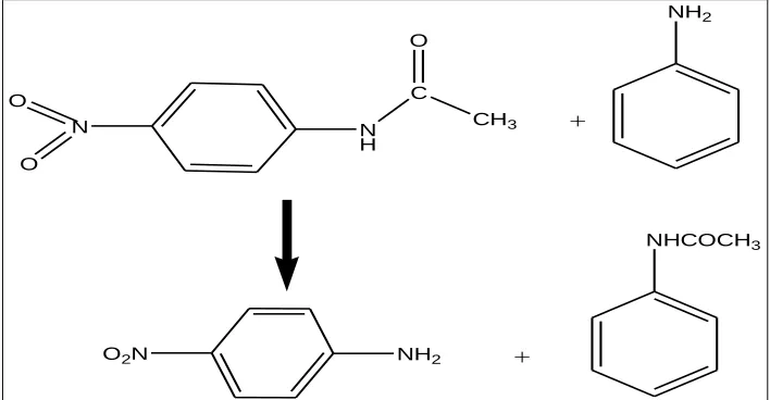
The presence of aniline product due to amylamine acetyltransferase reaction from p-nitroactanilide and it has low ionization potential, 5.5217 eV and low electron affinity 0.79512 eV, by DFT calculations; therefore it acts as an electron donor more than guanine. Hence the presence of aniline protects the DNA from the electron transfer from the nucleus.
On the other hand compound E (N-acetylanthranilic acid) (NAA) is metabolizedto 5-hydroxyanthranilic acid and anthranilamide in the rat liver and caused dilated congested central vein and capillaries with prominent vonkupffer cells in their lumens. Neovascularization between hepatic lobules were seen. These signs previously described by Saadoun et al 24 in non-cirrhotic intrahepatic portal hypertension. They might explain as sinusoidal outflow obstruction secondary to direct injury to hepatic endothelial cell.
[image:11.612.129.483.55.239.2]
FIG. 8: MINIMUM ENERGY STRUCTURE OF COMPOUND E (NAA).
HSP70 is a general antiapoptotic protein that protects the tissues from cytotoxicity induced by oxidative stress and other stress conditions by Tas et al., 8. The level of HSP70 was significantly enhanced in stressful situation by Wang et al. 25. In the present study, significant elevation in the expression of HSP70 positive cells in all Paracetamol derivatives was noticed. The sharp increase in its expression in compound B more than the others might be due to different histopathological pathways other than apoptosis. The increasing levels of HSP70 expression might be attributed to the oxidative effect of Paracetamol, which enhance its expression as a cellular defense. Thus, HSP70 expression is correlated with apoptosis, this in agreement with Morimoto et al. 26 who reported that HSP protects other proteins from unfolding, or refolds denatured proteins. It was proved that HSP70 could correct the apoptosis through inhibition of Bax activation and prevention of release of pro-apoptotic factors from the mitochondria so it was unable to affect apoptosis once caspase-3 or caspase-9 activation had occurred by Stankiewicz et al 27.
This was also supported by the present findings where our results showed positive correlation between HSP70 and caspase 3 expressions. On the other hand some authors had reported that it could act as death determinants causing decrease cytochrome C release and increase the activation of caspase-3 showed by Xanthoudakis and Nicholson
28
It was found that extracellular Hsp70 can stimulate innate immune mechanisms by promoting the expression of tumor necrosis factor α and interleukin-6 and downstream nuclear factor kappa B signaling, all of which are biologically important components of early liver regeneration as reported by Joshua et al 29.
CONCLUSION: The increased caspase-3
positivity and expression of HSP70 in rat liver tissues treated with paracetamol may be ultimately interlinked in the pathogenic network of its toxicity. The low level of these parameters was assessed as immunohistochemical evidence to support the possibility of getting safe paracetamol like compounds without hepatic damage. P-nitroacetanilide had almost no degenerative effects with more or less normal level caspase-3 and HSP70 while other compunds especially n-acetylanthranilic acid compound caused variable degree of appoptosis. Under the light of these aspects p-nitroacetanilide, compound C, could be used safely and considered as a good alternative to paracetamol. Further studies should be done to confirm this opinion at the biochemical point of view.
REFERENCES:
1. Khandelwal N, James LP, Sanders C, Larson AM, Lee WM. Unrecognized acetaminophen toxicity as a cause of indeterminate acute liver failure. Hepatology 2011; 53:567–576
2. Ucheya RE, Igweh JC. Histological changes in kidney structure following a long-term administration of paracetamol (acetaminophen) in pregnant Sprague– Dawley rats. Niger J Physiol Sci 2006; 21:77–81.
3. Al-Belooshia T, John A, Tariq S, Al-Otaiba A, Raza H. Increased mitochondrial stress and modulation of mitochondrial respiratory enzyme activities in acetaminophen-induced toxicity in mouse macrophage cells. Food Chem Toxicol 2010; 48:2624–2632.
4. Lavrik IN: Systems biology of apoptosis signaling networks. Curr Opin Biotechnol 2010; 21: 551–555. doi: 10.1016/j.copbio.2010.07.001
5. Janicke, R.U., Spregart, M.L., Wati, M.R., Porter, A.G., Caspase-3 is required for DNA fragmentation and morphological changes associated with apoptosis. J. Biol. Chem.1998; 273, 9357–9360.
6. Meki A.R., Esmail E.E.D.F., Hussein A.A., Hassanein H.M. Caspase-3 and heat shock protein-70 in rat liver treated with aflatoxin B1: Effect of melatonin. Toxicon. 2004; 43:93–100. doi: 10.1016/j.toxicon. 2003. 10.026.
7. Turturici, G., Sconzo, G., & Geraci, F. Hsp70 and its molecular role in nervous system diseases. Biochemistry research international, 2011.
8. Tas U, Ogeturk M, Meydan S, Kus I, Kuloglu T, Ilhan N, Kose E, Sarsilmaz M. Hepatotoxic activity of toluene
inhalation and protective role of melatonin Toxicol Ind Health. 2011; 27(5):465-73.
9. Steel R., Doherty J. P., Buzzard K., Clemons N., Hawkins C. J. and Anderson R. L.HSP72 inhibits apoptosis upstream of the mitochondria and not through interactions with Apaf-1. J. Biol. Chem. 2004; 279, 51490–51499. 10. Anwar El-Shahawy: DFT-Cancer Energy Barrier and
spectroscopic Studies of Aspirin Paracetamol and Some Analogues, Computational Chemistry, 2014; 2, 6-17. 11. Bancroft JD, Gamble M. Theory and practice of
histological techniques. 6th ed. Philadelphia: Churchill Livingstone, Elsevier; 2008; pp. 126, 150, 440.
12. Issa NM, El-Sherif NM: Histological and Immunohistochemical Study on the Toxic Effects of Anthracene on the lung and liver of Adult Male Albino Rats and the Possible Protective Role of Ocimum gratissimum Extract. J Cell Biol Histol 2015; 1(1): 103. 13. Saito C, Zwingmann C, Jaeschke H. Novel mechanisms of
protection against acetaminophen hepatotoxicity in mice by glutathione and N- acetylcysteine. Hepatology 2010; 51:246–254.].
14. Madkour, F. F., & Abdel-Daim, M. M: Hepatoprotective and antioxidant activity of dunaliella salina in paracetamol-induced acute toxicity in rats. Indian journal of pharmaceutical sciences, 2013; 75(6): 642.
15. Paget GE, Barnes JM Laurence DR, Bacharach AL. Toxicity test for evaluation of drug activities. Pharmacometrics. 19641st ed London and New York Academic Press: 135. In:, editors. P.
16. Wafaa BY, Sanna K, Salwa K. Effect of prolonged acetaminophen (Panadol) ingestion on the mouse liver, kidney and testis histology. Saudi J Bio Sci. 1999; 6:168– 178
17. Payasi A, Chaudhary M, Singh BM, Gupta A, Sehgal R. Sub-acute toxicity studies of paracetamolinfusion in albino Wistar rats. Int J Pharm Sci Drug. 2010; 2:142–145. 18. Oz HS, McClain CJ, Nagasawa HT, Ray MB, De Villiers
WJ, Chen TS. Diverse antioxidants protect against acetaminophen hepatotoxicity. J Biochem Mol Toxicol. 2004; 18: 361 – 368.
19. Galal RM, Zaki HF, Seif El-Nasr MM, Agha AM. Potential protective effect of honey against paracetamol-induced hepatotoxicity. Arch Iran Med. 2012; 15(11): 674 –680.
20. Murat Polat, Serkan Cerrah, Bulent Albayrak, et al., “Assessing the Effect of Leptin on Liver Damage in Case of Hepatic Injury Associated with Paracetamol Poisoning,” Gastroenterology Research and Practice, vol. 2015, Article ID 357360, 8 pages, doi: 10.1155/ 2015/357360
21. Catala A. Lipid peroxidation of membrane phospholipids generates hydroxy-alkenals and oxidized phospholipids active in physiological and/or pathological conditions. Chemistry and Physics of Lipids. 2009; 157: 1 – 11. 22. Kon K, Ikejima K, Okumura K, Aoyama T, Arai K, Takei
Y, Lemasters JJ, Sato N. Role of apoptosis in acetaminophen hepatotoxicity. J Gastroenterol Hepatol. 2007; 22 Suppl 1:S49–S52
23. Hathway, D. E., Foreign Compound Metabolism in Mammals, Royal Society of Chemistry, 1970-Medical 24. Saadoun, D., Cazals-Hatem, D., Denninger, M. H.,
Boudaoud, L., Pham, B. N., Mallet, V& Valla, D. et al. Association of idiopathic hepatic sinusoidal dilatation with the immunological features of the antiphospholipid syndrome. Gut 2004; 53(10), 1516-1519.
hepatocellular carcinoma cell line BEL-7402. World Journal of Gastroenterology 2005; 11: 5561–5564 26. Morimoto, R.T., Tissieres, A., Georgopoulos, C.: The
biology of heat-shock proteins and molecular chaperones. Cold Spring Harb. Monogr. Ser. 1994; 26, 496.
27. Stankiewicz A. R., Lachapelle G., Foo C. P., Radicioni S. M. and Mosser D. D: Hsp70 inhibits heat-induced apoptosis upstream of mitochondria by preventing Bax translocation. J. Biol. Chem. 2005; 280, 38729–38739
28. Xanthoudakis S, Nicholson DW. Heat-shock proteins as death determinants. Nat Cell Biol. 2000; Sep; 2(9):E163-5.
29. Joshua H. Wolf, Tricia R. Bhatti, Suomi Fouraschen, Shourjo Chakravorty, Liqing Wang, Sunil Kurian, Daniel Salomon, Kim M. Olthoff, Wayne W. Hancock, and Matthew H. Levine: Heat Shock Protein 70 Is Required for Optimal Liver Regeneration After Partial Hepatectomy in Mice liver transplantation 2014; 20:376–385.
All © 2013 are reserved by International Journal of Pharmaceutical Sciences and Research. This Journal licensed under a Creative Commons Attribution-NonCommercial-ShareAlike 3.0 Unported License.
This article can be downloaded to ANDROID OS based mobile. Scan QR Code using Code/Bar Scanner from your mobile. (Scanners are available on Google Playstore)
How to cite this article: