Copyright © 2002, American Society for Microbiology. All Rights Reserved.
PreservCyt Transport Medium Used for the ThinPrep Pap Test Is a
Suitable Medium for Detection of
Chlamydia trachomatis
by the
COBAS Amplicor CT/NG Test: Results of a Preliminary Study
and Future Implications
Anne Bianchi,
1François Moret,
2Jean-Marc Desrues,
3Thierry Champenois,
3Yves Dervaux,
4Orlane Desvouas,
3André Oursin,
3Dominique Quinzat,
5Roger Dachez,
5Christian Bathelier,
3and Christophe Ronsin
3*
Laboratoire Départemental de Seine-Saint-Denis, Conseil Ge´ne´ral, Bondy,
1Roche Diagnostics, Meylan,
2LCL Service de Biologie
Moléculaire, Ivry sur Seine,
3Cytyc France SARL, Villepinte,
4and Laboratoire Claude Lévy, Paris,
5France
Received 13 August 2001/Returned for modification 22 November 2001/Accepted 24 January 2002
The commercial COBAS Amplicor CT/NG test (Roche Diagnostic Systems, Meylan, France) is a sensitive
and specific method for detection of
Chlamydia trachomatis
infections. This test currently consists of using a
nucleic acid amplification method to detect
C. trachomatis
in first-void urine specimens and in endocervical
swabs collected in 2-sucrose-phosphate (2SP) transport medium. We conducted a prospective study to
deter-mine whether the automated COBAS Amplicor CT/NG test can detect
C. trachomatis
in cervical specimens
collected in PreservCyt transport medium (ThinPrep Pap Test; Cytyc Corporation, Boxborough, Mass.).
PreservCyt medium is used to preserve cervical samples before the preparation of ThinPrep slides. We
collected 1,000 cervical specimens from young women (age range, 15 to 25 years) during routine Pap smear
tests. Only specimens with normal cytology and in which the gynecologist found no clinical evidence of
urogenital infections were selected. The samples were stored in PreservCyt transport medium at 15 to 20°C.
C.
trachomatis
was detected in 22 of the 1,000 cervical specimens that had been stored in PreservCyt. To confirm
the positive samples, the test was repeated on new endocervical swab specimens collected in 2SP transport
medium. Only 9 of the 22 positive patients agreed to undergo this control, but all 9 retested positive. To
evaluate the influence of storage conditions on the sensitivity of the
C. trachomatis
PCR test, all of the positive
samples were stored at 15 to 20°C in PreservCyt transport medium and were retested every 2 weeks for 6 weeks.
C. trachomatis
was successfully amplified from all 22 specimens for the whole 6-week period. The prevalence of
C. trachomatis
infection was 2.2% in our study population. These results demonstrate that PreservCyt transport
medium is a suitable transport medium for detection of
C. trachomatis
by the COBAS Amplicor CT/NG test.
The ThinPrep Pap Test may enable gynecologists to monitor for both cervical lesions and
C. trachomatis
infections with a single endocervical specimen.
Chlamydia trachomatis
infection is one of the most common
sexually transmitted diseases and represents a major public
health problem (48). In women, untreated
C. trachomatis
in-fections cause cervicitis and pelvic inflammatory disease, which
may lead to infertility and ectopic pregnancy, and expose
new-born infants to
C. trachomatis
(37). In the early 1990s,
molec-ular diagnostic methods based on nucleic acid amplification
tests such as PCR, ligase chain reaction, and
transcription-mediated amplification were developed. These techniques
have gradually replaced traditional techniques for detection of
C. trachomatis
(5, 32, 49). Commercial DNA amplification
tests are now routinely used for diagnosis of
C. trachomatis
infections. Although these commercial tests are based on
dif-ferent nucleic acid amplification technologies, many studies
have shown that nucleic acid amplification-based tests are
more sensitive than culture-based tests (17, 28, 33, 35, 39, 40)
and that they offer the opportunity to use noninvasive clinical
samples (2, 3, 8, 12, 30, 31, 36). Therefore, nucleic acid
ampli-fication tests are considered to be a more suitable method for
screening and diagnosing chlamydial infections (6, 14, 20, 30,
35, 44, 45, 46). A large proportion of infected individuals are
asymptomatic (50% of infected men and 70 to 80% of infected
women), and these individuals serve as a reservoir for the
transmission of
C. trachomatis
(5). Thus, it is important that
C.
trachomatis
be diagnosed at an early stage and that infected
persons be treated to prevent the spread of this disease.
Var-ious
C. trachomatis
screening programs have shown that the
prevalence of
Chlamydia
infections has decreased in recent
years (1, 15, 41).
High-risk populations, such as sexually active young women,
should be screened for
C. trachomatis
and monitored for
pre-cancerous lesions of the cervix; however, the major problem is
the technical feasibility of this kind of screening program. The
ideal situation would be use of a single specimen for both
cytological monitoring and detection of a number of pathogens
so that the patient does not have to be examined repeatedly.
Papanicolaou (Pap) smear tests are recommended for
adoles-cent and young women for detection of abnormal cervical
cytology (34). In 1996, the U.S. Food and Drug Administration
approved a liquid method for collecting and preparing cervical
* Corresponding author. Mailing address: L.C.L., Service de
Biolo-gie Moléculaire, 78 Avenue de Verdun, BP 110, 94208 Ivry sur Seine,
France. Phone: 01-49-59-17-10. Fax: 01-49-59-17-98. E-mail: c.ronsin
@lablcl.com.
1749
on May 15, 2020 by guest
http://jcm.asm.org/
samples for cytological screening (ThinPrep Pap Test; Cytyc
Corporation, Boxborough, Mass.). The ThinPrep Pap Test
preserves endocervical cells in a liquid alcohol (PreservCyt
transport medium; Cytyc Corporation) to ensure optimal
mor-phological preservation. These samples are subsequently used
to prepare Pap slides, which are stained for microscopic
exam-ination. The ThinPrep Pap Test is significantly more effective
than the conventional Pap smear test for the detection of
precancerous lesions (16, 18, 22, 24, 26, 27, 29). We carried out
a prospective study to evaluate the ability of the automated
PCR COBAS Amplicor CT/NG test (9, 21) to detect
C.
tra-chomatis
in the PreservCyt transport medium used for the
ThinPrep Pap Test.
MATERIALS AND METHODS
Preliminary assays.Firstly, to assess the presence of endogenous amplification inhibitors and to determine the technical feasibility of using PreservCyt as a storage medium, 13 endocervical PreservCyt samples were selected. Seven of these specimens contained blood and were thus treated with 0.5 to 1 ml of an acetic acid solution to lyse red blood cells. These samples were processed as described below (“Pretreatment of PreservCyt specimens”). The COBAS Am-plicor CT/NG test was performed in duplicate on each lysate. Secondly, we compared the in vitro sensitivities of the PreservCyt transport medium and the classical 2SP transport medium (0.2 M sucrose–0.02 M phosphate; QUELAB Laboratories Inc., Montréal, Canada) for detection ofC. trachomatis. Four separate batches of theC. trachomatisserotype D reference strain (ATCC VR 885; stored at⫺80°C) were serially diluted with 2SP and with PreservCyt trans-port medium such that there were 103to 10⫺2inclusion-forming units (IFU)/ml.
This reference strain stock was previously used to compare the sensitivities of all
C. trachomatisdiagnostic tests commercially available in France (4). Negative controls, consisting of the 2SP medium or the PreservCyt transport medium alone, were included. Each dilution was tested in duplicate by the COBAS Amplicor CT/NG test.
Patient population.Specimens were obtained between May 2000 and Novem-ber 2000 from consenting patients registered with different general practitioners, dermatologists, gynecologists, and family planning centers. We collected 1,000 PreservCyt specimens from young women (age range, 15 to 25 years) during Pap screening (ThinPrep Pap Test). Samples were selected according to two criteria: (i) absence of clinical signs of urogenital infection, including genital or pelvic pain, dysuria, and urethritis, and (ii) absence of atypical cells such as inflamma-tory cells and cervical cancer cells or precursor lesions (ASCUS [atypical squa-mous cells of undetermined significance], low-grade squasqua-mous intraepithelial lesions, or high-grade squamous intraepithelial lesions).
Preparation of ThinPrep slides from PreservCyt specimens.PreservCyt spec-imens were collected by inserting a cytobrush into the endocervical canal. The cytobrush was immediately placed in a vial of PreservCyt transport medium. These specimens were stored at 15 to 20°C and transported to the laboratory within 24 h of collection. In the cytology laboratory (Laboratoire Claude Lévy, Paris, France), a ThinPrep 2000 processor (Cytyc Corporation) was used to prepare slides (one slide per patient). The ThinPrep 2000 processor automati-cally prepares cytological slides from cells suspended in PreservCyt transport medium. It disperses the samples and transfers cells onto a microscopic glass slide in a 20-mm-diameter circle. The slide is then automatically deposited into a 95% ethanol bath before being stained with Papanicolaou stain. ThinPrep slides were observed by two independent cytopathologists.
Cytological reports were formulated according to the Bethesda System, in-cluding evaluation of correct sampling of the transformation zone (presence of metaplastic and/or columnar cells). PreservCyt specimens were stored at 15 to 20°C for as long as 6 weeks, in case the sample had to be retested.
Pretreatment of PreservCyt specimens.PreservCyt specimens were tested by PCR within 1 week of collection. The PreservCyt transport medium containing residual endocervical cells was vortexed vigorously. For each specimen, two 1-ml samples were transferred into two 2-ml polypropylene tubes, each containing 1 ml of PreservCyt transport medium. The tubes were centrifuged at 12,500⫻gfor 15 min at 20°C. Supernatants were discarded, each cellular pellet was resus-pended in 500l of 2SP transport medium, and cellular suspensions were pooled in a single tube (final volume, 1 ml). One hundred microliters of this suspension was added to 100l of CT/NG Lysis Buffer. After a 10-min incubation at room temperature, 200l of CT/NG Specimen Diluent was added to the lysate. The
contents were mixed well by vortexing, and tubes were incubated at 90°C for 10 min. Lysates were centrifuged at 12,500⫻gfor 10 min. A COBAS Amplicor CT/NG test was performed with 50l of lysate, according to the manufacturer’s instructions.
Control endocervical swab specimens in 2SP transport medium.To confirm the positive results obtained with the PreservCyt transport medium, patients with positive results (C. trachomatis A660,⬎0.8) were asked by their gynecologists to
give a new endocervical swab (alginate swabs) specimen collected by standard procedures into 2SP medium. The swabs were stored at 2 to 8°C and tested within 3 days of collection. One hundred microliters of CT/NG Lysis Buffer was mixed with 100l of the sample in 2SP transport medium and incubated at room temperature for 10 min. Two hundred microliters of CT/NG Specimen Diluent was added to each tube, and tubes were incubated at room temperature for 10 min. The COBAS Amplicor CT/NG test was performed with 50l of lysate, according to the manufacturer’s instructions.
Amplification and detection ofC. trachomatisby the COBAS Amplicor CT/NG PCR test.We transferred 50l of lysate to A-tubes containing 50l of Working Master Mix. The A-tubes were placed in a COBAS Amplicor thermocycler. An internal control (IC) was included with each amplification reaction to monitor inhibition and was coamplified with theC. trachomatistarget DNA. AmpErase was incorporated to prevent carryover contamination by uracil-N-glycosylase. The COBAS Amplicor system automatically denatures the amplified DNA tar-get, hybridizes the amplicons with the target-specific probes bound to magnetic microparticles, and detects the amplicon/probe complexes by a colorimetric reaction. TheC. trachomatistarget and the IC were detected in separate reac-tions with specific oligonucleotide capture probes.
Interpretation of results.Specimens yieldingC. trachomatissignals above the positive cutoff (C. trachomatisA660,⬎0.8) were interpreted as positive.
Speci-mens in which theC. trachomatissignal was below the negative cutoff were interpreted as negative if the IC signal was above the assigned cutoff (C. tracho-matisA660,⬍0.2; ICA660,⬎0.2). Specimens for which theC. trachomatisresults
were between the negative and positive cutoffs (0.2⬍C. trachomatisA660⬍0.8)
were considered to be in a gray zone if the ICA660was⬎0.2. Specimens yielding
signals below the cutoff values (A660⬍0.2) for bothC. trachomatisand the IC
were considered to contain inhibitory substances. For specimens containing inhibitory substances or in the gray zone, another aliquot of the original speci-men was retested in duplicate. These specispeci-mens were considered to be positive if at least one of the duplicates yielded aC. trachomatisA660of⬎0.8. They were
interpreted as negative if the two repeat tests yielded aC. trachomatisA660of
⬍0.2 and the IC signals were above the assigned cutoff (ICA660,⬎0.2). If the two
repeat tests yielded aC. trachomatisA660of⬍0.2 and the IC signal was below the
assigned cutoff (ICA660,⬍0.2), the specimens were considered to contain
in-hibitory substances and were excluded.
Evaluation of storage conditions in PreservCyt vials.PreservCyt specimens in which theC. trachomatissignal was above the positive cutoff (C. trachomatisA660
of⬎0.8) were stored at 15 to 20°C for 6 weeks. For each positive PreservCyt specimen, the PCR pretreatment was repeated as described above and the lysates were retested by the COBAS Amplicor CT/NG test 2, 4, and 6 weeks after collection (days 15, 30, and 45).
RESULTS
Preliminary assays: preparation of PreservCyt specimens
and in vitro sensitivity assay.
Some preliminary tests were
necessary to evaluate the technical feasibility of conducting a
prospective study to detect
C. trachomatis
by the ThinPrep Pap
test. One major problem with nucleic acid amplification tests is
the presence of potential inhibitors in the biological samples,
which can lead to false-negative results. Firstly, PreservCyt
transport medium is an alcohol-based solution that may inhibit
PCR. Secondly, for an optimal cytological examination, any
bloody PreservCyt specimens must be pretreated with 0.5 to 1
ml of a pure acetic acid solution to lyse the red blood cells
(hemoglobin is known to be a potential inhibitor of PCR). We
evaluated the efficiency of the pretreatment procedure at
re-moving potential PCR inhibitors from PreservCyt specimens.
The IC was detected in all 13 PreservCyt specimens tested by
the COBAS Amplicor CT/NG test (IC
A
660,
⬎
0.8). These
encouraging results led us to compare the detection sensitivity
on May 15, 2020 by guest
http://jcm.asm.org/
of the PreservCyt transport medium with that of the 2SP
trans-port medium in a dilution assay using a titrated solution of
purified infectious elementary bodies of
C. trachomatis
(sero-var D, VR 885).
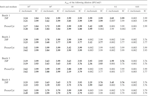
The results obtained were highly reproducible for duplicate
assays performed with each of the four batches (same assay
run) for the 10
3- to 10-IFU/ml dilutions in both types of
trans-port medium. At these dilutions, all assays performed with 2SP
and with PreservCyt gave positive signals for
C. trachomatis
(Table 1). At the 1-IFU/ml dilution, one batch gave positive
signals for
C. trachomatis
with both types of transport medium.
The other three batches all gave negative signals for
C.
tracho-matis
with the PreservCyt medium, and all but one gave
pos-itive signals with the 2SP medium (Table 1).
None of the assays performed at the 0.01-IFU/ml dilution
gave positive signals for
C. trachomatis
. The IC was detected in
all samples, and all of the negative controls gave negative
results. These conditions allowed us to determine the
thresh-old of sensitivity (the highest dilution tested that gave positive
duplicate results with the four batches of dilutions) to be the
10-IFU/ml dilution in PreservCyt medium.
Results of the COBAS Amplicor CT/NG test on PreservCyt
specimens.
C. trachomatis
was detected in 22 of the 1,000
specimens (2.2%) (Table 2). No inhibition was observed in any
of the 978 specimens with negative results as assessed by the IC
of the COBAS Amplicor CT/NG test.
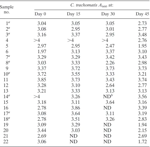
Storage conditions.
For each positive PreservCyt specimen,
a COBAS Amplicor CT/NG test was performed 15, 30, and 45
days after collection. All of the 22 positive specimens yielded
positive results at each of these time points (
C. trachomatis
A
660,
⬎
0.8) (Table 3). Most of the PreservCyt specimens
re-tested gave very similar optical density values at each time
point. Optical density values decreased by one
A
660unit for
just five PreservCyt specimens (samples 4, 5, 19, 20, and 22)
(Table 3). The storage conditions (45 days at 15 to 20°C) could
be responsible for a slight degradation in DNA, which would
affect the sensitivity of PCR.
Confirmation of infection by testing of independent
endo-cervical swabs and prevalence of urogenital chlamydial
infec-tion.
Only 9 of the 22 positive patients agreed to be retested.
All nine endocervical swab specimens were positive (
C.
tracho-matis
A
660,
⬎
0.8). Because PreservCyt transport medium is
conventionally used to monitor endocervical specimens for
cervical cancer and because all specimens with abnormal
cy-tology or in which there was evidence of urogenital infection
were excluded from this study, we can consider our population
asymptomatic. The prevalence of
C. trachomatis
in this
popu-TABLE 1. Comparison of the sensitivities of 2SP and PreservCyt (ThinPrep Pap test) for detection of
C. trachomatis
by the COBAS
Amplicor CT/NG test
Batch and medium
A660of the following dilution (IFU/ml)a:
103 102 10 1 0.1 0.01
C. trachomatis IC C. trachomatis IC C. trachomatis IC C. trachomatis IC C. trachomatis IC C. trachomatis IC
Batch 1
2SP
3.24
3.84
3.54
3.99
3.99
3.99
3.99
3.99
2.03
3.99
0.003
3.99
3.21
3.99
3.62
3.99
3.85
3.99
3.99
3.99
0.007
3.99
0.003
3.99
PreserCyt
3.17
3.84
3.72
3.99
3.99
3.99
3.42
3.99
0.001
3.99
0.001
3.99
3.28
3.48
3.84
3.84
3.99
1.00
3.99
0.002
3.99
0.002
3.99
Batch 2
2SP
3.30
3.99
3.78
3.99
3.99
3.99
0.002
3.99
0.002
3.99
0.002
3.76
3.23
3.99
3.60
3.77
3.99
3.77
3.12
3.77
0.002
3.77
0.002
3.77
PreserCyt
3.42
3.99
3.99
3.99
3.42
3.99
0.002
3.99
0.002
3.99
0.003
3.99
3.62
3.99
3.84
3.99
3.99
3.99
0.003
3.99
0.002
3.99
0.002
3.93
Batch 3
2SP
3.19
3.99
3.63
3.99
3.63
3.93
3.93
3.93
1.99
3.76
0.002
3.76
3.19
3.93
3.63
3.63
3.93
3.76
2.36
3.93
0.001
3.76
0.001
3.76
PreserCyt
3.62
3.99
3.99
3.99
3.80
3.99
0.002
3.99
0.001
3.99
0.001
3.99
3.62
3.99
3.80
3.99
2.19
3.79
0.002
3.77
0.001
3.77
0.003
3.77
Batch 4
2SP
3.33
3.93
3.63
3.63
3.75
3.93
2.25
3.76
3.45
3.76
0.002
3.76
3.33
3.93
3.39
3.93
3.75
3.76
3.99
3.63
0.002
3.76
0.002
3.76
PreserCyt
3.62
3.99
3.79
3.79
3.99
3.99
0.003
3.99
0.002
3.79
0.002
3.79
3.49
3.99
3.99
3.79
3.79
3.79
0.003
3.99
0.002
3.79
0.003
3.79
aResults for duplicate assays are shown. Positive results are boldfaced..
on May 15, 2020 by guest
http://jcm.asm.org/
[image:3.587.45.547.92.415.2]lation was 2.2% (Table 2), with the highest prevalence in
women aged 19 to 25 years (2.3%; 20 out of 884).
DISCUSSION
The aim of this study was to evaluate the technical feasibility
of using a routine PCR test to detect
C. trachomatis
in cervical
specimens collected in the PreservCyt transport medium, a
collection medium for the Pap smear (ThinPrep Pap Test). We
evaluated the technical feasibility (DNA isolation procedure)
and the in vitro sensitivity of
C. trachomatis
detection in
Cyt transport medium in preliminary assays. Because
Preserv-Cyt transport medium is an alcohol-based solution and because
endocervical cells are collected with cytobrushes, which induce
bleeding, we adapted the treatment of PreserCyt specimens in
an attempt to remove potential PCR inhibitors (COBAS
Am-plicor CT/NG test). The COBAS AmAm-plicor IC was detected in
all PreservCyt specimens tested (bloody and nonbloody
sam-ples), indicating that there were no inhibitors after the sample
preparation procedure.
We used four separate batches of the
C. trachomatis
sero-type D reference strain that had been serially diluted in 2SP
and in PreservCyt transport medium to compare the in vitro
sensitivity and reproducibility of the PCR COBAS Amplicor
CT/NG test for detection of
C. trachomatis
. The threshold of
sensitivity obtained with titrated dilutions in PreservCyt
me-dium was 10 IFU/ml. This means that exactly 0.125 IFU of
C.
trachomatis
was tested in the assay. In France, a group of
experts used the same
C. trachomatis
strain and protocol to
compare the sensitivities of all of the commercially available
C.
trachomatis
diagnostic tests in 1997. They concluded that the
sensitivity threshold does not have to be higher than 100
IFU/ml (4). It is difficult to explain the difference in sensitivity
observed between the two types of medium.
It is possible that the pretreatment step (centrifugation and
resuspension in 2SP) carried out before the dilutions in
Preser-Cyt influenced the sensitivity. This pretreatment step was
car-ried out because we wanted to treat the PreservCyt dilutions in
the same way as the PreservCyt specimens. The pretreatment
may reduce the amount of material present before the nucleic
acid extraction step, which would in turn reduce the number of
DNA copies in the amplification mixture. This might explain
the loss of reproducibility observed with the higher dilutions.
The dilutions prepared in 2SP medium are not subjected to
this pretreatment step (they are lysed immediately). We are
currently studying the pretreatment step involved in the
prep-aration of PreservCyt specimens using both quantitative
real-time PCR and the COBAS Amplicor CT/NG test for detection
of
C. trachomatis
.
Based on the results of our preliminary assays, we decided to
screen 1,000 PreservCyt specimens with the COBAS Amplicor
CT/NG test. PreservCyt specimens were collected from a
pop-ulation of women (age range, 15 to 25 years) during routine
Pap screening. PreservCyt specimens were selected according
to two criteria: normal cytology and the absence of clinical
symptoms of infection mentioned by the practitioners. Thus,
this population could be considered homogeneous and
asymp-tomatic. Of the 1,000 endocervical specimens collected in
PreservCyt transport medium, 22 were positive (prevalence,
2.2%). The specificity of diagnosis of chlamydial infection
could be confirmed for only nine of these women, who agreed
to give another endocervical sample under classical conditions
(alginate swabs, 2SP transport medium). The COBAS
Ampli-cor test confirmed that all nine of these patients were positive
for
Chlamydia
. For all 1,000 PreservCyt specimens tested, no
inhibition was observed, as assessed by the IC of the COBAS
Amplicor test.
The prevalence of
C. trachomatis
infection in this
asymptom-atic population of women was 2.2%, with a higher prevalence
for women aged 19 to 25 years. Our laboratory routinely
diag-TABLE 2. Performance of COBAS Amplicor CT/NG test for
detection of
C. trachomatis
in ThinPrep Pap test samples
aAge (yr) of female population (no. of persons)b
No. of PCR results Specimens with inhibition Positive Negative
15 (
n
⫽
5)
1
4
None
16 (
n
⫽
13)
13
None
17 (
n
⫽
31)
1
30
None
18 (
n
⫽
67)
67
None
19 (
n
⫽
76)
76
None
20 (
n
⫽
130)
5
125
None
21 (
n
⫽
132)
4
128
None
22 (
n
⫽
117)
4
113
None
23 (
n
⫽
150)
2
148
None
24 (
n
⫽
124)
3
121
None
25 (
n
⫽
155)
2
153
None
Total (
n
⫽
1,000)
22
978
None
aNone of the women tested showed clinical signs of urogenital infection, and
cytological results were normal for all specimens.
bC. trachomatisPCR results were positive for 2 of the 116 15- to 18-year-olds
[image:4.587.44.284.94.246.2]tested (prevalence, 1.7%) and for 20 of the 884 19- to 25-year-olds tested (prevalence, 2.3%).
TABLE 3. Impact of storage conditions on the performance of the
COBAS Amplicor CT/NG test
Sample no.
C. trachomatis A660at:
Day 0 Day 15 Day 30 Day 45
1
a3.04
3.05
3.05
2.73
2
a3.08
2.95
3.01
2.77
3
a3.16
3.37
2.95
3.48
4
⬎4
⬎4
⬎4
2.76
5
2.97
2.95
2.47
1.95
6
1.97
3.13
3.37
3.10
7
a3.29
3.29
3.42
3.43
8
a3.03
3.33
2.26
2.98
9
3.37
3.72
3.73
3.73
10
a3.72
3.55
3.33
3.21
11
3.85
3.73
3.43
3.74
12
3.28
3.10
2.64
2.77
13
3.21
3.33
3.13
3.13
14
a⬎4
3.26
ND
b3.56
15
3.18
3.11
3.64
3.16
16
2.78
3.86
ND
3.39
17
a3.08
3.64
3.11
3.19
18
a2.78
3.51
3.26
2.83
19
3.09
3.29
ND
1.94
20
3.44
3.03
ND
2.15
21
2.69
ND
ND
2.69
22
3.06
ND
ND
1.72
aFor this sample, a second independent PCR test was performed on an
endocervical swab specimen collected in 2SP.
bND, not determined.
on May 15, 2020 by guest
http://jcm.asm.org/
[image:4.587.44.284.468.703.2]noses
C. trachomatis
from endocervical swab specimens
col-lected in 2SP transport medium. The endocervical swab
spec-imens tested routinely and the PreservCyt specspec-imens tested in
this study were collected from women living in the same
geo-graphic areas. Last year, 7,335 endocervical swab specimens
were routinely analyzed by the COBAS Amplicor CT/NG test;
232 of the specimens tested were positive (3.01%). Between
January and June 2001, 121 specimens out of 6,400 were
pos-itive (1.92%). These prevalences are similar to that observed in
our study on PreservCyt specimens and are in agreement with
those reported previously in France (7, 15, 47). Although the
prevalence observed in our study is similar to that reported in
different studies in the same geographic area, we are aware
that in a low-risk population (prevalence of
⬍
5%), the positive
predictive value decreases and a nonculture test must be
con-firmed by a second method (Clinical Practice Guidelines). We
are currently examining more samples from the youngest
pa-tients (age range, 15 to 20 years) to see if the prevalence
changes.
Because PreservCyt specimens are stored for 6 weeks at 15
to 20°C in the cytopathology laboratory in case the results are
ambiguous and new slides need to be made, we evaluated the
influence of storage conditions on PCR sensitivity. We found
that PreservCyt specimens can be stored for 6 weeks under
these conditions without affecting the sensitivity of DNA
am-plification. For all positive PreservCyt specimens,
C.
trachoma-tis
target DNA could be successfully amplified even after 6
weeks of storage. These results confirm the findings of a
vious study showing that PreservCyt transport medium
pre-serves the integrity of nucleic acids (10). These authors used
cord blood lymphocytes, cancer cell lines, or clinical cervical
samples collected in PreservCyt transport medium to show that
RNA integrity was still intact after storage at room
tempera-ture or at 4°C for 24 h. Even after 1 month at 4°C in PreservCyt
transport medium, Northern blotting detected intact 28S and
18S RNAs.
Although our results prove that PreservCyt transport
me-dium is a suitable collection meme-dium for routine screening of
C. trachomatis
infections with the COBAS Amplicor CT/NG
test, other recent studies have reported the technical feasibility
of detecting
C. trachomatis
from the ThinPrep Pap Test by use
of molecular methods (DNA probe or ligase chain reaction
test) and by use of a direct fluorescence assay (19, 25).
More-over, many studies have evaluated the detection of human
papillomavirus (HPV) DNA in the residual sample volume of
liquid-based Pap tests (11, 42, 43). In clinical practice, HPV is
currently detected by molecular methods (in situ hybridization,
Hybrid Capture Test, or PCR) to identify women infected with
high-risk HPVs or women with equivocal cervical lesions (50).
Finally, a recent study has demonstrated that PCR methods
can be used to detect herpes simplex virus in ThinPrep cervical
cytologic specimens and that this test is useful when the
mi-crocopic diagnosis is equivocal (13).
This is the first evaluation of the COBAS Amplicor CT/NG
test for detection of
C. trachomatis
by the fluid-based ThinPrep
Pap method. Our preliminary data suggest that the PCR test
for detection of
C. trachomatis
can be used to screen
Preserv-Cyt specimens from populations of women who undergo
an-nual Pap smear tests. Cervical dysplasia can occur during
ad-olescence, especially when women have multiple sexual
partners. This population is exposed to sexually transmitted
diseases such as
Chlamydia
and HPV infections, especially
types 16 and 18, which are a high risk factor for invasive cancer
in adults (23, 37, 38).
The ThinPrep Pap Test can be used both to monitor for
cervical cancer and to screen for
C. trachomatis
with the same
specimen, without repeatedly examining the patient. The
com-bination of DNA amplification tests is a new and promising
approach, which can be used to detect multiple pathogens
(such as HPV, herpes simplex virus,
C. trachomatis
,
Neisseria
gonorrhoeae
, and
Mycoplasma
spp.) from a single PreservCyt
specimen. This will lead to important cost savings and benefit
to the patient.
ACKNOWLEDGMENTS
We thank Cytyc France SARL and Roche Diagnostics, Meylan,
France, for supplying the
Chlamydia
kits.
REFERENCES
1.Addiss, D. G., M. L. Vaughn, D. Ludka, J. Pfister, and J. P. Davis.1993. Decreased prevalence ofChlamydia trachomatisinfection associated with a selective screening programme in family planning clinics in Wisconsin. Sex. Transm. Dis.20:28–34.
2.Bassiri, M., H. Y. Hu, M. A. Domeika, J. Burczak, L. O. Svensson, H. H. Lee, and P. A. Mardh.1995. Detection ofChlamydia trachomatisin urine speci-mens from women by ligase chain reaction. J. Clin. Microbiol.33:898–900. 3.Bauwens, J. E., A. M. Clark, M. J. Loeffelholz, S. A. Herman, and W. E. Stamm.1993. Diagnosis ofChlamydia trachomatisurethritis in men by poly-merase chain reaction assay of first-catch urine. J. Clin. Microbiol.31:3013– 3016.
4.Bianchi, A., B. De Barbeyrac, C. Bebear, C. Buffet-Janvresse, F. Eb, C. Janot, P. Maisonneuve, M. L. Migueres, J. Orfila, C. Scieux, and J. M. Alonso.
1998. Multi-laboratory comparison of 28 commercially availableChlamydia trachomatisdiagnostic tests, p. 587–590.InR. S. Stephens et al. (ed.), Pro-ceedings of the Ninth International Symposium on Human Chlamydial In-fection. International Chlamydia Symposium, San Francisco, Calif. 5.Black, C.1997. Current methods of laboratory diagnosis ofChlamydia
tra-chomatisinfections. Clin. Microbiol. Rev.10:160–184.
6.Buimer, M., G. J. J. van Doornum, S. Ching, P. G. H. Peerbooms, P. K. Plier, D. Ram, and H. H. Lee.1996. Detection ofChlamydia trachomatisand
Neisseria gonorrhoeaeby ligase chain reaction-based assays with clinical spec-imens from various sites: implications for diagnostic testing and screening. J. Clin. Microbiol.34:2395–2400.
7.Chauffert, O., E. Laurent, P. Sednaoui, P. Gouëzel, and V. Goulet.1997. Surveillance des infections àC. trachomatispar un réseau de laboratoires (RENACHLA 1995). B. E. H.15:64–65.
8.Chernesky, M. A., D. Jang, H. Lee, J. D. Burczak, H. Hu, J. Sellors, S. J. Toamzic-Allen, and J. B. Mahony.1994. Diagnosis ofChlamydia trachomatis
infections in men and women by testing first-void urine by ligase chain reaction. J. Clin. Microbiol.32:2682–2685.
9.DiDomenico, N., H. Link, R. Knobel, T. Caratsch, W. Weschler, Z. G. Loewy, and M. Rosenstraus.1996. COBAS AMPLICOR: a fully automated RNA and DNA amplification and detection system for routine diagnostic PCR. Clin. Chem.42:1915–1923.
10.Dimulescu, I., E. R. Unger, D. R. Lee, W. C. Reeves, and S. D. Vernon.1998. Characterization of RNA in cytologic samples preserved in a methanol-based collection solution. Mol. Diagn.3:67–71.
11.Ferenczy, A., E. Franco, J. Arseneau, T. C. Wright, and R. M. Richart.1996. Diagnostic performance of Hybrid Capture human papillomavirus deoxyri-bonucleic acid assay combined with liquid-based cytologic study. Am. J. Obstet. Gynecol.175:651–656.
12.Ferrero, D. V., H. N. Meyers, D. E. Schultz, and S. A. Willis.1998. Perfor-mance of the Gen-Probe AMPLIFIED Chlamydia TrachomatisAssay in detectingChlamydia trachomatisin endocervical and urine specimens from women and urethral and urine specimens from men attending sexually trans-mitted disease and family planning clinics. J. Clin. Microbiol.36:3230–3233. 13.Fiel-Gan, M. D., C. F. Villamil, S. R. Mandavilli, M. E. Ludwig, and G. J. Tsongalis.1999. Rapid detection of HSV from cytologic specimens collected into ThinPrep fixative. Acta Cytol.43:1034–1038.
14.Goessens, W. H. F., J. W. Mouton, W. I. van der Meijden, S. Deelen, T. H. van Rijsoort-Vos, N. Lemmens-den Toom, H. A. Verbrugh, and R. Verk-ooyen.1997. Comparison of three commercially available amplification as-says, AMP CT, LCx, and COBAS AMPLICOR, for detection ofChlamydia trachomatisin first-void urine. J. Clin. Microbiol.35:2628–2633.
15.Goulet, V., E. Laurent, and A. Bianchi.1999. Les Chlamydioses uro-génitales en France en 1997 Reseau RENACLA. B. E. H.16:61–63.
on May 15, 2020 by guest
http://jcm.asm.org/
16.Guidos, B. J., and S. M. Selvaggi.1999. Use of the Thin Prep Pap Test in clinical practice. Diagn. Cytopathol.20:70–73.
17.Hook, E. W., III, K. Smith, C. Mullen, J. Stephens, L. Rinehardt, M. S. Pate, and H. Lee.1997. Diagnosis of genitourinaryChlamydia trachomatis infec-tions by using the ligase chain reaction on patient-obtained vaginal swabs. J. Clin. Microbiol.35:2133–2135.
18.Hutchinson, M. L., P. Agarwal, T. Denault, B. Berger, and E. S. Cibas.1992. A new look at cervical cytology. ThinPrep multicenter trial results. Acta Cytol.36:499–504.
19.Inhorn, S. L., P. J. Wand, T. C. Wright, K. D. Hatch, J. Hallum, and B. B. Lentrichia. 2001.Chlamydia trachomatis and Pap testing from a single, fluid-based sample. A multicenter study. J. Reprod. Med.46:237–242. 20.Johnson, R. E., T. A. Green, J. Schachter, R. B. Jones, E. Hook, C. M. Black,
D. H. Martin, M. E. St Louis, and W. E. Stamm.2000. Evaluation of nucleic acid amplification tests as reference tests forChlamydia trachomatis infec-tions in asymptomatic men. J. Clin. Microbiol.38:4382–4386.
21.Jungkind, D., S. DiRenzo, K. G. Beavis, and N. S. Silverman.1996. Evalu-ation of automated COBAS AMPLICOR PCR system for detection of several infectious agents and its impact on laboratory management. J. Clin. Microbiol.34:2778–2783.
22.Koss, L. G.2000. Utility of liquid-based cytology for cervical carcinoma screening. Cancer90:67–69.
23.Lavin, C., E. Goodman, S. Perlman, L. S. Kelly, and S. J. Emans.1997. Follow-up of abnormal Papanicolaou smears in a hospital-based adolescent clinic. J. Pediatr. Adolesc. Gynecol.10:141–145.
24.Lee, K. R., R. Ashfaq, G. G. Birdsong, M. E. Corkill, K. M. McIntosh, and S. L. Inhorn.1997. Comparison of conventional Papanicolaou smears and a fluid-based, thin-layer system for cervical cancer screening. Obstet. Gynecol.
90:278–284.
25.Lentrichia, B. B., S. S. Hecht, D. Lapen, and M. K. Corkill.1998. Potential for routine concurrent determination ofChlamydiaand cervical abnormal-ities by single fluid-based sampling. Prim. Care Update Ob. Gyns.5:149–150. 26.Linder, J., and D. Zahniser.1997. The ThinPrep Pap test. A review of
clinical studies. Acta Cytol.41:30–38.
27.Linder. J., and D. Zahniser.1988. ThinPrep Papanicolaou testing to reduce false-negative cervical cytology. Arch. Pathol. Lab. Med.122:139–144. 28.Loeffelholz, M. J., C. A. Lewinski, S. R. Silver, A. P. Purohit, S. A. Herman,
D. A. Buonagurio, and E. A. Dragon.1992. Detection ofChlamydia tracho-matisin endocervical specimens by polymerase chain reaction. J. Clin. Mi-crobiol.30:2847–2851.
29.Monsonego. J., A. Autillo-Touati, C. Bergeron, R. Dachez, J. Liaras, J. Saurel, L. Zerat, P. Chatelain, and C. Mottot.2001. Liquid-based cytology for primary cervical cancer screening: a multi-centre study. Br. J. Cancer
84:360–366.
30.Morre, S. A., I. G. M. Van Valkengoed, R. M. Moes, A. J. P. Boeke, C. J. L. M. Meijer, and A. J. C. Van den Brule.1999. Determination of
Chlamydia trachomatisprevalence in an asymptomatic screening population: performances of the LCx and COBAS Amplicor tests with urine specimens. J. Clin. Microbiol.37:3092–3096.
31.Mouton, J. W., R. Verkooyen, W. I. van der Meijden, T. H. van Rijsoort-Vos, W. H. F. Goessens, J. A. J. W. Kluytmans, S. D. A. Deelen, A. Luijendijk, and H. A. Verbaugh.1997. Detection ofChlamydia trachomatisin male and female urine specimens by using the amplifiedChlamydia trachomatistest. J. Clin. Microbiol.35:1369–1372.
32.Ostergaard, L.1999. Diagnosis of urogenitalChlamydia trachomatis infec-tion by use of DNA amplificainfec-tion. APMIS Suppl. Rev.89:5–36.
33.Pasternack, R., P. Vuorinen, and A. Miettinen.1997. Evaluation of the Gen-ProbeChlamydia trachomatistranscription-mediated amplification as-say with urine specimens from women. J. Clin. Microbiol.35:676–678.
34.Perlman, S. E.1999. Pap smears: screening, interpretation, treatment. Ado-lesc. Med.10:243–254.
35.Puolakkainen, M., E. Hiltunen-Back, T. Reunala, S. Suhonen, P. Lähteen-mäki, M. Lehtinen, and J. Paavonen.1998. Comparison of performances of two commercially available tests, a PCR assay and a ligase chain reaction test, in detection of urogenitalChlamydia trachomatisinfection. J. Clin. Microbiol.36:1489–1493.
36.Quinn, T. C., L. Welsh, A. Lentz, K. Crotchfelt, J. Zenilman, J. Newhall, and C. Gaydos.1996. Diagnosis by AMPLICOR PCR ofChlamydia trachomatis
infection in urine samples from women and men attending sexually trans-mitted disease clinics. J. Clin. Microbiol.34:1401–1406.
37.Reddy, S. P., S. R. Yeturu, and R. Slupik.1997.Chlamydia trachomatisin adolescents: a review. J. Pediatr. Adolesc. Gynecol.10:59–72.
38.Roye, C. F.1992. Abnormal cervical cytology in adolescents: a literature review. J. Adolesc. Health13:643–650.
39.Schachter, J., W. E. Stamm, T. C. Quinn, W. W. Andrews, J. D. Burczak, and H. H. Lee.1994. Ligase chain reaction to detectChlamydia trachomatis
infection of the cervix. J. Clin. Microbiol.32:2540–2543.
40.Schepetiuk, S., T. Kok, L. Martin, R. Waddell, and G. Higgins.1997. De-tection of Chlamydia trachomatisin urine samples by nucleic acid tests: comparison with culture and enzyme immunoassay of genital swab speci-mens. J. Clin. Microbiol.35:3355–3357.
41.Scholes, D., A. Stergachis, F. Heidrich, H. Andrilla, K. K. Holmes, and W. Stamm.1996. Prevention of pelvic inflammatory disease by screening for cervical chlamydial infection. N. Engl. J. Med.334:1362–1366.
42.Sherman, M. E., M. H. Schiffman, A. T. Lorincz, R. Herrero, M. L. Hutchin-son, C. Bratti, D. Zahniser, J. Morales, A. Hildesheim, K. Helgesen, D. Kelly, M. Alfaro, F. Mena, I. Balmaceda, L. Mango, and M. Greenberg.1997. Cervical specimens collected in liquid buffer are suitable for both cytologic screening and ancillary human papillomavirus testing. Cancer81:89–97. 43.Sherman, M. E., M. Mendoza, K. R. Lee, R. Ashfaq, G. G. Birdsong, M. E.
Corkill, K. M. McIntosh, S. L. Inhorn, D. J. Zahniser, G. Baber, C. Barber, and M. H. Stoler.1998. Performance of liquid-based, thin-layer cervical cytology: correlation with reference diagnoses and human papillomavirus testing. Mod. Pathol.11:837–843.
44.Toye, B., R. W. Peeling, P. Jessamine, P. Claman, and I. Gemmill.1996. Diagnosis ofChlamydia trachomatisinfections in asymptomatic men and women by PCR assay. J. Clin. Microbiol.34:1396–1400.
45.Van Der Pol, B., T. C. Quinn, C. A. Gaydos, K. Crotchfelt, J. Schachter, J. Moncada, D. Jungkind, D. H. Martin, B. Turner, C. Peyton, and R. B. Jones.
2000. Multicenter evaluation of the AMPLICOR and automated COBAS AMPLICOR CT/NG tests for detection ofChlamydia trachomatis. J. Clin. Microbiol.38:1105–1112.
46.Vincelette, J., J. Schirm, M. Bogard, A. M. Bourgault, D. S. Luijt, A. Bianchi, P. C. V. Vader, A. Butcher, and M. Rosenstraus.1999. Multicentre evalua-tion of the fully automated COBAS Amplicor PCR for the detecevalua-tion of
Chlamydia trachomatisin urogenital specimens. J. Clin. Microbiol.37:74–80. 47.Warszawski. J., L. Meyer, and P. Weber.1997. Prévalence deC. trachomatis
dans une clientèle de gynécologues libéraux en région parisienne. B. E. H.
15:65–66.
48.WHO Initiative on HIV/AIDS and Sexually Transmitted Infections.21 Sep-tember 2000, revision date. An overview of selected curable sexually trans-mitted diseases. [Online.] World Health Organization, Geneva, Switzerland. http://www.who.int/HIV_AIDS/figures/global_report.html.
49.Wolcott, M. J.1992. Advances in nucleic acid-based detection methods. Clin. Microbiol. Rev.5:370–386.
50.Zuna, R. E., W. Moore, and S. T. Dunn.2001. HPV DNA testing of the residual sample of liquid-based Pap test: utility as a quality assurance mon-itor. Mod. Pathol.14:147–151.