This is an open access journal, and articles are distributed under the terms of the Creative Commons Attribution-Non Commercial-ShareAlike 4.0 License, which allows others to remix, tweak, and build upon the work non-commercially, as long as appropriate credit is given and the new creations are licensed under the identical terms.
© 2019 Journal of Advanced Pharmacy Education & Research | Published by SPER Publication
9
Expression of Micro RNAs 206 and 133b and serum IL-17 levels
in preeclamptic females
Ayman M Hany
1*, Yasser H Nasser
1, Hanan H Ahmed
1, Laila A Rashed
1, Mostafa M Mostafa
1, Walaa
Ibrahim
1, Salah A Sanad
21Medical Biochemistry and Molecular Biology Department, Kasr Al Ainy School of Medicine, Cairo University, Kasr Al Ainy St., ElManial, Cairo 11562, Egypt. 2Obstetrics
and Gynecology Department, Kasr Al Ainy School of Medicine, Cairo University, Kasr Al Ainy St., ElManial, Cairo 11562, Egypt.
Correspondence: Ayman M Hany, Medical Biochemistry and Molecular Biology Department, Kasr Al Ainy School of Medicine, Cairo University, Kasr Al Ainy St., ElManial, Cairo 11562, Egypt. E mail: [email protected]
ABSTRACT
Kidney diseases are common health problems around the world. Preeclampsia is defined by American college of obstetrics and gynecologists (ACOG) as a pregnancy specific hypertensive disease with multisystem disorder. It usually occurs after 20th week gestation most often near term and can be superimposed on another hypertensive disorder. Micro RNAs (miRs) are little, non-coding RNAs with 22 nucleotides in length with important functions in adjusting many biological processes. The gene for human miR- 206 is centralized on chromosome 6 in a bicistronic group jointly with the gene for miR-133b; gene IL-17 is downstream of 206/133b cluster. The aim of our study is to determine the change in the expression of miRNAs 206 and 133b in the placentae of preeclamptic females and in serum IL-17 and correlate them to the severity of preeclampsia. The present study was conducted on 80 pregnant females divided into 2 groups: Group I: which included 40 preeclamptic females and Group II controls which included 40 normotensive pregnant females. Both miRNA 206 and miRNA 133b are highly expressed in the placentae of preeclamptic Egyptian patients compared to the controls. Micro RNA 206 with 95% sensitivity and specificity, while Micro RNA 133b sensitivity was 90% and specificity was 95%, thus providing promising evidence that the expression of both parameters can be used as diagnostic markers for preeclampsia. The placental expression of miRNA 206 and miRNA does not seem to correlate to the severity of the condition. Similarly, serum level of interleukin 17 which was found to be increased in preeclampsia patients, and does not seem to correlate to the severity of the condition.
Keywords: Preeclampsia microRNA Interlukin17 Micro RNA206 Micro RNA133b.
Introduction
Preeclampsia is defined by American college of obstetrics and gynecologists (ACOG) as a pregnancy specific hypertensive disease with multisystem disorder. It usually occurs after 20th
week gestation most often near term and can be superimposed on another hypertensive disorder [1]. The disease is mainly due
to defective placentation that leads to poor uterine and placental perfusion that contributes to the release of some antiangiogenic factors as a result of oxidative stress, which
leads to microangiopathy and endothelial dysfunction [2].
Micro RNAs (miRs) are little, non-coding RNAs, 22 nucleotides in length, with significant functions in adjusting considerable biological processes inclusive apoptosis, cell discrimination, and cell advancement. They perform their
actions by binding to the 3’ untranslated region either partially or completely promoting either degradation of mRNA transcripts or translational inhibition [3].
In preeclampsia many of these processes are impaired, so miRNAs can be used for better understanding the pathology of preeclampsia [4]. During early development of myocytes, the
cluster of mi RNA 206/133b plays an important role in homeostatic maintenance of skeletal muscle [5]. The gene for
human miR-206 is centralized on chromosome 6 in a bicistronic group jointly to the gene for miR- 133b [6]. IL-17 is
a potent cytokine that is produced by T helper cells which increases in women suffering from preeclampsia [7]. The gene
of IL-17 is downstream of 206/133b cluster [8]. The aim of our
study is to determine the change in the expression of miRNAs 206 and 133b in the placentae of preeclamptic females and in
Access this article online
Website: www.japer.in E-ISSN: 2249-3379
How to cite this article: Ayman M Hany, Yasser H Nasser, Hanan H Ahmed, Laila A Rashed, Mostafa M Mostafa, Walaa Ibrahimand et al.
Expression of Micro RNAs 206 and 133b and serum IL-17 levels in preeclamptic females. J Adv Pharm Edu Res 2019;9(4):9-13.
serum IL-17 and correlate them to the severity of preeclampsia.
Materials and Methods:
The present study was conducted on 80 pregnant females divided into 2 groups:
Group I: which included 40 preeclamptic females attending Kasr Al Ainy Hospital, Obstetrics and Gynecology Department and Group II controls which included 40 normotensive pregnant females.
In this study we tried to evaluate the usefulness of the differential expression of miRNAs 206 and 133b in the placentas of preeclamptic females and serum IL-17 and correlate them to the severity of preeclampsia A blood sample was withdrawn from all subjects between the 32nd and 38th week of gestation.
Two milliliters of blood were delivered into centrifuge tubes, left to stand for ten minutes, centrifuged at 3500 rpm for 10
min at room temperature and stored at−80 °C until use.
Measurement of serum IL-17 by enzyme-linked immunosorbent assay (ELISA) Human IL-17 ELISA Kit was provided by Chongqing Biospes Co., Ltd, China, Catalog No.: BEK1123. miRNAs 206 and 133b expression and quantitation will be detected by real time PCR
1. miRNA extraction from samples:
a) miRNA was isolated using miRNA extraction kit supplied by mirVanaTM PARISTM Kit, ambion, USA.
b) Sample preparation by adding 300µL of Binding Buffer solution (L3) to tissue sample, then mixed well by vortexing and 300µL 70% ethanol was added.
Assessment of quality and concentration of
isolated RNA
Nanodrop® spectrophotometer was used to measure the absorbance of isolated RNA at 260 nm, 280nm and 230nm. Detection and quantification of the amplified miRNAs 206 and 133b using real time PCR Reverse Transcription – Polymerase Chain Reaction (RT-PCR) Quantification using the TaqMan® MicroRNA Assays was done using two-stepRTPCR
1. In the reverse transcription (RT) step, cDNA was reverse transcribed from total RNA samples using specific micRNA primers from the TaqMan® MicroRNA Assays and reagents from the TaqMan® MicroRNA Reverse Transcription Kit.
2. In the PCR step, PCR products were amplified from cDNA samples using the TaqMan®MicroRNA Assay together with the TaqMan ®Universal PCR Master Mix.
Results:
Placental miRNA206, miRNA 133b and
serum IL-17 are increased in patients with
preeclampsia
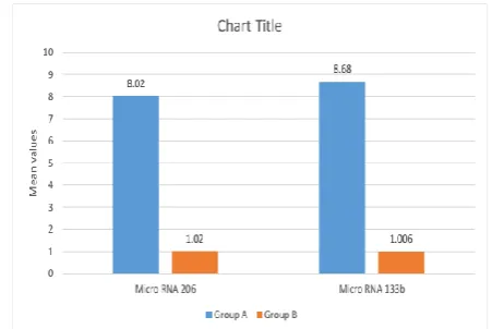
The mean placental miRNA 206 and the mean placental miRNA 133b are significantly higher in preeclamptic females than in normotensive females with p value equals to (0.0001) Figure (1).
Also, the mean serum IL-17 is significantly higher in preeclamptic females than in the normotensive females with a p value equals to (0.002) Figure (2).
Figure 1: mean values of placental micro RNA 206 and micro
RNA 133b in the studied groups.
Figure 2.Mean values of serum IL_17 in both groups.
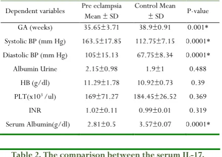
There was considerable development in the average standards of the systolic BP, diastolic BP and serum Albumin in the preeclamptic females compared to the control group while there was a significant decrease in the mean levels of the gestational age in the preeclamptic females compared to the control group, as regards albuminuria, serum haemoglobin, platlet count and INR there was no statistical difference between the two groups (table 1).
Table 1: Comparison between the demographic, clinical and laboratory data of studied groups
Dependent variables Pre eclampsia Mean ± SD
Control Mean ± SD P-value GA (weeks) 35.65±3.71 38.9±0.91 0.001* Systolic BP (mm Hg) 163.5±17.85 112.75±7.15 0.0001* Diastolic BP (mm Hg) 105±15.13 67.75±8.34 0.0001* Albumin Urine 2.15±0.98 1.9±1 0.488
HB (g/dl) 11.29±1.78 10.92±0.73 0.39 PLT(x103 /ul) 169±71.27 184.45±26.52 0.369
INR 1.02±0.11 0.99±0.01 0.319
Serum Albumin(g/dl) 2.81±0.5 3.57±0.07 0.0001*
Table 2. The comparison between the serum IL-17, placental micro RNA 206 and placental Micro RNA 133b
according to the clinical severity in the preeclamptic group
Mean Preeclampsia
Mean Preeclampsia with
severe
p
IL17 367.5942 316.2394 0.222003
MicroRNA206 7.5656 8.4055 0.725194
MicroRNA133b 8.8122 8.5727 0.927741
An examination of the region down the curve worth founded on receiver operating advantage curves for each miRNA detected that placental miRNAs 206 yielded significant results, with AUC of 0.916 (p value < 0.0001) with sensitivity 95% and 95%specificity, in differentiating preeclampsia cases from healthy pregnant females which strongly suggest placental miRNAs 206 as efficient preeclampsia diagnostic biomarker. An examination of the region down the curve wroth founded on receiver operating advantage curves for each miRNA detected that placental miRNA133b yielded significant results, with AUC of 0.967 (p value < 0.0001) with sensitivity 90% and 95% specificity, in differentiating preeclampsia cases from healthy pregnant females which strongly suggest placental miRNAs 133b as efficient preeclampsia diagnostic biomarker. Examination of the area under the curve (AUC) wroth founded on receiver operating advantage curves for serum IL-17 detected that it is sensitive by 65% and specific by 75% and AUC was 0.774 with p value equals 0.002.
Using Chi square to detect the direction and Eta square to determine a relationship between severity and each of the placental micro RNAs 206 or 133b, there was no significant relationship between severity and placental microRNA 206 (X2 =0.134 p=0.715) and placental micro RNA 133b (X2
=0.009p=0.925).
Using chi square and Eta square, there was only a significant relationship between systolic and diastolic blood pressure (p=0.012), and also albumin in urine (p=0.004) and gestational age (p=0.005) with the severity of the condition.
Discussion:
As regards gestation algae, McKinney et al. (2016) found that the gestation algae were significantly deceased in their patients than in control group, which was matching with our results that show the mean gestational age in preeclampsia was (35.65±3.71) while in control group was (38.9±0.91) with p value =0.001 [9].
Our results showed that there was a statistically significant up-regulation in placental expression of microRNA 206 and microRNA 133b (p<0.0001) as well as statistically significant increase in serum IL-17 in patients with preeclampsia rather than the control group (p=0.002).
The finding that expression of miRNA 206 is significantly higher in preeclamptic females than normal pregnant females was in agreement with [10], who showed that in preeclampsia
miR-206 was greater in each of the circulations and the placenta and placental IGF-1 mRNA expression was lower-controlled in placental tissue. It is well determined that the pregnancy maternal IGF-1 has significant development influences on the fetus and myometrial vasculature, which are closely connected processes to the reasons of preeclampsia [11, 12]. That research has experimentally supported reciprocal
actions with a number of genes recognized to be immediately included in the pathology of preeclampsia, especially at the placenta: VEGF [13] NOTCH-3 [14], IGF-1 [11] and
hypoxia-inducible agent 1 α (HIF-1α) [15]. This could be daring to be a
physiological response to hypoxia in the placenta.
To the best of our knowledge there are no reports or studies on the efficacy of miRNA 133b in preeclampsia patients as diagnostic marker for diagnosis of preeclampsia.
A number of studies illustrated that aberrant expression level of miRNA in the placenta is associated with preeclampsia [16, 17]. Some of the miRNAs are having been determined to be up
and down-adjusted in preeclamptic placentas. Thumping expression of several miRNAs in placenta gives rise to the pathogenesis of PE with change key processes in placental advancement as lead to limited reproduction and insignificant infestation of trophoblast cells, additionally inadequate remodeling of maternal spiral arteries and defective angiogenesis [18].
How miRNA expression is regulated in human placenta is not well understood. But researches documented that different reasons can control miRNA expression in placenta, including epigenetic regulation, oxygen tension, signaling molecules and environmental toxins. C19MC miRNAs expression pattern is associated with methylation status of a distal CpG-rich region located at 17.6 kb upstream of the miRNA cluster [19].
Concerning to preeclampsia, the genomic shape in which miR-206 is established of attention. Moving in the direction in which a stream of miR-206 is the interleukin 17a (IL17a) gene that expresses IL-17, a potent cytokine mediating pro-inflammatory responses [20]. T- Helper cells that have been
produced IL-17 (TH17) observed to be accessed in the circulation of females with preeclampsia. In addition to the same research, the cytokine itself was also found to be accessed in the circulation of a rodent model of preeclampsia
As regards serum IL-17 in our study, it has been increased (mean level in preeclampsia was 339.34±91.73 while in control group was 246.02±88.34).
The previous information on circulating IL-17 levels in preeclampsia was argumentative.
Our findings are harmonic with the previous researches confirming a greater prevalence of IL-17 levels in preeclampsias [22, 23].
Excessed circulating IL-17 levels showed in preeclampsia could be contributing to the advancement of popularizing intravascular inflammatory responses advantage of the maternal syndrome of the illness. IL-17 induces pro-inflammatory reactions by triggering the expression of other cytokines (IL-6, granulocyte colony-activating agent, tumor necrosis factor-α), chemokines (CXCL1, CXCL2, CCL20), inflammatory effectors (serious phase proteins, supplement) and antimicrobial proteins [22, 24]. Concerning, preeclampsia has
previously been clearly showing the existence to be connected with great quantity of pro-inflammatory cytokines, chemokines, serious phase proteins, and supplement activation products in the maternal circulation [25]. Cornelius DC et al.
(2014) reported that TH17 cells, likely via their secretion of IL-17 induce placental and renal oxidative stress which leads to production of agonistic autoantibodies to the angiotensin II type I receptor and placental vascular dysfunction resulting in the development of hypertension [26]. Molvarec A et al. (2015)
previously observed that serum IL-17 levels were considerably greater in preeclamptic patients than in healthy non-pregnant and pregnant females [7]. They did not find any connection
among serum IL-17 concentrations of preeclamptic patients and their clinical advantages and serum sFlt-1 and PlGF levels or sFlt-1/PlGFrates. Meanwhile, the greatest serum IL-17 level and sFlt-1/PlGF rate were reported to be additive for the danger of preeclampsia, as reported by the substantially increased strange rates of their incorporation than of either alone.
Most recently Darmochwal-Kolarz et al. (2017) reported that there were higher concentrations of IL-17 and lower concentrations of TGF-β in patients with placental insufficiency when compared to control [23]. In the group of
women patients with placental insufficiency, the levels of IL- 17 positively relate with systolic blood pressure (r = 0.42, p < 0.01). Researchers concluded that it appears potential that the excesses concentrations of IL-17 and the shortage of TGF-β in pregnancy complicated by FGR and PE can be answerable for the activation of inflammatory response showed in PE cases. Yang X et al. (2017) illustrated that the levels of miR-155 and IL-17 expression were established to be greater in preeclamptic placentas and serum, compared to the control group [27]. The results from this research explained a
connection among miR-155 and IL-17 in proteinuria through slow onset of PE.
However, Ozkan et al. (2014) found that IL-35 and IL-17 levels of preeclamptic women were significantly lower compared to normal pregnant women [28]. They attributed this
to the fact that the gestational age in their control group was
safety greater than that of the preeclamptic group. As circulating IL-17 levels have been reported to excess with improving gestation, the variation in gestational age might discompose their findings.
More research studies were done to measure IL-17 levels in serum of 13 preeclampsia women and 14 normal pregnant women with multiplex bead array, and no considerable variation was spotted [29].
Talking about the relationship between serum IL-17 and severity of the disease our results found no statistical difference between preeclampsia and preeclampsia with severe features which was in concordance with Toldi et al. (2011) who found no statistically significant differences in the proportion of IL-17-producing lymphocytes between patients with preeclampsia and preeclampsia with severe features [22].
Also, Molvarec et al. (2015) could not find any relationship between serum IL-17 concentrations of preeclamptic patients
[7].
From the above results, it seems probable that miRNAs as mi206 and miRNA 133b could potentially aid the early detection of clinical conditions associated with pregnancy or fetal development. A substantial amount of miRNAs has been discovered in different body fluids in the form of cell free. These cell free miRNAs are stable and their expression profiles are various among different types of fluid, depending on tissue and biological stages. The other advantages of using miRNA biomarkers in non-invasive prenatal diagnosis is that simple detection and amplification methods, lower complexity, tissue-restricted expression profiles, and sequence conservation between humans and model organisms [30].
Conclusion:
Both miRNA 206 and miRNA 133b are highly expressed in the placentae of preeclamptic Egyptian patients compared to the controls. Micro RNA 206 with 95% sensitivity and specificity, while Micro RNA 133b sensitivity was 90% and specificity was 95%, thus providing promising evidence that the expression of both parameters can be used as diagnostic markers for preeclampsia. The placental expression of miRNA 206 and miRNA does not seem to correlate to the severity of the condition. Similarly, serum level of interleukin 17 which was found to be increased in preeclampsia patients, and does not seem to correlate to the severity of the condition.
Perspectives:
Moreover, in vitro and in vivo studies utilizing particular miRNA weak cells are guaranteed to comprehensively comprehend the rates of the aberrantly expressed miRNAs in the beginning or advancement of preeclampsia. Whilst, it would be of considerable clinical benefit to over and above exam these little RNAs as possibility biomarkers for diagnosis of preeclampsia in a greater and prospective cohort.
1. ACOG (2013) Task force on hypertension in pregnancy. Hypertension in Pregnancy American College of Obstetricians and Gynecologists.
2. Steegers EA, von Dadelszen P, Duvekot JJ et al. (2010): Preeclampsia. Lancet; 376:631–644.
3. Ha M, Kim VN. (2014): Regulation of microRNA biogenesis. Nat Rev Mol Cell Biol.;15: 509– 524. 4. Fu G, Brki J, Hayder H, et al. (2013): MicroRNAs in
human placental development and pregnancy complications. Int J MolSci; 14: 5519-5544.
5. Williams AH, Liu N, van Rooij E, Olson EN (2009): MicroRNA control of muscle development and disease. CurrOpin Cell Biol. 2009, 21: 461-469.
6. Kozomara A and Griffiths-Jones S (2011): miRBase: integrating microRNA annotation and deep-sequencing data. Nucleic acids research; 39: D152-7.
7. Molvarec A, Czegle I, Szijártó J, et al. (2015): Increased circulating interleukin17 levels in preeclampsia. J ReprodImmunol.2015; 112:53-7. 8. Jin W, Dong C. (2013): IL-17 cytokines in immunity
and inflammation. Emerg Microbes Infect 2013; 2. 9. David McKinney, Heather Boyd, Amanda Langager, et
al. (2016): The impact of fetal growth restriction on latency in the setting of expectant management of preeclampsia American Journal of Obstetrics & Gynecology, Volume 214, Issue 3, 395.e1 - 395.e 10. Akehurst C, Small HY, Sharafetdinova L, Forrest R, et
al. (2015): Differential expression of microRNA-206 and its target genes in preeclampsia. J Hypertens. 33(10): 2068–2074.
11. Corcoran JJ, Charnock JC, Martin J, et al. (2012): Differential effect of insulin like growth factor-I on constriction of human uterine and placental arteries. J ClinEndocrinolMetab; 97:E2098–E2104.
12. Vatten LJ, Nilsen TI, Juul A, et al. (2008): Changes in circulating level of IGF-I and IGFbinding protein-1 from the first to second trimester as predictors of preeclampsia. Eur J Endocrinol ; 158:101–105. 13. Zhao W, Zhang J, Lu Y, et al. (2001): The vasorelaxant
effect of H(2)S as a novel endogenous gaseous K(ATP) channel opener. EMBO J. ;20(21):6008–6016. 14. De Falco M, Cobellis L, Giraldi D, et al. (2007):
Expression and distribution of notch protein members in human placenta throughout pregnancy. Placenta; 28:118–126.
15. Yue JQ, Guan J, Wang XY, et al. (2013): MicroRNA-206 is involved in hypoxia-induced pulmonary hypertension through targeting of the HIF-1 alpha/Fhl-1 pathway. Lab Invest 2; 93:748–759.
16. Luo SS, Ishibashi O, Ishikawa G, et al. (2009): Human villous trophoblasts express and secrete placenta-specific microRNAs into maternal circulation via exosomes. BiolReprod; 81: 717-729.
17. Fu G, Ye G, Nadeem L. (2013): MicroRNA-376c impairs transforming growth factor-beta and nodal
signaling to promote trophoblast cell proliferation and invasion. Hypertension.;61(4):864–872.
18. Zhang Y, Fei M, Xue G, et al. (2012): Elevated levels of hypoxia-inducible microRNA-210 in pre-eclampsia: New insights into molecular mechanisms for the disease. J Cell Mol Med; 16: 249-259.
19. Rippe V, Dittberner L, Lorenz VN, et al. (2010): The two stem cell microRNA gene clusters c19mc and mir-371-3 are activated by specific chromosomal rearrangements in a subgroup of thyroid adenomas. Center for Human Genetics, 5(3): PMC2831057. 20. Jin W, Dong C. (2013): IL-17 cytokines in immunity
and inflammation. Emerg Microbes Infect 2013; 2. 21. Wallace K, Dhillon P, Richards S, et al. (2011):
Interleukin 17 increases blood pressure during pregnancy: a link between autoimmunity and preeclampsia? FasebJ; 25.
22. Toldi G., Rigó, J., Stenczer, B., et al. (2011): Increased Prevalence of IL-17- Producing Peripheral Blood Lymphocytes in Pre-eclampsia. American Journal of
Reproductive Immunology, 66: 223–229.
doi:10.1111/j.1600-0897.2011.00987.
23. Darmochwal-Kolarz D, Michalak M, Kolarz B et al. (2017): The Role of Interleukin-17, Interleukin-23, and Transforming Growth Factor-β in Pregnancy Complicated by Placental Insufficiency. Biomed Res Int. 2017; 2017:6904325. doi: 10.1155/2017/6904325. Epub.
24. Onishi RM and Gaffen SL (2010): Interleukin-17 and its target genes: mechanisms of interleukin-17 function in disease. Immunology. 129(3): 311–321.
25. Derzsy, Z., Prohaszka, Z., Rigo, J., et al. (2010): Activation of the complement system in normal pregnancy and preeclampsia. MolImmunol 47, 1500- 1506.
26. Cornelius DC and Lamarca B. (2014): TH17- and IL-17- mediated autoantibodies and placental oxidative stress play a role in the pathophysiology of pre-eclampsia. Minerva Ginecol. 2014; 66(3): 243-9. 27. Yang X, Zhang J, and Ding Y. (2017): Association of
microRNA-155, interleukin 17A, and proteinuria in preeclampsia. Medicine (Baltimore); 96(18): e6509. 28. ZehraSemaOzkan, Mehmet Simsek, FulyaIlhan, et al.
(2014): Plasma IL-17, IL-35, interferon-γ, SOCS3 and TGF-β levels in pregnant women with preeclampsia, and their relation with severity of disease The Journal of Maternal-Fetal & Neonatal Medicine Vol. 27, Iss. 15. 29. Jonsson, Y., Ruber, M., Matthiesen, L., et al. (2006):
Cytokine mapping of sera from women with
preeclampsia and normal pregnancies. J
ReprodImmunol 70, 83-91