R E S E A R C H
Open Access
Platelet-derived growth factor
(PDGF)-BB-mediated induction of monocyte chemoattractant
protein 1 in human astrocytes: implications for
HIV-associated neuroinflammation
Crystal Bethel-Brown
1, Honghong Yao
1, Guoku Hu
2and Shilpa Buch
1,3*Abstract
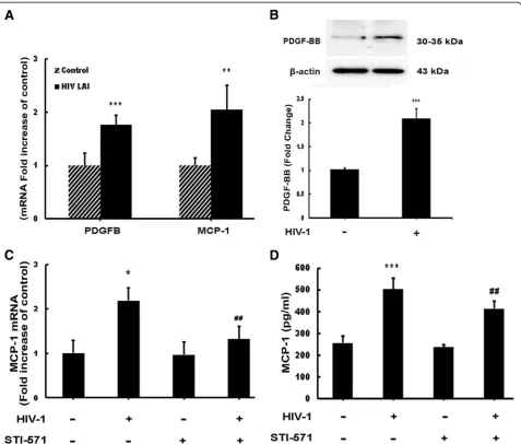
Chemokine (C-C motif) ligand 2, also known as monocyte chemoattractant protein 1 (MCP-1) is an important factor for the pathogenesis of HIV-associated neurocognitive disorders (HAND). The mechanisms of MCP-1-mediated neuropathogenesis, in part, revolve around its neuroinflammatory role and the recruitment of monocytes into the central nervous system (CNS) via the disrupted blood-brain barrier (BBB). We have previously demonstrated that HIV-1/HIV-1 Tat upregulate platelet-derived growth factor (PDGF)-BB, a known cerebrovascular permeant; subsequently, the present study was aimed at exploring the regulation of MCP-1 by PDGF-BB in astrocytes with implications in HAND. Specifically, the data herein demonstrate that exposure of human astrocytes to HIV-1 LAI elevated PDGF-B and MCP-1 levels. Furthermore, treating astrocytes with the human recombinant PDGF-BB protein significantly increased the production and release of MCP-1 at both the RNA and protein levels. MCP-1 induction was regulated by activation of extracellular-signal-regulated kinase (ERK)1/2, c-Jun N-terminal kinase (JNK) and p38 mitogen-activated protein (MAP) kinases and phosphatidylinositol 3-kinase (PI3K)/Akt pathways and the
downstream transcription factor, nuclear factorκB (NFκB). Chromatin immunoprecipitation (ChIP) assays
demonstrated increased binding of NFκB to the human MCP-1 promoter following PDGF-BB exposure. Conditioned media from PDGF-BB-treated astrocytes increased monocyte transmigration through human brain microvascular endothelial cells (HBMECs), an effect that was blocked by STI-571, a tyrosine kinase inhibitor (PDGF receptor (PDGF-R) blocker). PDGF-BB-mediated release of MCP-1 was critical for increased permeability in anin vitroBBB model as evidenced by blocking antibody assays. Since MCP-1 is linked to disease severity, understanding its modulation by PDGF-BB could aid in understanding the proinflammatory responses in HAND. These results suggest that astrocyte activation by PDGF-BB exaggerates monocyte recruitment into the brain via MCP-1 and underscores the critical role astrocytes play in HAND.
Background
HIV-associated neurocognitive disorders (HAND) re-main a common complication of HIV infection affecting up to 60% of infected individuals despite the use of anti-retroviral therapy (ART) [1]. With the advancement of ART the prevalence of HAND has actually increased,
partly due to both increased survival rates of HIV-infected individuals and to the reduced ability of most of these drugs to cross the blood–brain barrier (BBB). Among the factors involved in the pathogenesis of HAND, influx of HIV-infected monocytes in response to the chemokine monocyte chemoattractant protein 1 (MCP-1) via a breached endothelial barrier, plays a crit-ical role in disease pathogenesis. MCP-1 plays a vital role in the recruitment of monocytes into the brain contrib-uting to neuroinflammation and BBB disruption [2,3]. This chemokine has been extensively studied and is expressed by a number of cell types including astrocytes, * Correspondence:[email protected]
1Department of Pharmacology and Experimental Neuroscience, University of
Nebraska Medical Center, Omaha, NE 68198, USA
3Department of Pharmacology and Experimental Neuroscience, 985880
Nebraska Medical Center (DRC 8011), University of Nebraska Medical Center, Omaha, NE 68198-5880, USA
Full list of author information is available at the end of the article
microglia and neurons [4,5]. Elevated expression of MCP-1 has been demonstrated in various diseases in-cluding multiple sclerosis, amyloid lateral sclerosis, lupus nephritis, peripheral neuropathy and Alzheimer’s disease [6-13]. While increased expression of MCP-1 has been shown to correlate with HIV-associated central nervous system (CNS) complications, regulation of this chemo-kine in the context of HIV disease remains less clear. Understanding the molecular mechanisms modulating MCP-1 may thus provide insights into development of therapeutic targets for many neurodegenerative diseases including HAND.
Platelet-derived growth factor (PDGF) is a well known and potent inducer of MCP-1. The PDGF family of pro-teins is very closely related to the vascular endothelial growth factor (VEGF) family and is highly conserved throughout the animal kingdom [14]. These proteins are usually expressed as dimers: PDGF-A and PDGF-B can form homodimers or heterodimers, and PDGF-C and PDGF-D form homodimers. For the sake of clarity, in this study, PDGF-B refers to the RNA expression, whereas PDGF-BB refers to the protein expression of these genes. Many studies on PDGF have focused pri-marily on its mitogenic effects [15-17], however, diver-gent effects of PDGF are rapidly emerging. For example, recent studies by Lawrence et al. have demonstrated PDGF to be a cerebrovascular permeant that can disrupt BBB integrity during ischemic stroke conditions [18]. Along similar lines, it has been shown that PDGF-BB can disrupt BBB via the modulation of molecules im-portant in maintaining tight junctions such as ZO-1 and adhesion molecules [19].
Since astrocytes are a major source of MCP-1 in the brain and PDGF-BB has been shown to be an inducer of MCP-1, the purpose of this study was to explore the modulation of MCP-1 by PDGF-BB released from HIV-treated astrocytes. We hypothesize that PDGF-BB induced by HIV-1/HIV-1 Tat can result in astrocytic ac-tivation and release of MCP-1 and BBB disruption. The data demonstrate that the exposure of human astrocytes to HIV-1 LAI resulted in the induction of PDGF at both the mRNA and protein levels. To explicate the mechan-ism/s involved in PDGF-BB/MCP-1 interaction, human astrocytes were then treated with PDGF-BB and moni-tored for expression of MCP-1. Utilizing pharmaco-logical and genetic approaches we demonstrate the involvement of extracellular-signal-regulated kinase (ERK)1/2, c-Jun N-terminal kinase (JNK) and p38 mitogen-activated protein kinases (MAPKs), Phosphati-dylinositol 3-kinase (PI3K)/Akt pathways and the tran-scription factor nuclear factor κB (NFκB) in PDGF-BB-mediated induction of MCP-1 in astrocytes. Because both PDGF-BB and MCP-1 are known to affect the BBB and since astrocytic end-feet processes are in close
contact with the endothelia, we also addressed the func-tional implications this may have on the BBB. Using pharmacological and neutralizing antibody approaches, we reveal that both PDGF-BB and MCP-1 play critical roles in reducing the integrity of the BBB. These data highlight the role of PDGF-BB in astrocytic release of MCP-1, which in turn, is critical for recruitment of monocytes across the BBB. Taken together, these studies underscore the role of PDGF signaling as a potential therapeutic target of HAND.
Materials and methods
Materials
Recombinant human PDGF-BB was purchased from R&D Systems (Minneapolis, MN, USA). All experiments involving the treatment of cells with exogenous PDGF-BB were conducted under serum-free conditions because serum induces PDGF. STI-571, an inhibitor of tyrosine kinase receptors, was obtained from Novartis (Basel, Switzerland). The specific phosphatidinylinositol-3’ kin-ase (PI3K) inhibitor LY294002, mitogen-activated pro-tein (MAP) kinase kinase (MEK) inhibitor U0126 and p38 mitogen activated kinase (p38) inhibitor SB203558 were purchased from Promega (Madison, WI, USA). The JNK inhibitor SP600125 was purchased from Assay Designs (Ann Arbor, MI, USA). MCP-1 neutralizing antibody was obtained from eBioscience (San Diego, CA, USA). The C-C chemokine receptor type 2 (CCR2) antagonist, RS 102895, was purchased from Sigma (St Louis, MO, USA). The chromatin immunoprecipitation (ChIP) assay kit was obtained from Upstate, (Billerica, MA, USA).
Cell culture and cell lines
penicillin (100 U/ml), streptomycin (100 μg/ml), essen-tial amino acids, and vitamins according to our previous publication [21].
Cell treatment
Human astrocytes were serum starved overnight prior to treatment. The HIV-1 LAI used in this study was propa-gated in stimulated peripheral blood mononuclear cells (PBMCs) [22] (supplied by Dr Howard Gendelman, Uni-versity of Nebraska Medical Center, Omaha, NE, USA). The rationale for using a C-X-C chemokine receptor type 4 (CXCR-4)-tropic virus is based upon previous studies demonstrating the susceptibility of astrocytes to CXCR4-tropic viruses [23,24]. In the pharmacological inhibitor studies, the cells were pretreated with inhibi-tors specific for MEK (U0126, 1 μM), JNK (SP600125, 10 μM), P38 (SB20358, 1 μM) and PI3K (LY294002, 1
μM) for 1 h prior to PDGF-BB exposure. The inhibitors were not removed from the astrocytes during PDGF-BB treatment and concentrations utilized in this study were based upon our previous studies [25].
MCP-1 protein analysis by enzyme-linked immunosorbent assay (ELISA)
MCP-1 levels were examined using an MCP-1 ELISA kit purchased from R&D Systems. Samples were analyzed for MCP-1 protein according to the manufacturer’s instructions in triplicates determined in at least three in-dependent experiments.
Reverse transcription and real-time RTPCR (RT-PCR)
Total RNA was extracted using Trizol reagent according to the manufacturer’s protocol (Invitrogen). RNA (1μg) was used for cDNA production according to manufac-turer’s instructions (Thermo Scientific, Waltham, MA, USA). The sequences of primers used for human MCP-1 (GenBank accession number NM 002982) were as
fol-lows: sense: CAGCAGGTGTCCCAAAGAAGCTGT
antisense: CCATTCCTTATTGGGGTCAGCACAGA.
The sequences of primers used for glyceraldehyde-3-phosphate dehydrogenase (GAPDH) (GenBank accession number NM 002046) were as follows: sense: TGCACC ACCAACTGCTTAGC; antisense: GGCATGGACTGTG GTCATGAG. The primers for CCR2 (GenBank acces-sion number NM 001123396) were as follows: first round sense: TCTGGAGACCTCAACCAAATG; anti-sense: GGAAATGCGTCCTTGTTCAA. The primers used in the second round were as follows: sense: CCCTGTAT
CTCCGCCTTCACT; antisense: TTCAGCTTGTGGC
TTGTCTCA. Quantitative analyses of mRNA were con-ducted using an ABI 7500 Fast Real-Time PCR system (Applied Biosystems, Foster City, CA, USA). The primers for PDGF-B, MCP-1 and 18S were purchased from SA Biosciences (Frederick, MD, USA). Data were normalized
using Ct values for GAPDH or 18S in each sample. To cal-culate relative amounts mRNA, the average Ct values were subtracted from GAPDH or 18S values for each target gene to provide changes in Ct value. Fold change in ex-pression was calculated as log2relative units.
Flow cytometry
Untreated human primary astrocytes and A172 human astrocytes were collected in cold phosphate-buffered sa-line (PBS) and ethylenediaminetetra-acetic acid (EDTA) (5 mM) followed by incubation with anti-PDGF receptor
α(PDGF-Rα) (CD140α; 1:5) and anti-PDGF-Rβ(CD140β; 1:5) BD LSR II (BD Biosciences, San Jose, CA, USA) was used for fluorescence acquisition, and data were analyzed with FACS Diva software (BD Biosciences).
Western blotting
PDGF-BB treated astrocytes were lysed using the Mam-malian Cell Lysis kit (Sigma) and the NE-PER Nuclear and Cytoplasmic Extraction kit (Pierce, Rockford, IL, USA) as per manufacturer’s instructions. Cell lysates were subjected to separation by 12% sodium dodecyl sul-fate polyacrylamide gel electrophoresis (SDS-PAGE) (30
μg protein per well) and transferred to polyvinylidene difluoride (PVDF) membranes. The blots were blocked with 5% non-fat dry milk in PBS. Western blots were then probed with antibodies recognizing phosphorylated forms of ERK1/2, JNK, p38 and Akt and total forms of ERK1/2, JNK, and Akt (Cell Signaling, Danvers, MA, USA; 1:500); NFκB-p65 and phosphorylated inhibitor of
κB (pIκB) Cell Signaling, (1:1,000); Histone (1:1,000) and
β-actin antibodies (Santa Cruz Biotechnologies, Santa Cruz, CA, USA; 1:5,000). Signals were detected by chemiluminescence (Pierce).
Transduction with adenoviral constructs
A172 cells were infected with adenoviral constructs con-taining either wild-type (WT) or dominant-negative (DN) forms of Akt (a kind gift from Dr K Walsh, Tufts University School of Medicine, Boston, MA, USA). In addition, cells were also transduced with recombinant adenoviral vectors expressing full-length p65/RelA or p65/RelA mutant (RelA 1–300) (a kind gift from Dr S Maggiwar, University of Rochester Medical Center, Rochester, NY, USA) used at a multiplicity of infection (MOI) of 50 as previously described [26]. Astrocytes infected for 24 h with adenoviral constructs were subse-quently treated with PDGF-BB, followed by assessment by western blotting or ELISA.
Short interfering RNA transfection of astrocytes
density of 4 × 10⁴ cells per well 1 day prior to trans-fection. Cell culture medium was replaced with 250 ml pre-warmed OPTI-MEM I culture medium. Lipofectamine-2000 reagent (Invitrogen) was then combined with serum-free medium for 5 minutes at room temperature. The PDGF-Rβ siRNA was then added into the mixture described above to a final concentration of 5μM. Then, siRNA and the reagent mixture were incubated for 20 minutes at room temperature, after which, the combined mixture was added to the cells. The cell culture plate was shaken gently for 5 s and incubated for 24 h at 37°C. Knockdown efficiency (Figure 1D) of the transfected astrocytes were (54%) as determined by RT-PCR.
Transfection with plasmid constructs
DN and WT constructs of MEK were provided by Dr Young Han Lee (Konkuk University, Korea). A172 cells were transfected with plasmid constructs containing ei-ther WT or DN forms of MEK as described above. The transfection efficiency was 42% as determined by immu-nostaining (data not shown).
ChIP assay
The ChIP assay was performed according to the manu-facturer’s instructions (Upstate, Billerica, MA, USA) with slight modifications. After treatment of the cells, 18.5% fresh formaldehyde was added directly into the medium at a final concentration of 1% formaldehyde and incu-bated for 10 minutes at room temperature, followed by quenching with 125 mM glycine. The cells were then scraped using 2 ml pre-chilled PBS containing 1 × prote-ase inhibitor mixture. The cell pellet was harvested by spinning at 800gat 4°C, and lysis buffer was added (pro-vided in the kit) to harvest nuclei. DNA was then sheared by sonication. A total of 50 μl of the sheared crosslinked chromatin was then mixed with 20 ml pro-tein A magnetic beads and 5 mg of immunopreciptating Abs against NFκB p65, acetyl histone H3 (as a positive control), and normal rabbit IgG (as a negative control) diluted in 450 ml dilution buffer overnight at 4°C. The magnetic beads binding Ab-chromatin complex was then washed with 0.5 ml each of a series of cold wash buffers in the order of low salt buffer, high salt buffer, LiCl buffer, and Tris-EDTA buffer. The crosslinking of protein-DNA complexes were reversed to free DNA by incubation at 62°C for 2 h and purified using DNA puri-fication spin columns following the manufacturer’s instructions. Finally, the purified DNA was amplified (35 cycles) via PCR to identify the promoter region contain-ing NFκB binding site ‘GGGCCTTTCC’. The sequence of the primers used to identify the MCP-1 promoter bound to NFκB were as follows: sense: GCATCAGAGC ATTGACCCTCA; antisense: AGGTCAGTGCTGGCG TGAGA.
Monocyte isolation and transmigration
Primary HBMECs seeded on 6.5 mm polyester transwell inserts (5.0 μm pore size) were grown to confluence. Following confluency, conditioned media from primary astrocytes treated with PDGF-BB was added to the bot-tom chamber of the transwell and allowed to incubate at 37°C in a humid atmosphere of 5% CO2for 24 h. MCP-1 neutralizing antibody was added to PDGF-BB treated conditioned media prior to HBMEC exposure for 24 h. Monocytes were obtained from HIV-1, HIV-2 and hepa-titis B seronegative donor leukopacks, and separated by countercurrent centrifugal elutriation as previously described [27]. Monocytes were washed with PBS and fluorescently labeled with 10 μM Cell tracker green (Invitrogen) for 10 minutes at room temperature. Labeled cells (2 × 10⁵ cells) were added to the upper compart-ments of transwell inserts and allowed to transmigrate at 37°C in a humid atmosphere of 5% CO2for 24 h. Trans-migrated monocytes were quantified using florescent plate reader (435 nm/528 nm excitation/emission).
Statistical analysis
Statistical analysis was performed using one-way analysis of variance (ANOVA) with a post hoc Student’s t test. Results were judged statistically significant ifP <0.05 by analysis of variance.
Results
HIV-1-mediated upregulation of PDGF-B and MCP-1 in astrocytes
Figure 1Engagement of platelet-derived growth factor receptor (PDGF-R) is critical for PDGF-BB induced monocyte chemoattractant protein 1 (MCP-1) expression in astrocytes.(A) Human primary as well as A172 primary astrocytes express both PDGF-αand PDGF-β receptors. Untreated human primary and A172 astrocytes were subjected to flow cytometric analyses using antibodies against human PDGF-αR (CD140α) and PDGF-βR (CD140β). (B,C) PDGF-BB-mediated induction of MCP-1 mRNA and protein expression in human primary and A172 astrocytes. (B) Human primary and A172 astrocytes were exposed to PDGF-BB for 6 h. Total RNA isolated from human A172 astrocytes was subjected to real-time reverse-transcriptase polymerase chain reaction (RT-PCR) analysis using primers for human MCP-1. (C) Supernatant fluid from astrocytes treated with PDGF-BB for 24 h were assessed for release of chemokine MCP-1 using the human MCP-1 enzyme-linked
expression. Blocking the PDGF-R significantly reduced HIV-mediated induction of MCP-1 RNA (Figure 2C) and protein (Figure 2D).
PDGF-BB-mediated upregulation of MCP-1 in astrocytes
All experiments involving the treatment of cells with ex-ogenous PDGF-BB protein were conducted under serum-free conditions since PDGF promoter is known to have serum response elements [28,29]. To investigate the role of PDGF on MCP-1 expression, A172 cells were treated with recombinant PDGF-BB (20 ng/ml) for the
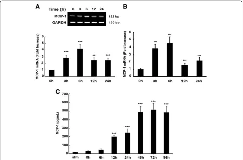
indicated times and mRNA levels were assessed by RT-PCR and real-time RT-RT-PCR. The concentration of PDGF-BB used was based on previous studies [25,30]. Following exposure of A172 astrocytes to PDGF-BB, there was a time-dependent upregulation of MCP-1 mRNA compared with untreated cells (Figure 3A) with a peak induction at 6 h post treatment (4.5-fold) and a subsequent downregulation at 12 h. Similar results were obtained via real-time PCR (Figure 3B).
astrocytes, we next examined the MCP-1 protein levels. Cells were serum-starved as described and treated with PDGF-BB for 0 to 96 h. Supernatant fluids were col-lected and MCP-1 protein levels were assessed by ELISA. As shown in Figure 3C, PDGF-BB upregulated MCP-1 protein levels in a time-dependent manner with a continual increase that plateaued at 48 h post treat-ment. Cumulatively, these data clearly demonstrate that PDGF-BB treatment mediated the induction of MCP-1 RNA and protein in astrocytes.
Engagement of PDGF-Rβis critical for PDGF-induced MCP-1 expression in astrocytes
Because PDGF-BB mediates its effects via binding to its cognate receptors PDGF-Rα and PDGF-Rβ, our next step was to examine the role of receptor binding in PDGF-BB-induced MCP-1 release by astrocytes. How-ever, before proceeding with this, it was important to
first examine whether astrocytes indeed expressed PDGF-Rα and PDGF-Rβ. Flow cytometric analysis of A172 and human primary astrocytes revealed a stark dif-ference between the two types of cells. As shown in Figure 1A, of the A172 cells examined, endogenous levels of PDGF-Rαand PDGF-Rβwere 0.17% and 19.2%, respectively. Human primary astrocytes expressed higher levels of PDGF-Rα (4.6%) and lower PDGF-Rβ (10.9%) levels compared to A172 astrocytes. Despite the differ-ence in relative distribution of the PDGF receptors, PDGF-Rβ was the major receptor form expressed in both types of astrocytes utilized in this study. Similar results were observed by RT-PCR (data not shown).
assessment, respectively. As shown in Figure 1B, PDGF-BB upregulated MCP-1 mRNA levels in both human A172 as well as human primary astrocytes. MCP-1 pro-tein ELISA analysis also corroborated this upregulation, as shown in Figure 1C.
To determine the role that the PDGF-Rs played in PDGF-BB-mediated MCP-1 release from astrocytes, human A172 astrocytes were transfected with PDGF-Rβ siRNA followed by treatment of PDGF-BB and MCP-1 levels were assessed 24 h later. As shown in Figure 1D, transfection of human A172 cells with PDGF-RβsiRNA resulted in efficient knockdown of PDGF-Rβas demon-strated by RT-PCR. Cells were transfected with either PDGF-Rβ siRNA or non-specific (Non) siRNA control followed by treatment with BB for 24 h. PDGF-BB-mediated induction of MCP-1 mRNA was attenuated in cells transfected with PDGF-RβsiRNA but not in the Non siRNA controls cells (Figure 1E). These results were further confirmed by examining PDGF-BB-mediated in-duction of MCP-1 protein levels. As shown in Figure 1F and as expected, PDGF-BB treatment resulted in enhanced release of MCP-1 protein in Non siRNA trans-fected cells but not in cells transtrans-fected with PDGF-Rβ siRNA. Taken together, these findings confirmed the in-volvement of PDGF-Rs in PDGF-BB-mediated induction of MCP-1 in astrocytes.
PDGF-BB-mediated induction of MCP-1 involves MAPK and PI3K/Akt cell signaling pathways
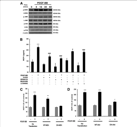
Having determined PDGF-BB-mediated induction of MCP-1, we next sought to elucidate the signaling path-ways involved in this process. Since PDGF-BB is a mito-gen and a pro-survival protein, we examined the involvement of MAPK and phosphoinositide-3-kinase/ Akt pathways. Human A172 cells were exposed to PDGF-BB and phosphorylation of MAPK and PI3K/Akt signaling molecules was assessed by western blotting. Treatment of astrocytes with PDGF-BB resulted in a time-dependent increase in phosphorylation of ERK1/2, JNK, p38 and Akt, with maximal activation at 30 min-utes following treatment (Figure 4A). Next, to address the functional implication of MAPK and PI3K/Akt path-ways in PDGF-mediated induction of MCP-1 expression, A172 cells were pretreated with inhibitors specific for the respective signaling pathways followed by PDGF-BB treatment for 24 h and subsequently assessed for expres-sion of MCP-1. As shown in Figure 4B, treatment of cells with the MAPK and PI3K/Akt inhibitors resulted in amelioration of PDGF-BB-mediated induction of MCP-1 protein levels.
Further validation of the involvement of the MAPK pathway in this process was confirmed by transfecting cells with either the WT or DN constructs of MEK followed by 24 h treatment with PDGF-BB.
PDGF-BB-mediated induction of MCP-1 was attenuated by DN-MEK, but not by WT-MEK constructs (Figure 4C). To confirm the role of PI3K/Akt in PDGF-BB-mediated MCP-1, A172 cells were transduced with adenoviral constructs containing either WT or DN forms of Akt. As shown in Figure 4D, cells transduced with DN Akt construct failed to upregulate MCP-1 unlike the cells transduced with the WT Akt construct. Taken together, these findings confirm the involvement of both MAPK and PI3K/Akt cascades in PDGF-BB-mediated induction of MCP-1 in astrocytes.
Involvement of PDGF-Rβin the regulation of MAPKs and PI3K/Akt cell signaling pathways
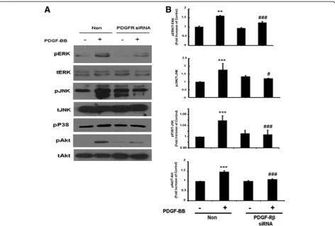
Since PDGF-BB acts through its cognate receptor, the next logical step was to link the MAPK and PI3K/Akt pathways to PDGF-R. To achieve this, siRNA was again employed. PDGF-Rβ expression was knocked down using the siRNA approach and ERK, JNK, p38, JNK and Akt phosphorylation levels were assessed. Briefly, A172 cells were transfected with PDGF-RβsiRNA for 24 h fol-lowed by treatment with PDGF-BB for 1 h. As shown in Figure 5A,B, cells transfected with PDGF-Rβ siRNA failed to demonstrate PDGF-BB-mediated activation of ERK, JNK, p38 and Akt. These results underpin the role of PDGF-Rβin PDGF-BB-mediated activation of MAPK and PI3K/Akt pathways.
Involvement of NFκB in PDGF-BB-induced expression of MCP-1 in astrocytes
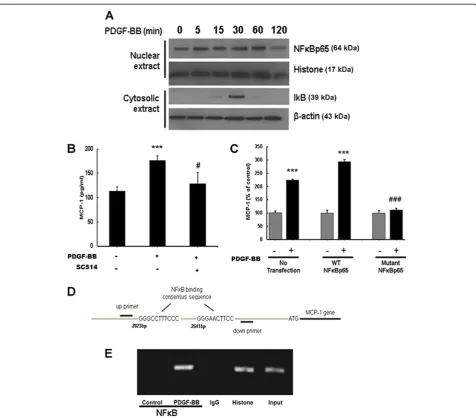
Having determined the role of MAPK and PI3K/Akt in PDGF-BB-mediated induction of MCP-1 expression, we rationalized the involvement of NFκB in this process since this transcription factor is downstream of the aforementioned signaling mediators [31-33]. Exposure of astrocytes to PDGF-BB resulted in a time-dependent in-crease of p65 subunit of NFκB in the nucleus with a concomitant transient increase in cytosolic pIκB (Figure 6A). Validation of the role of NFκB was further determined by pretreating astrocytes with the IκB path-way inhibitor (SC514), followed by PDGF-BB treatment for 24 h. As shown in Figure 6B, SC514 inhibited PDGF-BB-mediated induction of MCP-1, thereby underscoring the role of NFκB in this process.
Since NFκB is a transcription factor that mitigates its effects on target genes by binding to promoter regions, we next sought to confirm the binding of NFκB with MCP-1 promoter in its natural chromatin context by ChIP assay to reveal active sites accessible to NFκB. A172 cells were treated with PDGF-BB for 1 h followed
by RNA extraction and processed using a ChIP analysis kit. These experiments revealed increased binding of NFκB to the MCP-1 promoter in A172 cells treated with PDGF-BB (Figure 6D,E) and, along with preceding data, substantiate the role of NFκB in PDGF-BB-mediated regulation of MCP-1 in astrocytes.
MAPK and PI3K/Akt cell signaling pathways lie upstream of PDGF-BB induced NFκB in astrocytes
Having determined the involvement of ERK1/2, JNK and p38 MAPKs and NFκB in PDGF-BB-mediated MCP-1 the next logical step was to examine whether there existed a link that could tie together the activation of MAP kinase and Akt pathways with NFκB. Astrocytes were transfected with either the WT or DN constructs of MEK prior to PDGF-BB treatment as described before. Nuclear fractions were extracted and NFκB phosphorylation was assessed via western blotting. PDGF-BB-mediated induction of NFκB was attenuated by DN-MEK, but not by WT-MEK construct (Figure 7A). To confirm the role of PI3K/Akt in PDGF-BB-mediated NFκB, A172 cells were transduced with adenoviral con-structs containing either WT or DN forms of Akt, trea-ted with PDGF-BB and assessed for NFκB expression. As shown in Figure 7B, cells transduced with DN Akt con-struct failed to upregulate NFκB unlike the cells trans-duced with the WT Akt construct. These findings thus
linked PDGF-BB-mediated activation of MAP kinase and Akt pathways to the downstream activation of NFκB.
MCP-1 released from astrocytes increases monocyte transmigration in human brain microvascular endothelial cells
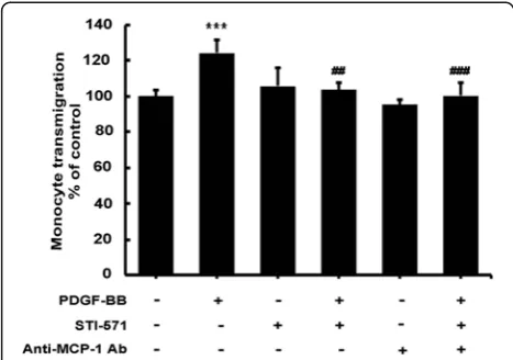
cells treated with PDGF-BB resulted in a significant in-crease in monocyte transmigration of endothelial cells, an effect that was blocked by PDGF-R blocker STI-571.
Since monocytes express the MCP-1 receptor CCR2 [34,35], the next step was to determine whether increased MCP-1 was also able to enhance monocyte migration. Also shown in Figure 8A, conditioned media from primary astrocytes cells treated with PDGF-BB resulted in a dramatic increase in monocyte transmigra-tion of endothelial cells and this effect was ameliorated in conditioned media treated with the MCP-1 neutralizing
antibody. These findings thus confirm that MCP-1 was involved in PDGF-BB-mediated disruption of the endo-thelial barrier permeability and not only underpin the role of MCP-1 in BBB breakdown, but reveal a vital role that activated astrocytes play in BBB disruption and HAND pathogenesis.
Discussion
Antiretroviral therapies have proven highly effective in controlling systemic viral infection, thus leading to increased longevity in patients with AIDS. The inability Figure 6Platelet-derived growth factor (PDGF)-BB-mediated induction of monocyte chemoattractant protein 1 (MCP-1) expression involves nuclear factorκB (NFκB) activation.(A) Exposure of A172 cells to PDGF-BB resulted in a time-dependent increase in phosphorylation of the p65 subunit of NFκB in the nuclear cellular fraction. Reciprocally, PDGF-BB exposure resulted in a time-dependent increase in inhibitor of
of some of these drugs to cross the BBB results in slow and smoldering infection in the CNS. Subsequently, the brain becomes a sanctuary of virus-induced toxicity leading to increased prevalence of HAND in HIV-infected individuals. One of the hallmark features of HAND is increased astrogliosis comprising of increased numbers of activated astrocytes, culminating ultimately into increased neuronal degeneration. It is well recog-nized that activation of astrocytes leads to the release of a barrage of inflammatory mediators such as PDGF-BB. PDGF-BB has been implicated in a variety of patho-logical conditions; however, its role in HIV pathogenesis remains less clear.
In the present study we demonstrate that HIV-1-mediated induction of MCP-1, a potent chemokine vital to the sustained proinflammatory response in HIV-1 pathogenesis, is regulated by PDGF-BB. To determine the implication of increased PDGF-BB we demonstrated that PDGF-BB treatment of human astrocyte cell line and primary cultures resulted in induction of MCP-1 and this process was mediated via the binding of PDGF-BB to its cognate PDGF-Rβ. These findings are consist-ent with the reports on PDGF-mediated induction of
MCP-1 in other cell types such as the fibroblast cell line and smooth muscle cells [36,37]. In our efforts to dissect the upstream signaling events that are involved in MCP-1 release from astrocytes, using both the pharmaco-logical as well as genetic approaches we demonstrated the role of MAPK and PI3K/Akt in PDGF-BB-mediated induction of MCP-1 from astrocytes. The involvement of MAPK and PI3K/Akt pathways in the induction of MCP-1 expression are in agreement with the role these pathways play in induction of this chemokine in other cell types including osteoblasts, mesangial cells and endothelial cells [38-40]. The transcription factor, NFκB is known to play a key role in PDGF-BB signaling and also in the expression of proinflammatory cytokines/ chemokines including MCP-1 [31,33,41]. Consistent with other reports, our studies also revealed that PDGF-BB-mediated induction of MCP-1 involved both NFκB acti-vation and its binding to the MCP-1 promoter.
integrity. Intriguingly, conditioned media from PDGF-BB treated astrocytes did indeed increase monocyte transmigration and this effect was attributable to MCP-1 as demonstrated in the blocking antibody experiments. This role of MCP-1 is in agreement with the findings reported by Eugenin et al. who have demonstrated that HIV-infected leukocyte transmigration across a tissue culture model of human BBB involved MCP-1 [42]. In addition to disrupting the barrier permeability, MCP-1 also manifested increased monocyte recruitment. This latter function is in keeping with its known roles both as a chemoattractant and as a biomarker of HIV neuro-pathogenesis [42,43].
The function of MCP-1 demonstrated in this study can have ramifications in the pathogenesis of HAND. Based on the proximity of astrocytes to the endothelium and their ability to secrete both PDGF-BB and the che-mokine MCP-1 as well as their abundance in the CNS, it can be argued that during HIV-1 infection, viral proteins can initiate a toxic cascade that can be self-perpetuating. HIV-1/HIV-1 Tat can trigger increased expression of PDGF-BB, which in turn, can lead to increased MCP-1 expression that can manifest as an amplified influx of monocytes into the CNS. PDGF-BB has already been shown to disrupt the BBB [19] and a similar function has been demonstrated for MCP-1 [42]. Therefore the release of both these mediators can independently
disrupt the endothelial barrier while enhancing neuroin-flammation [19,42], which can have serious ramifications in HAND.
In summary, our studies have mapped out a detailed molecular pathway of PDGF-BB-mediated MCP-1 ex-pression in astrocytes involving ERK1/2, JNK MAPK ac-tivation, with the subsequent activation of NFκB resulting in increased MCP-1 expression, ultimately leading to increased monocyte transmigration and increased permeability in the brains of individuals infected with HIV.
Competing interests
The authors declare that they have no competing interests.
Authors’contributions
CB-B designed research, performed research and wrote the manuscript; HY designed the research and wrote the manuscript; GH performed research; SB planned and designed the research and wrote the manuscript. All authors have read and approved the final version of the manuscript.
Acknowledgements
We thank Dr Randall Davis (Oklahoma State University, Stillwater, OK, USA) for providing the A172 cell line and Dr Monique Stins (Johns Hopkins University, Baltimore, MD, USA) for providing the HBMECs. We thank Dr Howard Gendelman (University of Nebraska Medical Center, Omaha, NE, USA) for providing us the HIV-1 LAI. We thank Dr Sanjay Maggirwar (University of Rochester Medical Center, Rochester, NY, USA) for providing us with recombinant adenovirus vectors of full-length RelA and mutant RelA. We also thank Dr Young Han Lee (Konkuk University, Korea) for providing the constitutively active and dominant-negative constructs of MEK and Dr K Walsh (Tufts University School of Medicine, Boston, MA, USA) for providing the adenoviral vectors expressing the WT and DN forms of Akt.
Author details
1Department of Pharmacology and Experimental Neuroscience, University of
Nebraska Medical Center, Omaha, NE 68198, USA.2Department of Medical Microbiology and Immunology, Creighton Medical Center, Omaha, NE 68178, USA.3Department of Pharmacology and Experimental Neuroscience, 985880 Nebraska Medical Center (DRC 8011), University of Nebraska Medical Center, Omaha, NE 68198-5880, USA.
Received: 29 March 2012 Accepted: 15 November 2012 Published: 1 December 2012
References
1. Heaton RK, Clifford DB, Franklin DR Jr, Woods SP, Ake C, Vaida F, Ellis RJ, Letendre SL, Marcotte TD, Atkinson JH, Rivera-Mindt M, Vigil OR, Taylor MJ, Collier AC, Marra CM, Gelman BB, McArthur JC, Morgello S, Simpson DM, McCutchan JA, Abramson I, Gamst A, Fennema-Notestine C, Jernigan TL, Wong J, Grant I, CHARTER Group:HIV-associated neurocognitive disorders persist in the era of potent antiretroviral therapy: CHARTER Study. Neurology2010,75:2087–2096.
2. Weiss JM, Nath A, Major EO, Berman JW:HIV-1 Tat induces monocyte chemoattractant protein-1-mediated monocyte transmigration across a model of the human blood–brain barrier and up-regulates CCR5 expression on human monocytes.J Immunol1999,163:2953–2959. 3. Fuentes ME, Durham SK, Swerdel MR, Lewin AC, Barton DS, Megill JR,
Bravo R, Lira SA:Controlled recruitment of monocytes and macrophages to specific organs through transgenic expression of monocyte chemoattractant protein-1.J Immunol1995,155:5769–5776.
4. Lee SC, Liu W, Dickson DW, Brosnan CF, Berman JW:Cytokine production by human fetal microglia and astrocytes. Differential induction by lipopolysaccharide and IL-1 beta.J Immunol1993,150:2659–2667. 5. Rock RB, Hu S, Sheng WS, Peterson PK:Morphine stimulates CCL2
production by human neurons.J Neuroinflammation2006,3:32. 6. Ransohoff RM, Hamilton TA, Tani M, Stoler MH, Shick HE, Major JA, Estes ML,
Thomas DM, Tuohy VK:Astrocyte expression of mRNA encoding
Figure 8Platelet-derived growth factor (PDGF)-BB-induced monocyte transmigration and permeability is mediated by monocyte chemoattractant protein 1 (MCP-1).Human brain microvascular endothelial cells (HBMECs) were exposed for 24 h to supernatants from A172 cells treated with PDGF-BB with or without STI-571. MCP-1 neutralizing antibody was added to PDGF-BB-treated media prior to HBMEC exposure for 24 h. Human monocytes were added to the top transwell for 24 h followed by assessment of transmigration. Exposure of HBMECs to supernatants from A172 cells treated with PDGF-BB resulted in increased permeability and enhanced monocyte transmigration, which was abrogated by MCP-1 neutralizing antibody. All the data are presented as mean ± SD of three individual experiments. ***P<0.001 versus control group; ##P
cytokines IP-10 and JE/MCP-1 in experimental autoimmune encephalomyelitis.FASEB J1993,7:592–600.
7. Mahad DJ, Ransohoff RM:The role of MCP-1 (CCL2) and CCR2 in multiple sclerosis and experimental autoimmune encephalomyelitis (EAE). Semin Immunol2003,15:23–32.
8. Van Der Voorn P, Tekstra J, Beelen RH, Tensen CP, Van Der Valk P, De Groot CJ:Expression of MCP-1 by reactive astrocytes in demyelinating multiple sclerosis lesions.Am J Pathol1999,154:45–51. 9. Tanuma N, Sakuma H, Sasaki A, Matsumoto Y:Chemokine expression by
astrocytes plays a role in microglia/macrophage activation and subsequent neurodegeneration in secondary progressive multiple sclerosis.Acta Neuropathol2006,112:195–204.
10. Berman JW, Guida MP, Warren J, Amat J, Brosnan CF:Localization of monocyte chemoattractant peptide-1 expression in the central nervous system in experimental autoimmune encephalomyelitis and trauma in the rat.J Immunol1996,156:3017–3023.
11. Yamamoto M, Horiba M, Buescher JL, Huang D, Gendelman HE, Ransohoff RM, Ikezu T:Overexpression of monocyte chemotactic protein-1/CCL2 in beta-amyloid precursor protein transgenic mice show accelerated diffuse beta-amyloid deposition.Am J Pathol2005,
166:1475–1485.
12. Marks SD, Shah V, Pilkington C, Tullus K:Urinary monocyte chemoattractant protein-1 correlates with disease activity in lupus nephritis.Pediatr Nephrol2010,25:2283–2288.
13. Groh J, Heinl K, Kohl B, Wessig C, Greeske J, Fischer S, Martini R:
Attenuation of MCP-1/CCL2 expression ameliorates neuropathy in a mouse model for Charcot-Marie-Tooth 1X.Hum Mol Genet2010,
19:3530–3543.
14. Andrae J, Gallini R, Betsholtz C:Role of platelet-derived growth factors in physiology and medicine.Genes Dev2008,22:1276–1312.
15. Kohler N, Lipton A:Platelets as a source of fibroblast growth-promoting activity.Exp Cell Res1974,87:297–301.
16. Ross R, Glomset J, Kariya B, Harker L:A platelet-dependent serum factor that stimulates the proliferation of arterial smooth muscle cellsin vitro. Proc Natl Acad Sci U S A1974,71:1207–1210.
17. Bowen-Pope DF, Malpass TW, Foster DM, Ross R:Platelet-derived growth factor in vivo: levels, activity, and rate of clearance.Blood1984,
64:458–469.
18. Su EJ, Fredriksson L, Geyer M, Folestad E, Cale J, Andrae J, Gao Y, Pietras K, Mann K, Yepes M, Strickland DK, Betsholtz C, Eriksson U, Lawrence DA:
Activation of PDGF-CC by tissue plasminogen activator impairs blood–brain barrier integrity during ischemic stroke.Nat Med2008,
14:731–737.
19. Yao H, Duan M, Buch S:Cocaine-mediated induction of platelet-derived growth factor: implication for increased vascular permeability.Blood
2011,117:2538–2547.
20. Davis RL, Dertien J, Syapin PJ:Ethanol-induced modulation of inducible nitric-oxide synthase activity in human A172 astrocytoma cells. Alcohol Clin Exp Res2002,26:1404–1411.
21. Yao H, Kim K, Duan M, Hayashi T, Guo M, Morgello S, Prat A, Wang J, Su TP, Buch S:Cocaine hijacks sigma1 receptor to initiate induction of activated leukocyte cell adhesion molecule: implication for increased monocyte adhesion and migration in the CNS.J Neurosci2011,31:5942–5955. 22. Gorantla S, Santos K, Meyer V, Dewhurst S, Bowers WJ, Federoff HJ,
Gendelman HE, Poluektova L:Human dendritic cells transduced with herpes simplex virus amplicons encoding human immunodeficiency virus type 1 (HIV-1) gp120 elicit adaptive immune responses from human cells engrafted into NOD/SCID mice and confer partial protection against HIV-1 challenge.J Virol2005,79:2124–2132.
23. Bezzi P, Domercq M, Brambilla L, Galli R, Schols D, De Clercq E, Vescovi A, Bagetta G, Kollias G, Meldolesi J, Volterra A:CXCR4-activated astrocyte glutamate release via TNFalpha: amplification by microglia triggers neurotoxicity.Nat Neurosci2001,4:702–710.
24. Kaul M, Garden GA, Lipton SA:Pathways to neuronal injury and apoptosis in HIV-associated dementia.Nature2001,410:988–994.
25. Bethel-Brown C, Yao H, Callen S, Lee YH, Dash PK, Kumar A, Buch S:HIV-1 Tat-mediated induction of platelet-derived growth factor in astrocytes: role of early growth response gene 1.J Immunol2011,186:4119–4129. 26. Kaul M, Ma Q, Medders KE, Desai MK, Lipton SA:HIV-1 coreceptors CCR5
and CXCR4 both mediate neuronal cell death but CCR5 paradoxically can also contribute to protection.Cell Death Differ2007,14:296–305.
27. Gendelman HE, Orenstein JM, Martin MA, Ferrua C, Mitra R, Phipps T, Wahl LA, Lane HC, Fauci AS, Burke DS:Efficient isolation and propagation of human immunodeficiency virus on recombinant colony-stimulating factor 1-treated monocytes.J Exp Med1988,167:1428–1441.
28. Dhillon NK, Peng F, Ransohoff RM, Buch S:PDGF synergistically enhances IFN-gamma-induced expression of CXCL10 in blood-derived
macrophages: implications for HIV dementia.J Immunol2007,
179:2722–2730.
29. Perez-Albuerne ED, Schatteman G, Sanders LK, Nathans D:Transcriptional regulatory elements downstream of the JunB gene.Proc Natl Acad Sci U S A1993,90:11960–11964.
30. Peng F, Yao H, Bai X, Zhu X, Reiner BC, Beazely M, Funa K, Xiong H, Buch S:
Platelet-derived growth factor-mediated induction of the synaptic plasticity gene Arc/Arg3.1.J Biol Chem2010,285:21615–21624. 31. Masai N, Tatebe J, Yoshino G, Morita T:Indoxyl sulfate stimulates
monocyte chemoattractant protein-1 expression in human umbilical vein endothelial cells by inducing oxidative stress through activation of the NADPH oxidase-nuclear factor-kappaB pathway.Circ J2010,
74:2216–2224.
32. Ueda A, Okuda K, Ohno S, Shirai A, Igarashi T, Matsunaga K, Fukushima J, Kawamoto S, Ishigatsubo Y, Okubo T:NF-kappa B and Sp1 regulate transcription of the human monocyte chemoattractant protein-1 gene. J Immunol1994,153:2052–2063.
33. Panicker SR, Sreenivas P, Babu MS, Karunagaran D, Kartha CC:Quercetin attenuates Monocyte Chemoattractant Protein-1 gene expression in glucose primed aortic endothelial cells through NF-kappaB and AP-1. Pharmacol Res2010,62:328–336.
34. Frade JM, Mellado M, del Real G, Gutierrez-Ramos JC, Lind P, Martinez AC:
Characterization of the CCR2 chemokine receptor: functional CCR2 receptor expression in B cells.J Immunol1997,159:5576–5584. 35. Fantuzzi L, Borghi P, Ciolli V, Pavlakis G, Belardelli F, Gessani S:Loss of CCR2
expression and functional response to monocyte chemotactic protein (MCP-1) during the differentiation of human monocytes: role of secreted MCP-1 in the regulation of the chemotactic response.Blood1999,
94:875–883.
36. Freter RR, Alberta JA, Hwang GY, Wrentmore AL, Stiles CD:Platelet-derived growth factor induction of the immediate-early gene MCP-1 is mediated by NF-kappaB and a 90-kDa phosphoprotein coactivator.J Biol Chem
1996,271:17417–17424.
37. Poon M, Hsu WC, Bogdanov VY, Taubman MB:Secretion of monocyte chemotactic activity by cultured rat aortic smooth muscle cells in response to PDGF is due predominantly to the induction of JE/MCP-1. Am J Pathol1996,149:307–317.
38. Han N, Zhang DY, Wang TB, Zhang PX, Jiang BG:Calcitonin gene-related peptide induces proliferation and monocyte chemoattractant protein-1 expression via extracellular signal-regulated kinase activation in rat osteoblasts.Chin Med J (Engl)2010,123:1748–1753.
39. Ding GX, Zhang AH, Huang SM, Pan XQ, Chen RH:SP600125, an inhibitor of c-Jun NH2-terminal kinase, blocks expression of angiotensin II-induced monocyte chemoattractant protein-1 in human mesangial cells.World J Pediatr2010,6:169–176.
40. Shimada H, Rajagopalan LE:Rho-kinase mediates lysophosphatidic acid-induced IL-8 and MCP-1 production via p38 and JNK pathways in human endothelial cells.FEBS Lett2010,584:2827–2832.
41. Remppis A, Bea F, Greten HJ, Buttler A, Wang H, Zhou Q, Preusch MR, Enk R, Ehehalt R, Katus H, Blessing E:Rhizoma Coptidis inhibits LPS-induced MCP-1/CCL2 production in murine macrophages via an AP-1 and NFkappaB-dependent pathway.Mediators Inflamm2010,2010:194896. 42. Eugenin EA, Osiecki K, Lopez L, Goldstein H, Calderon TM, Berman JW:
CCL2/monocyte chemoattractant protein-1 mediates enhanced transmigration of human immunodeficiency virus (HIV)-infected leukocytes across the blood–brain barrier: a potential mechanism of HIV-CNS invasion and NeuroAIDS.J Neurosci2006,26:1098–1106. 43. Dhillon NK, Williams R, Callen S, Zien C, Narayan O, Buch S:Roles of MCP-1
in development of HIV-dementia.Front Biosci2008,13:3913–3918.
doi:10.1186/1742-2094-9-262