61 IJRHR (2016), 1(1): 61-72
Study of Peste Des Petits Ruminants (PPR) in Some Border Areas of
Iran by Nested- PCR
Rezazadeh, F1*; Madadgar, O2; Poureini, F3
1- Department of clinical Sciences, Faculty of Veterinary Medicine, University of Tabriz, Tabriz- Iran 2- Department of Microbiology, Faculty of Veterinary Medicine, University of Tehran, Tehran- Iran 3- Graduated of Veterinary Medicine , Faculty of Veterinary Medicine, University of Tabriz, Tabriz- Iran
*
Corresponding author: Rezazadeh, F
Email: [email protected], Tel: +9841336378742, Fax: +9841336378743
Abstract
In order to study on Peste Des Petits Ruminants (PPR) in some border areas of Iran, totally 70 blood samples were
taken via jugular vein from Kamyaran, Piranshahr, Lahijan, Ghorveh and Bilesavar district, in a tube with EDTA.
The sampling was in the flocks located in Iran borders. The samples were sent near the ice container to laboratory.
The blood samples included 28 goat and 42 sheep which totally consisted of 45 and 25 female and male respectively.
Samples collected from Ghorveh, Lahijan, Billesavar, Kamyaran and Piranshahr were 13, 10, 18, 13, and 16
respectively. In the above mentioned areas, non-specific symptoms of PPR such as high mortality rate, diarrhea that
resistant to the treatment, abortion and observation of ulcer in the penis of the male animals were observed.
Sampling was not only from suspected animals but also was done with animals with no specific clinical signs
(apparently normal sheep and goats). Blood samples centrifuged at 4° C in 3000 rpm for 10 minutes and serum was
separated quietly. Buffy coat was used for extracting PPRV (RNA). RT-PCR was performed by using 448bp that is
specific for F gene. In the second step, all samples were tested with Nested-PCR. Live attenuated vaccine was used as
a positive control in PCR. Statistical analysis of data was done by SPSS and analysis of variance. Out of 70 samples,
N=10 (15%) were positive in the PCR tests. The infection in sheep and goat was N=4 (%10) and N=6 (%22)
respectively. No significant differences (P>0/05), were observed between the gender and infection. In the age group of
less than 1, 1-2, 2-3 and over 3 years-old, infection rate was %14, 5 %34, 2 %12 and %0, respectively presenting no
significant difference (P>0/05). The infection rate in Ghorveh, Lahijan, Billesavar, Kamyaran and piranshahr, was
%30, %0, %30, %20 and %20, respectively. The analysis showed that there were no significant differences between
the flocks in the locations (P>0/05). Other studies are suggested to be done in different borders of Iran to distinguish
infection of PPR.
Key words: PPR, Sheep, Goat, Iran
Introduction
Peste des petits ruminants (PPR) is a highly
contagious viral disease of sheep and goat which
was first reported in West Africa in early 1940s
(Gargadennec and Lalanne, 1942). The PPR belongs
to genus Morbillivirus in the family of
Paramyxoviridae (Gibbs and Taylor, 1979).
Morbilli viruses are antigenically related to viruses
such as: Measles, Rinderpest and Canine Distemper
virus (Barrett et al. 1996, Bazarghani et al. 2006).
This disease occurs in two forms of acute and
sub-acute (Anderson and Mckay, 1994) in domestic and
62 IJRHR (2016), 1(1): 61-72
high fever, occulo-nasal discharge, pneumonia,
necrosis stomatitis, enteritis and diarrhea (Gibbs et
al. 1979). This disease is one of the major reports of
World Organization for Animal Health (OIE)
(Lefevre and Diallo 1990). In areas where it
happens in epizootic form, it makes wretched
lesions, so that morbidity rate rises up to 90% and
mortality rate increases up to 80% (Lefevre &
Diallo 1990). Goat is more sensitive than sheep
(Lefevre & Diallo 1990). It does not have any cure
but prevention and control is effective. It has been a
while that PPR like other countries of middle East
conquered the borders of Iran. Due to this, highly
important disease in animal health and economy,
reviewing its various aspects is essential. In
addition, Iran is a neighboring country to Iraq,
Turkey, Afghanistan and Pakistan which are major
centers of animal and human diseases (Toplu,
2004). PPR is confirmed with virus isolation
(Bazarghani et al. 2006). Due to importance of
differentiating PPR with other diseases which have
the same clinical signs such as Rinderpest,
laboratory test should be conducted. These tests can
detect virus's antibody or antigen in the sample. In
this study, Nested-PCR was used to detect the
presence of PPR virus in border areas and all of the
tests were conducted in Virology lab of Faculty of
Veterinary Medicine of University of Tehran. This
disease is highly contagious when it first happens in
native population. Morbidity rate reaches 90% and
mortality rate reaches 50% to 80% (Abubakr.M et
al. 2008). In areas where PPR is enzootic, it's
prevalence is low and usually in these areas,
animals become infected every 2 to 3 years. It
occurs mostly in infants and not much in elders.
Between various techniques of detecting PPRV,
PCR with using primers of gene F is the most
acceptable technique for diagnosis and
epidemiological studies (Brindha et al. 2001).
RT-PCR with general primer of phosphoprotein (P) and
some special primers of gene F was introduced by
Barrett et al. 1993 and Couacy et al. 2002 to
diagnose and differentiate PPR from RP.
Materials and Methods
Seventy sheep and goats (48 sheep and 22 goats)
were selected from border areas of Iran regardless
of their breed, age and sex. Five ml of blood
obtained from each animal using Venujects
(Mediplus-K2EDTA-4ml China) was sent to
laboratory near ice. The reason for selecting these
border areas was due to the presence of signs like
extreme mortality rate, resistance to cure, abortion
and lesions in penile mucosa; however, all of the
samples were taken from visually healthy animals.
In the laboratory, samples were centrifuged in 4'c
with 3000 r/min for 10 minutes; then, serum was
isolated and stored in -20'c during the test time.
After centrifuging, the middle part of samples which
contained Buffy coat was used to extract PPR
virus's RNA.
Areas which were studied:
1) Gorveh, is in Kordestan province between
Sanandaj and Hamedan. (Latitude 35 and
Longitude 47).
2) Rood Boneh, part of Lahijan city from
Gilan Province (Latitude 37 and Longitude
50).
3) Piranshahr, in southern part of west
Azerbaijan province which has borders with
63 IJRHR (2016), 1(1): 61-72
4) Border town of Kamyaran in Kordestan
province (Latitude 34 and Longitude 35).
5) Bilesavar town in Ardabil province which
has borders with Azerbaijan from west and
North-West parts (Latitude 39 and
Longitude 48).
RNA extraction
The RNA was extracted from blood Buffy
coats using the viral gene-spin kit (Intron
Biotechnology, South Korea) according to the
manufacturer’s instructions. Briefly, 300 μL of blood buffy coats and 500 μL lysis buffer were
mixed by vortex. After adding proteinase-k,
samples were incubated at 55 ˚C for 10 min and
centrifuged for 1 min at 13,000 rpm. A volume
of 700 μL binding buffer was added and shaken gently; then, 500 μL washing buffer-A was
added to suspension and centrifuged for 1 min
at 13,000 rpm. This step was repeated by
washing buffer-B. Finally, 30 μL elution buffer
was added and after centrifuging, extracted
nucleic acid was collected.
Reverse transcription
Reverse transcription was performed on
ribonucleic acids (RNA) extracted from blood
buffy coats by 2-steps RT-PCR kit (Vivantis,
Malaysia). A volume of 8 μL RNA, 1 μL
random hexamer primer (50 ng concentrations)
and 1 μL dNTP mix (10 mM) were mixed and incubated in 65 ˚C for 5 min and were placed on ice. Then, 0.5 μL M-MuLV reverse transcriptase enzyme (100 unit) and 2.5 μL of
10X Buffer M-MulV and 7.5 μL nuclease-free
water were added and placed in 42 ˚C for 60 min and 85 ˚C for 10 min.
RT-PCR and Nested RT-PCR
Reverse transcriptase polymerase chain reaction
(RT-PCR) and Nested RT-PCR were performed by
the method suggested by Forsyth and Barrett,
(1995). Primer sequences were targeted for F gene
and sequences were: PPRV F1
5’-ATCACAGTGTTAAAGCCTGTAGAGG-3’at
position 777-801 and PPRV F2 5’-
GAGACTGAGTTTGTGACCTACAAGC-3’ at
position1124-1148 and primers for second step of
Nested PCR were: PPRV F1A
5’-ATGCTCTGTCAGTGATAACC-3’ at position
802-821 and PPRV F2A
5’-TTATGGACAGAAGGGACAAG-3’ at position
1092-1110 which amplify specific 371 and 308 bp
PCR products, respectively. Briefly, 2.5 μL of 10X
PCR buffer, 0.75 μL Mgcl2 (50 mM), 0.25 μL Taq
DNA polymerase, 1 μL dNTP mix (10 mM) and 1 μL from each primers (10 mM) were added and then total volume reached to 22 μL with distilled water. Finally, 3 μL cDNA or PCR product (for Nested
RT-PCR) was added to it and was placed in the
thermocycler. For RT-PCR, after an initial
denaturation period of 5min at 95 ˚C, reactions were subjected to 30 cycles of 1 min at 94 ˚C, 1 min at 50 ˚C and 1 min at 72 ˚C and final extension of 7 min at 72 ˚C. Then, RT-PCR products were subjected
for Nested RT-PCR with condition including an
initial denaturation period of 5min at 95 ˚C, then 30 cycles of 1 min at 94 ˚C, 1 min at 65 ˚C and 1min at 72 ˚C and final extension of 10 min at 72 ˚C. Each 10 μL of reaction products with 2 μL of
loading buffer were electrophoresed through 1.5%
agarose gel and were stained with ethidium
bromide. The appropriate molecular weight markers
64 IJRHR (2016), 1(1): 61-72
used. The positive control included the extracted
nucleic acid of the commercial strains of the vaccine
(Nigeria 75.1, Spain) and the negative control
consisted of all the RT-PCR/PCR reagents except
for the nucleic acid; these were included in each
reaction (Forsyth and Barrett, 1995). Evaluation of
polymerase chain reaction for the detection and
characterization of rinderpest and peste des petits
ruminants viruses for epidemiological studies was
done. The statistical analysis of data was performed
with SPSS V.17. Ratio comparison and Chi-square
test were used to find the significant relation
between data.
Results
In this study, seventy blood samples of
sheep and goat from various areas of Iran were
collected. From all of these samples, 34 were from
Bilesavar and Piranshahr which were (taken from
suspicious animals) and 36 were from Gorveh,
Lahijan and kamyaran (taken from visually healthy
animals). From 34 suspicious samples, 5 were
positive and from 36 visually healthy samples 5
were positive (Table 1-3).
Table 1: Total number of samples based on sex and area of study
Breed & Sex Lahijan Gorveh Bilesouvar Kamyaran Piranshahr Total Percent
Sheep Female 4 5 10 0 10 29 41.42
Male 3 5 2 0 3 13 18.57
Goat Female 2 1 4 6 3 16 22.85
Male 1 2 2 7 0 12 17.14
Total 10 13 18 13 16 70 100
Table 2: Results of disease study in goat based on age
Results Under 1
year old
1 to 2
years old
2 to 3
years old
upper than
3 years old
Total Percent
Negative Female 5 3 2 3 13 46.42
Male 2 1 1 5 9 32.14
Positive Female 2 1 0 0 3 10.71
Male 0 2 1 0 3 10.71
Total 9 7 4 8 28 100
Table 3: Results of disease study in sheep based on age
Results Under 1
year old
1 to 2
years old
2 to 3
years old
upper than
3 years old
Total Percent
Negative Female 8 3 8 7 26 61
Male 4 4 4 1 12 28.57
Positive Female 1 2 0 0 3 7.14
Male 0 0 1 0 1 2.38
65 IJRHR (2016), 1(1): 61-72
Among 45 samples from female animals, 6
were positive (13.33%) and from 25 samples from
male animals, 4 were positive (16%); so, no
significant relation was found between sex and
infection. The infection rate based on animal age is
illustrated in figures 3-3 and 3-4. To check the
relation between age and infection rate, samples
were divided into 4 groups: Under one-year-old, one
to two years old, and two to three years old and
upper than three. The rates of infection in these
groups were respectively: %14, %34, %12, %0. But
in Chi-Square test, no significant relation was
observed between age and infection (P>0.05). In
figure 3-5 the total number of positive cases is
demonstrated. No significant relation was observed
concerning the area of the study either. Most
positive cases happened in Gorveh and Bilesouvar
(3 positive samples= 30%) and the least positive
cases were from Lahijan (0%). There was no
significant relation between the study areas and the
positive results (P>0.05). In figure 1-5, abundance
and percentage of positive cases are illustrated
respectively based on the study areas, which are
generally shown in table 1-3.
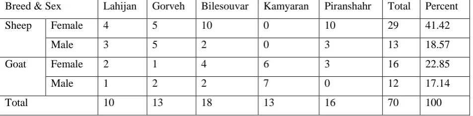
Figure 1: Results of multiplication of Gene F of PPR virus from sheep and goat blood samples Gorveh. MW is
100 pb., 1 positive control, 5, 11 and 14 positive samples, well 15 is negative control and well numbers 2, 3, 4, 6,
7, 8, 9, 10, 12, 13, are negatives.
66 IJRHR (2016), 1(1): 61-72
MW 1 7 11 18
Figure 2: Results of multiplication of Gene F of PPR virus from sheep and goat blood samples of Piranshahr.
MW is 100 bp., well number 1 is positive control., 7 and 11 and are positive samples, 18 well is negative and
other numbers are negatives.
MW 1 7 11 14
igure 3: Results of multiplication of Gene F of PPR virus from sheep and goat blood samples of Bilesavar. MW
67 IJRHR (2016), 1(1): 61-72
MW 1 7 11
Figure 4: Results of multiplication of Gene F of PPR virus from sheep and goat blood samples of Kamiaran and Piranshar.
MW is 100 bp., well number 1 is positive control. Well numbers 7and 11 are positive. Others are negative for PPR.
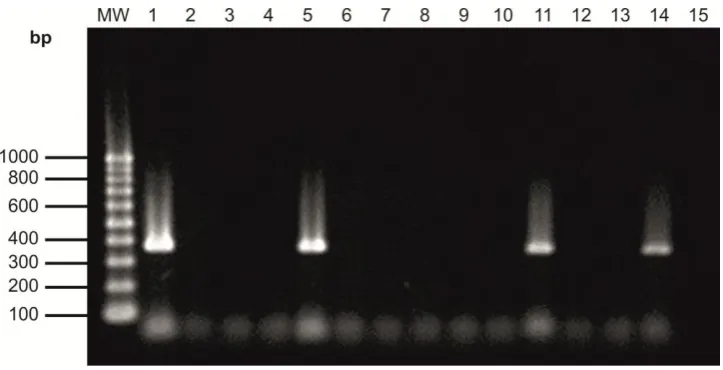
MW 1 2 3 4 5 6 7 8 9 10
Figure 5: RT-PCR results in samples on Agarose jell. MW is 100pb. Well numbers 1 and 2 are positive from
Kamiaran, 3, 4 and 5 positive samples from Gorveh, 6 and 7 positive samples from Piranshahr. Well numbers 8,
68 IJRHR (2016), 1(1): 61-72
Discussion
Among laboratory diagnostic methods,
Molecular techniques have the highest sensitivity
and specialty in detection of morbillivirus like
PPRV. In this study, Nested-PCR was used as a
diagnostic tool to detect PPR. Blood samples from
suspicious and visually healthy animals were first
analyzed with RT-PCR. They were then tested with
Nested PCR. The reason was to ensure the
Nested-PCR results and to increase sensitivity of virus
detection. Generally, Nested PCR is one of the
simplest, fastest and in the meantime one of the
most sensitive tests to confirm proliferated segments
in PCR reaction. In the preset study, out of 42 sheep
which were evaluated, 4 were positive (10%) and
out of 28 goats, 6 were positive (22%). Out of 34
samples which were suspicious, 5 were positive and
out of 36 samples which were visually healthy, 5
were positive. In the study of Mahajan et al. (2012),
in India, after evaluating 432 serum samples with
competitive ELISA, serum prevalence was 29.16%
in sheep and was 28.70% in goat. In another study,
by Munir et al. (2009) with ELISA, infection was
38.8% in sheep and was 25.6% in goat. Higher
serum prevalence in sheep could possibly be
because of its severe infection in sheep (Radostits,
2007). So, as a result of increasing serum
prevalence, more infected sheep survive (Bhaskar et
al. 2009). Shankar et al.'s (1998) study with ELISA
indicates that being infected with this disease,
mortality rate in sheep is less than goats and
therefore, the sheep survive. Sharma et al. (2007)
point out in their study that there is higher mortality
rate in goats than sheep which have PPR. In the
study conducted by Ahmad-Almajali et al. (2005) in
Jordan, out of 929 sheep blood sample and out of
400 goat blood sample, using competitive ELISA,
29% and 49%, respectively, were positive. In the
aforementioned article, blood samples were taken
from 122 different flocks in northern Jordan.
Keeping sheep and goat together is a risky factor in
which sheep gets the PPR infection but goat does
not. PPR is endemic in Pakistan and has high
morbidity and mortality rate. In Haider Ali-Khan's
study, 933 serum samples in Panjab province were
obtained from animals which had diarrhea and
severe respiratory disease. They were tested with
competitive ELISA to see if they had anti-PPR virus
antigen. 51.34% of samples were positive (P<0.432)
in small ruminants. The number of infected sheep
was higher than goat (in sheep 56.8% and in goat
48.24%). In animals with more than 2 years, old
prevalence of disease was higher than other age
groups. Females were more infected than males.
The reason for these findings may be due to
different geographical areas, topography and social
and financial status of farmers (Ali Khan et al.
2008).
However, in the current study, out of 45 female
samples, 6 were positive (13.33%) and out of 25
samples from male animals, 4 were positive (16%).
In Rahman et al.'s (2004) study, males were infected
more than females which confirms the present
study. Prevalence of PPR in Female goats is higher
than males but the difference is little. But based on
the study of Khan et al. (2008), it is significant. To
study the correlation between age and infection rate,
samples were divided 4 groups: Under one-year-old,
one to two years old, two to three years old and
upper than three. The most positive cases were in
69 IJRHR (2016), 1(1): 61-72
positive cases were inupper-than-three group (0%).
In the study of Mahajan et al. (2012) samples were
divided into three groups: 4 to 8 months, 8 to 12
months and upper than 12 months in which the
highest serum prevalence in sheep was from "upper
than 12 months" group (39.58%) and the least was
from "4 to 8 months" (20.83%). Ozkol et al. (2002)
indicated that animals smaller than six months old
and more than one-year old are more likely to be
facing disease. Taylor et al. (1979) mentioned that
infection ages to this virus is 4 to 24 months and
most of infection and mortality happens at this
period. This finding was approved by others (Abu
Elzein et al. 1990). Abubakar et al., (2009)
mentioned that with aging, prevalence of disease
increases. In Ahmad et al's report (2005), it was
mentioned that mortality rate in infants (one to three
months old) compared to lambs (four to twelve
months old) is higher and it is the same when lambs
are compared to adults. No significant relation
found when analyzing the data based on the study
areas. Most positive casese were in Gorveh and
Bilesouvar (3 positive samples=30%) and the least
positive cases were in Lahijan (0%).
The experimental study conducted by Hymann
et al. (2009) on goat reveals that with
Immunocapture ELISA and RT-PCR, we can
diagnose the illness before clinical signs arrive. He
advises developed countries to use Immunocapture
ELISA for early diagnosis of PPR. This disease was
first reported in Ilam province which has borders
with Iraq. In the same year, eight other provinces
reported the illness got by 39 flocks (1995). From
1997 to 2004, the illness spread widely in Iran.
During these years, there were different levels of
morbidity and mortality. In the aforementioned
period of time 1443 flocks were examined in which
Qom with 283 flocks and Semnan with 3 flocks had
the most and the least number of infected flocks,
respectively. Sheep and goats of Iran in that period
of time were being vaccinated by cell cultured PPR
vaccines and from 2005 on were being vaccinated
with African PPR. From the third week of March to
the third week of September 2005, 93 flocks which
were infected by PPR in 16 provinces of Iran were
determined; East Azerbaijan province had 19
infected flocks and Tehran and Chaharmahal
Bakhtiari and Qazvin had one infected flock
(Bazarghani et al. 2006). Despite vaccination and
Controlling efforts, the disease has spread in Iran
which may be due to the following reasons:
1) Higher sensitivity of Iranian goats and
sheep
2) Absence of effective and good quarantine
3) Traditional behaviors of animal owners
4) Absence of vaccination
Because this illness reduces meat and milk and
wool production, it needs a lot of attention
(Bazarghani et al. 2006)
Suggestions
In domestic areas, PPR is a major threat for
keeping small ruminants. So it has major impact on
farmers' financial status. As a consequence,
controlling PPR is a major step in fighting against
poverty. Considering that the virus is present at
border areas of Iran, it is suggested that more
complete studies be done to investigate the shape of
70 IJRHR (2016), 1(1): 61-72
such as Cows and Camels; so, enough attention
should be given to illness in these species especially
in situations where the vaccination has been
stopped. Additionally, it is suggested that, to control
the illness, we should have increased vaccination
efforts, controlled animal transportation and
disinfection of disease centers.
Acknowledgments
The author thanks from Dr. Farzin Azarpay due to
his kindly collaboration for preparation the
manuscript.
References
Abubakar, M., Ali, Q. and Khan, H.A. (2008).
Prevalence and mortality rate of peste des
petits ruminant (PPR): possible association
with abortion in goat. Trop. Anim. Health.
Prod. 40, 5, 317-321.
Abu-Elzein, E.M.E., Housawi, F.M.T., Bashareek,
Y., Gameel, A.A., Al-Afaleq, A.I. and
Anderson, E. (2004). Severe PPR infection in
gazelles kept under semi-free range conditions.
J. Vet. Med. B. Infect. Dis. Vet. Public. Health.
51, 68-71.
Ahmad, K., Jamal, S., Ali, Q. and Hussain, M.
(2005). An outbreak of peste des petits
ruminants in a goat flock in Okara, Pakistan.
Pakistan. Vet. J. 25, 3, 146-148.
Al-Majali, A.M. (2005).Seroepidemiology of
caprine brucellosis in Jordan. Small. Rumin.
Res. 58, 13-18.
Anderson, J., McKay, J.A., (1994). The detection of
antibodies against peste des petits ruminants
virus in cattle, sheep and goats and the possible
implications to rinderpest control programmes.
Epidem. Infection. 112, 225-231.
Bhaskar, S.R., Deshmukh, V.V., Chopade, N.A.,
Rautmare, S.S. (2009). Sero-prevalence of
peste des petits ruminants in Maharashtra.
Indian. J. Anim. Res. 43, 4, 285–287.
Bazarghani, T. T., Charkhkar, S., Doroudi, J. and
Bani Hassan, E. (2006). A review on peste des
petits ruminants (PPR) with special reference
to PPR in Iran. J. Vet. Med. B. Infect Dis Vet
Public Health. 53, 17-18.
Barrett, T., Rossiter, P.B. (1999). Rinderpest: the
disease and its impact on humans and animals.
Adv. Virus. Res. 53, 89–110.
Barrett, T., Taylor, W.P. (1996). The isolation of
peste des petits ruminants virus from northern
India. Vet. Microbiol. 51, 207-216.
Brindha, K., Raj, G.D., Ganesan, P.I., Thiagarajan,
V., Nainar, A.M.,Nachimuthu, K. (2001).
Comparison of virus isolation and
polymerasechain reaction for diagnosis of peste
des petits ruminants. Acta. Virol. 45, 3, 169–
172.
Couacy-Hymann, E., Roger, F., Hurard, C., Guillou,
J. P., Libeau, G. and Diallo, A. (2002). Rapid
and sensitive detection of peste des petits
ruminants virus by a polymerase chain reaction
assay. J. Virol. Methods. 100, 17-25.
Forsyth, M. A. and Barrett,T. (1995). Evaluation of
polymerase chain reaction for the detection and
characterization of rinderpest and peste des
petits ruminants viruses for epidemiological
studies. Virus. Res. 39, 151-163.
Gibbs EPJ, Taylor WP, Lawman MPJ, Bryant J.
(1979). Classification of peste des petits
ruminants virus as the fourth member of the
Genus Morbillivirus. Int. Virol. 11, 268-74.
Gargadennec, L., Lalanne, A. (1942). La peste des
71 IJRHR (2016), 1(1): 61-72
Technique et des Epizootie de l’Afrique
Occidental Franc.16–21.
Hymann CE, Bodjo SC, Koffi MY, Kouakou C,
Danho T. (2009). The early detection of
peste-des-petits-ruminants (PPR) virus antigens and
nucleic acid from experimentally infected goats
using RT-PCR and immunocapture ELISA
techniques. Res. Vet. Sci. (2009). 87, 2, 332-5.
Khan, H.A., Siddique, M., Abubakar, M., Arshad,
M.J., Hussain, M. (2008). Prevalence and
distribution of peste des petits ruminants virus
infectionin small ruminants. Small. Rumin.
Res. 79, 152–157.
Khan, M.A., Hussein, S.N., Bahadar, S., Ali, A.,
Shah, I.A. (2008). Anoutbreak of Peste des Petits Ruminants (PPR) in goats in district
Chitral, N.W.F.P., Pakistan. J. Biol. Sci. 3, 2, 19-22.
Lefvre, P.C., Diallo, A. (1990). Peste des petites
ruminants. Revue Scientifique Office
International des Epizooties. 9, 4, 951-965.
Libeau, G., Diallo, A., Calvez, D., Lefevre, P.C.
(1992). A competitive ELISAusing anti-N
monoclonal antibodies for specific detection of
RP antibodies in cattle and small ruminants.
Vet. Microbiol. 31, 147–160.
Libeau, G., Diallo, A., Colas, F., Guerre, L. (1994).
Rapid differential diagnosis of rinderpest and
peste des petits ruminants using an
immunocapture ELISA. Vet. Rec. 134, 300–
304.
Mahajan, S., Agrawal, R., Kumar, M., Mohan, A.
and Pande,N. (2012). Risk to seroconversion
to peste des petits ruminants (PPR) and
itsassociation with species, sex, age and
migration, Small. Rumin. Res. 104, 195-200.
Munir, M., Siddique, M., Ali, Q. (2009).
Comparative efficacy of standard AGID and
precipitinogen inhibition test with monoclonal
antibodiesbased competitive ELISA for the
serology of peste des petits ruminantsin sheep
and goats. Trop. Anim. Health. Prod. 41, 3,
413–420.
Ozkul, A., Akca, Y., Alkan, F., Barrett, T.,
Karaoglu, T., Dagalp, S.B., Anderson, J.,
Yesilbag, K., Cokcaliskan, C., Gencay, A.,
Burgu, I. (2002). Prevalence, distribution, and
host range of peste des petits ruminants virus,
Turkey. Emerg. Infect. Dis. 8, 7, 708–712.
Radostits, O. M., Gay, C.C., Blood, D.C.,
Hinchcliff, K.W. (2007). Vet. Med. Elsevier 9th edn, pp:1072–1079.
Rahman, A.U., Ashfaque, M., Rahman, S.U.,
Akhtar, M., Ullah, S. (2004). Pesti des petits
ruminants antigen in mesenteric lymph nodes
of goats slaughtered at D.I. Khan. Pakistan.
Vet. J. 2, 159–160.
Shankar, H., Gupta, V.K., Singh, N. (1998).
Occurrence of peste des petits ruminants like
disease in small ruminants in Uttar Pradesh.
Indian. J. Anim. Sci. 68, 1, 38–40.
Sharma, S., Mahajan, V., Kanwar, R.S., Filia, G.,
Kumar, H., Singh, R., Bal, M.S. (2007). Peste
des petits ruminants (PPR) outbreaks in sheep
and goats in Punjab. Indian J. Vet. Pathol. 31,
1, 32–35.
Taylor, W.P. (1979). Serological studies with the
virus of peste des petitsruminants in Nigeria.
Res.Vet. Sci. 26, 236–242.
Taylor, W.P., Abegunde, A. (1979). The isolation of
peste des petits ruminants virus from Nigerian
72 IJRHR (2016), 1(1): 61-72
Taylor, W.P., Diallo, A., Gopalakrishna, S. (2002).
Peste des petits ruminants has been widely
present in southern India since, if not before,
the late 1980s. Prev. Vet. Med. Jan. 22, 52,
305-12.
Toplu, N. (2004). Characteristic and
non-characteristic pathological findings in peste des
petits ruminants (PPR) of sheep in the Ege
district of Turkey. J. Comp. Pathol. 131,