IMPLEMENTATION OF BRAIN TUMOR IDENTIFICATION USING SVM AND
CLASSIFICATION USING BAYESIAN CLASSIFIER IN MRI IMAGES
Sree Sankar. J*, R.A. Isabel
*PG scholar Sivaji College of Engineering and Technology, Kanya Kumari dist. Tamil nadu
HOD of ECE Sivaji College of Engineering and Technology, Kanya Kumari dist. Tamil nadu
DOI: 10.5281/zenodo.51753
KEYWORDS
:
Support Vector Machine, Bayesian Classifier, Zernike moments.ABSTRACT
Brain tumors are one of the deadly diseases. Identifying the type of the brain tumor is very essential for the planning of treatment and surgery. Early detection and classification of the tumors will increase the chances of survival. In this paper we propose a methodology for the detection and classification of brain tumors from Magnetic Resonance Imaging (MRI) scans. Here for the tumor detection Support Vector Machine (SVM) is used and for classification of tumor Bayesian classifier is used. MRI segmentation is carried out using Watershed segmentation algorithm. The essential features from MRI are extracted using Zernike Moments.
INTRODUCTION
Brain tumors are serious diseases that are caused due to the abnormal cell formation within the brain. The brain tumors can be classified into two types: Malignant tumors and Benign tumors. The malignant tumors are cancerous tumors whereas benign tumors are non-cancerous tumors. Malignant tumors are mostly detected in children and benign tumors are found commonly in adults.
Malignant tumors are a group of diseases that involves the growth of cells in the brain that are capable of spreading to other parts of the body. Malignant tumors are classified into primary tumors and secondary tumors. The primary tumor starts within the brain. The secondary tumors have spread from somewhere else. Benign tumors involve the abnormal growth of cells which lacks the capability of spreading to other parts of the body.
However some of the benign tumors have the capacity to become as a malignant tumor, a non-cancerous tumor can become as a cancerous tumor, through a process called Tumor progression. While comparing the growth rate of the abnormal cell formation the benign tumors have a slower growth rate than the malignant tumors. The signs and symptoms of the brain tumors include frequent headaches. Usually the headaches are at the peak during morning and it goes away with vomiting. Other symptoms include mental changes, vision problems and when the disease starts progressing unconsciousness occurs.
The Magnetic Resonance Imaging (MRI) is a noninvasive medical procedure which is used by the physicians for diagnoses and to treat the medical condition. MRI uses powerful magnetic fields, radio frequency pulses and a computer to generate the details about the organs, soft tissues and bones.
PROPOSED METHODOLOGY
The details about the anatomy and the presence of any abnormalities in the brain are contained in the Magnetic Resonance Images. Here the MRI image is the input image and in order to investigate the presence of the tumor in the MRI, effective preprocessing and segmentation of the MRI images are necessary. The Figure 1(a) shows the proposed block diagram for Brain Tumor detection and classification.
boundaries of the tumor regions. The segmentation is followed by a morphological operation. Here in this paper we use morphological erosion.
In the proposed methodology the feature extraction is done using the Zernike moments. The usage of the Zernike moments in the MRI tumor investigation has some advantages. Zernike moments are actually orthogonal moments. It has simple rotational invariance property. It means that even when the input image is rotated at any angle the image maintains its originality. The output of the Zernike moments is the magnitude value (A) and the angle value (φ), which represents the orientation of the texture. The extracted feature contains the relevant information about the presence of tumor in the brain.
Figure 1(a) Proposed block diagram for Brain Tumor detection and classification.
After the Feature extraction using Zernike moment tumor detection is performed using Support Vector Machine (SVM). For the binary classification of images Support Vector Machine is a good classifier. In many binary classification the output obtained will be either yes or no. in the identification of Brain Tumor in MRI images SVM is chosen because of its admirable capability for deriving the facts about the presence or absence of tumor in the MRI image.
For the effective tumor classification we propose Bayesian classifier. Bayesian classifier is a statistical method used for the classification of images. There are mainly three types of learning methods. They are supervised learning, unsupervised learning and reinforcement learning. Bayesian classifier is a supervised learning method. Bayesian classifier is based on the Bayes theorem.
Where, P(H) is the priori probability, event before the evidence is observed and P(H/E) is the posterior probability, event after the evidence is observed. Bayesian classifier is capable of solving the problem that involves both categorical and continuous valued attributes. The Bayesian classifier is simple to implement and it requires only small amount of training data for classification. It is mostly used in medical applications for diagnoses and analysis of effective treatments. The Bayesian classifier will provide optimal decision making even when the Bayesian methods are computationally intractable.
EXPERIMENTAL RESULTS
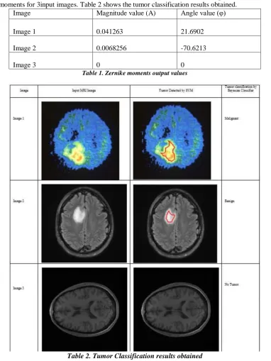
This section deals with the various simulation results obtained. Table 1 shows the output values obtained for the Zernike moments for 3input images. Table 2 shows the tumor classification results obtained.
Image Magnitude value (A) Angle value (φ)
Image 1 0.041263 21.6902
Image 2 0.0068256 -70.6213
Image 3 0 0
Table 1. Zernike moments output values
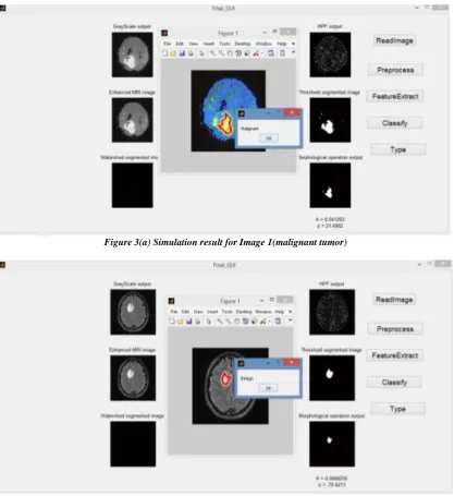
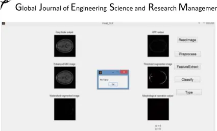
Here the simulation results obtained for the Image 1, Image 2 and Image 3 are shown. Figure 3(a) shows the simulation results obtained for Image 1 and it is classified as malignant tumor. Figure 3(b) shows the simulation results obtained for the Image 2 and it is classified as benign tumor. Figure 3(c) shows the simulation results obtained for Image 3 and it is classified as normal, it has no tumor present.
Figure 3(a) Simulation result for Image 1(malignant tumor)
Figure 3(c) Simulation result for Image 3 (No tumor)
CONCLUSION
In this paper we propose a novel method for the tumor detection and tumor classification. The proposed method is simple and easy to understand. The extraction of features using Zernike moments is very effective in the detection of tumor parts from different kinds of MRI brain tumor images invariant of its shape, size and intensity. The detection and classification of brain tumor using SVM and Bayesian classifier yields better performance.
REFERENCES
1. Ahmed kharrat, Karim Gasmi, et.al, “A Hybrid Approach for Automatic Classification of Brain MRI
Using Genetic Algorithm and Support Vector Machine,” Leonardo Journal of Sciences, pp.71-82, 2010. (Journal)
2. D.F. Specht, "Probabilistic Neural Networks for classification, mapping, or associative memory": Proceedings of IEEE International Conference on Neural Networks, Vol. I, IEEE Press, New York, pp. 525-532, June 1988.
3. Ahmed kharrat, KarimGasmi, et.al, “A Hybrid Approach for Automatic Classification of Brain MRI Using Genetic Algorithm and Support Vector Machine,”Leonardo Journal of Sciences, pp.71-82, 2010. (Journal).
4. Frank Z. Brill., Donald E. Brown., and Worthy N. Martin, “Fast Genetic Selection of features for Neural Network Classifiers,” IEEE Transaction on Neural Networks, Vol 3, No 2, pp 324-328, 1992. (IEEE Transaction).
5. Gunduz, C., B. Yener, and S. H. Gultekin “The cell graphs of cancer,” Bioinformatics. 20: i145-i151,
2004. (Journal or magazine citation).
6. G. Vijay Kumar et al., “Biological Early Brain Cancer Detection Using Artificial Neural Network,” International Journal on Computer Science and Engineering Vol. 02, No. 08, pp. 2721-2725, 2010.(Journal).
7. Jaya J, Thanushkodi K., Karnan M. Tracking algorithm for de-noising of MR brain images. International
Journal of Computer Science and Network Security 2009; 9:262-7.
8. Prastawa M, Bullitt E, Gerig G. Simulation of brain tumors in MR images for evaluation of segmentation efficacy. Medical Image Analysis 2009; 13: 297-311.
9. Ratan R, Sharma S, Sharma S. Multiparamter segmentation and quantization of brain tumor from MR