Original article
1: Department of Clinical Biochemistry, Faculty of Medicine, Tabriz University of Medical Sciences, Tabriz, Iran. 2: Department of Medical Genetics, Faculty of Medicine, Tehran University of Medical Sciences, Tehran, Iran. 3: Women's Reproductive Health Research Center, Tabriz University of Medical Sciences, Tabriz, Iran.
*Corresponding author: Mohammad Nouri; Tel: +98 4115541221; Fax: +98 4115541221; E-mail: [email protected]
Received: Oct 21, 2013; Accepted: Dec 10, 2013
www.RBMB.net
Identification of
Spata-19
New Variant with
Expression beyond Meiotic Phase of Mouse Testis
Development
Seyedmehdi Nourashrafeddin
1, Reza Ebrahimzadeh-Vesal
2,
Mohammad Hosein Modarressi
2, Ali Zekri
2, Mohammad Nouri*
1,3Abstract
Background:The study of specific genes expressed in the testis is important to understanding testis development and function. Spermatogenesis is an attractive model for the study of gene expression during germ cell differentiation. Spermatogenesis associated-19 (Spata-19) is a recently-identified important spermatogenesis-related gene specifically expressed in testis. Its protein product is involved in sperm cell development and reproduction. In this report we examined the expression of
Spata-19 mRNA in mouse testis, fetus, and cell lines.
Methods: Reverse transcription-polymerase chain reaction (RT-PCR), nested PCR, and PCR-restriction fragment length polymorphism (PCR-RFLP) were used to analyze Spata-19 mRNA expression in different stages of mouse testis development, mouse fetus, mouse embryonic fibroblasts (MEF), mouse embryonic stem cells (mESC), Sertoli cells, and NIH/3T3 cells.
Results: We identified a novel splice variant of Spata-19 in the mouse genome that it is expressed in the fetus and after the meiotic phase of spermatogenesis, and over-expressed in the post-meiotic stage of mouse spermatogenesis. This novel splice variant was absent in five days old mice testis, mESC, MEF, Sertoli, and NIH/3T3 cell lines.
Conclusion: The Spata-19 has a large novel splice variant in mouse testis that is expressed beyond meiotic phase of testis development. We suggest that this new Spata-19 mRNA variant might be involved in mitochondrial maintenance in sperm cells, and might be correlated with androgen secretion and male fertility.
Keywords: Fertility, Spata-19 new variant, Spermatogenesis, Testis
Introduction
Approximately 15% of couples are infertile (1), and the cause of contributed in 15%–30% of these cases are thought to be male genetic abnormalities (2).
Selectively-expressed genes in a certain organ may play physiological or functional roles in that particular tissue (3). The study and understanding of the mechanism of sperm formation, and identification and characterization of the genes specifically
expressed in the fetal or different stages of testis development may
aid in the discovery of previously uncharacterized genes related to testis function and spermatogenesis (4, 5), and understanding of the physiologic and pathogenic mechanisms of male infertility (6).
Mammalian spermatogenesis is an attractive model for the study of gene expression during male germ cell differentiation (3), and this process encompasses several rounds of mitotic division of
spermatogonia, meiotic division, and maturation of the haploid spermatid during spermatogenesis (7). Because meiosis in testicular germ cells differs from mitosis, gene expression differs in different stages of testis development (4, 8).
Some genes that control spermatogenesis in testis may not express in other organs (9). Some of these genes have been identified as spermatogenesis-associated genes. Abnormalities in the expression of these genes may cause male infertility (3, 10-12). One such gene is spermatogenesis associated-19 (Spata-19), also known as Spergene-1. Spata-19 is an important reproduction-related gene specifically expressed in testis and placenta (13). The Spata-19
protein product is located in the sperm mitochondrion outer membrane, is involved in sperm cell and multicellular organismal development and cell differentiation (14). Previous studies have revealed that Spata-19 has association with cancerous cell proliferation (15) and prostate cancer (16).
The purpose of this study was to analyze Spata-19
mRNA expression in pre-meiotic, meiotic, and post-meiotic stages of mouse testis development, and also in mouse adult testes, fetus, embryonic stem cell (mESC), embryonic fibroblast (MEF), Sertoli, and NIH/3T3 cell lines to understand of its role in cellular proliferation and development.
Materials and Methods
Animals
Male Balb/c mice, 5, 15, and 25 days old, representing pre-meiotic, meiotic, and post-meiotic stages of mouse testis development, respectively (17), and adult mice were purchased from the Pasteur Institute of Iran (Tehran, Iran). Their testes were surgically removed and stored at −80 °C.
Cell Line and Stem Cell Culture
Mouse embryonic fibroblast, Sertoli, and NIH/3T3 cell lines were purchased from the Pasteur Institute of Iran, and cultured in Dulbecco’s Modified Eagle’s Medium (DMEM) with 10% fetal bovine serum (FBS) at 37°C in 5% CO2. After two days, cells were
harvested and about 2x10⁶ cells were separated for RNA extraction, cDNA synthesis, and reverse-transcriptase polymerase chain reaction (RT-PCR).
The mouse embryonic stem cell line C57BL/6J with normal male (XY) karyotype (Invitrogen) was
cultured on 0.1% gelatin-coated plates inKnockout Dulbecco’s Modified Eagle’s Medium (GIBCO-BRL), supplemented with 12.5% (v/v) ES qualified FBS (GIBCO-BRL), 2 mmol L-Glutamine (GIBCO-BRL), 1X nonessential amino acids (NEAA; GIBCO-BRL), 50 µg/ml penicillin and streptomycin, 50 µmol β-mercaptoethanol and 10³ U/ml Leukemia Inhibitory Factor (LIF) (Millipore). The cells were grown for 2–3 weeks. Adherent cells were detached by trypsin/EDTA for RNA extraction.
Extraction of RNA and cDNA Synthesis
Total RNA was extracted from the cell lines and testis tissues using Tri-Pure isolation reagent (Molecular Bioscience) according to the manufacturer’s instructions. RNA was dissolved in DEPC-treated water and its concentration was determined with a NanoDrop 2000 spectrophotometer (Thermo Fisher Scientific). One µg of total RNA from each sample was used for cDNA synthesis with an M-MLV reverse transcription kit (Fermentas) and random hexamer and oligo (dT) primers (Pharmacia, Sweden).
Reverse Transcription-PCR
The housekeeping gene hypoxanthine phosphoribosyltransferase (HPRT) was used as a positive control to analyze the quality of the cDNAs. We designed a specific primer pair to amplify a 496 base pair (bp) fragment of Spata-19 mRNA. The forward1 primer is 5’gagccaaggagtccctctgta3’ and the reverse1 primer is 5’cttagcattctgagcaagaagtcc3’. The PCR amplification of Spata-19 was performed with an initial denaturation at 94 ◦C for 5 min, followed by 35 cycles of denaturation at 94 ◦C for 30 sec, annealing at 59 ◦C for 30 sec, extension at 72 ◦C for 35 sec, and an additional extension at 72 ◦C for 7 min. Reaction volumes were 25 µl containing 1 µl of cDNA sample, PCR set, and Smart Taq polymerase.
PCR-RFLP and Agarose Gel Electrophoresis
PCR amplification of full-length Spata-19 from total testicular cDNA using specific 5’ and 3’ end primers generated two amplicons. The amplified PCR products from the testes of 15 and 25 days old mice were digested with the restriction enzyme BamH1 (Fermentas, Germany). The enzyme digestion mixtures contained 7 l of PCR product, 4 units of enzyme, 2 l of the enzyme 10X buffer and H2O to a
Rep. Biochem. Mol. Biol, Vol. 2, No. 2, Apr 2014 90
final volume of 15 l. The samples were digested for 16 h at 37 ◦C. The digested and PCR products were subjected to electrophoresis in a 1.5% agarose gel and visualized under ultraviolet light after DNA staining with ethidium bromide.
cDNA Sequence Analysis and Bioinformatics tools
Bands were cut from the gel and DNAs were isolated using a Bioneer gel purification kit and sequenced on an ABI Prism 3100 sequencer (Applied Biosystems, USA). The sequence was then blasted in GenBank (http://www.ncbi.nlm.nih.gov) by the software BLAST to determine the homology among various species and locations in the chromosome. The nucleic and deduced amino acid sequences were also analyzed using NCBI and Ensemble databases. ClustalW2 - Multiple Sequence Alignment was used to align sequences similar to
Spata-19 in different species to study the
phylogenetic relationships.
Cellular Distribution of Spata-19 Precursor mRNA
The screening experiments identified a novel variant
of Spata-19 precursor mRNA in mouse testis. The
cellular distribution of the novel Spata-19 precursor mRNA was determined as follows: a primer pair specific to 5’ end of Spata-19 mRNA (forward primer 5’ctgaggatgatcattacaac3’) and intron one (reverse primer 5’accactaagaccaatgtcac3’) were designed to amplify a 198 bp fragment of the novel
Spata-19 precursor mRNA from cDNAs of mouse
fetus, mESC, MEF, NIH/3T3 and Sertoli cell lines. The PCR amplification of the novel Spata-19
precursor mRNA was performed with 35 cycles of denaturation at 94 ◦C for 30s, annealing at 55 ◦C for 25s, extension at 72 ◦C for 30s, and a 7 min final extension at 72 ◦C.
Results
Spata-19 in the genome of Mus musculus is located
on chromosome 9 (Acc: MGI: 1922719), is about 4890 bp in length, and contains seven exons. The full-length mouse Spata-19 mRNA is 697 nucleotides in length and contains a 465 bp open reading frame (ORF) that encodes a 154 amino acid protein.
In the present study, we used RT-PCR to analyze
Spata-19 gene expression during different stages of
mouse testis development and in mouse cell lines. A product of about 496 bp was amplified using
forward1 5’ and reverse1 3’ Spata-19 gene-specific primers.
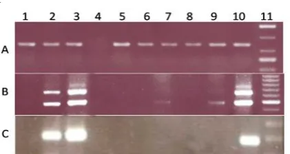
Fig. 1. RT-PCR and nested PCR result of mouse testis samples and cell lines. (A) All samples were controlled for the presence of cDNA using the housekeeping gene HPRT1. (B) Spata-19 gene expression in mouse testis samples, mESC, Sertoli, MEF, Fetus, and NIH3T3 cell lines. (C) Nested PCR was performed to expression analyze of Spata-19 new variant gene in mouse testis samples, mESC, Sertoli, Fetus, MEF, and NIH3T3 cell lines. Lane 1-3 is cDNA from 5, 15 and 25 days old testis, respectively. Lane 5-9 is cDNA from mESC, Sertoli, Fetus, MEF, and NIH/3T3 cell lines, respectively. Lane 4 is the negative control (water). Lane 10 is an adult testis as positive control .These experiments were repeated three independent times.
Control PCRs with HPRT primers amplified 148 bp fragments from all samples and verified the integrity of our cDNAs (Fig. 1A, Lanes 1-3 and 5-10). A negative control using water, as the template produced no bands, demonstrating that our reagents were not contaminated with genomic DNA (Fig. 1A, Lane 4).
A 496 bp Spata-19 RT-PCR product was amplified from 5, 15, 25 days old testes, fetus and adult mice testes (Fig. 1B, lanes 1-3, 7, and 10) and the amount of product increased with time, indicating an increase in the amount of Spata-19 mRNA (Fig 1B, Lanes 1-3). Spata-19 mRNA was strongly expressed in 15 and 25 days old mouse testis, as meiotic and post-meiotic stages of spermatogenesis respectively (Fig. 1B, Lanes 2 and 3). This Spata-19
transcript was detected in NIH/3T3 (Fig. 1B, Lane 9) but not in in mESC, Sertoli, or MEF cell lines (Fig. 1B, Lanes 5, 6 and 8). A 669 bp Spata-19 RT-PCR product was amplified from 15 and 25 days old and adult mice testes (Fig. 1B, Lanes 2, 3, and 10).
Two Spata-19 PCR products of 669 and 496 bp
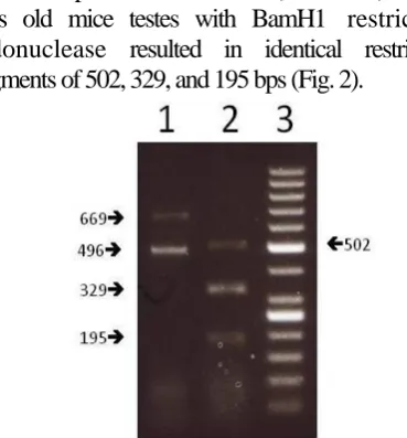
were amplified from the cDNAs of the testes from the 15 and 25 days old and adult mice (Fig 1B).To compare the products from the 15 and 25 days old mice, PCR-RFLP analysis was performed. Digestion
of the PCR products from the 15 (not shown) and 25 days old mice testes with BamH1 restriction endonuclease resulted in identical restriction fragments of 502, 329, and 195 bps (Fig. 2).
Fig. 2. Restriction fragment analysis of Spata-19 RT-PCR products from 25 days old mouse testis by agarose gel electrophoresis. Lane 1: Undigested Spata-19 PCR products from testis of 25 days old mouse. Lane 2: BamH1-digested products of Lane 1. These experiments were performed three independent times.
For further insight, we extracted the 669 bp fragment from the gel with a gel purification kit (Bioneer) and determined its DNA sequence. After BLASTing mouse genome sequence databases at the NCBI website, the sequence was localized to chromosome 9. The fragment was determined to be a novel precursor variant of Spata-19 mRNA that included intron one, which comprised 173 bps of the fragment. The nucleotide sequence of the novel
Spata-19 mRNA 669 bp variant is available from the
GenBank under the accession number JX089334. Protein sequence analysis with ExPASy translate tools (http://www.expasy.org/tools/) identified a stop codon at residue 77; therefore, the truncated Spata-19
mRNA variant encodes a protein of 76 amino acids (data not shown).
To further investigate the novel Spata-19 mRNA isoform, its expression was determined in all samples by nested PCR using primers to amplify a 198 bp fragment. We found that it was expressed in the 15 and 25 days old mice testis, representing the meiotic and post-meiotic stages of testis development and its expression was higher in the post-meiotic than the meiotic phase (Fig 1C, Lanes 2 and 3). The novel
Spata-19 mRNA isoform was also expressed in adult
testes (Fig 1C, Lane 10), but was not expressed in the
five day old testis, fetus and cell lines (Fig. 1C, Lanes 5-9).
Discussion
In eukaryotes, alternative splicing of precursor mRNA is the mane mechanism for increasing the information content from genome to transcriptome (18). Our results indicate that Spata-19 precursor mRNA is expressed after meiotic phase of testis development, and it is increased during mouse testis development by the time. However, its expression pattern is different in cell lines in comparison with Spata-19, because it has not any expression in fetus and NIH/3T3 cancerus cell lines. These findings are compatible with database
(http://www.ncbi.nlm.nih.gov/UniGene/ESTP rofile) and literature review that show the
Spata-19 expression is restricted to testis. The
analysis of gene expression in Sertoli cells after treatment by follicle-stimulating hormone (FSH) revealed that Spata-19 has no any expression in Sertoli cells (19). Also the expression analysis of genome-wide in the mouse embryonic stem cells revealed a significant inactivation of spermatogenesis related genes such as Spata-19 (20).
The NCBI Blast searches showed that the arrangement of exons and introns in Spata-19 are similar in mouse, rat, and human, and the 10 N-terminal amino acid residues in the Spata-19 protein are conserved (5). The function of the Spata-19
protein may be related to that of an adhesive molecule associated with mitochondria because of a mitochondria targeting signal sequence near the N-terminus (21, 22). Comparison and analysis of the amino acid sequence of Spata-19 with
new Spata-19 precursor mRNA variant
showed that the N-terminal region in exon one is identical between two cDNAs. According to our results that indicate the
Spata-19 precursor mRNA expression is
restricted to beyond the meiotic phase of germ cell development in testis, we suggest that
Spata-19 precursor mRNA contributes to
mitochondrial maintenance in sperm cells, and might be correlated with androgen secretion and male fertility.
Rep. Biochem. Mol. Biol, Vol. 2, No. 2, Apr 2014 92
According to a Suzuki-Toyota et al. (14) study,
the Spata-19 protein may function in developmental
and differentiation processes. Previous studies revealed that Spata-19 might be associated with cancerous cell proliferation (15) and prostate cancer disease (16). Regarding to the elevation of Spata-19 precursor mRNA during mouse testis development, and its absence in mESC and NIH3T3 cancerous cell line, these findings lead us to suggest that
Spata-19 new variant may play roles in male
germ cell development but it probably has not any roles in proliferation of cancerous cell.
In conclusion, our findings suggest that specific expression of Spata-19 new variant after meiotic phase of testis development might be correlated with spermatogenesis and male fertility, and might have some roles in the regulatory process of male gamete
development. But exact function of this variant remain unclear. We expect that these findings will be helpful to better understand the existing variants of Spata-19 during spermatogenesis.
Acknowledgements
This paper is a database of thesis from Tabriz University of Medical Sciences. The authors would like to thank Women’s Reproductive Health Research Center, Tabriz University of Medical Sciences for granting of this work, and also Department of Medical Genetic, Faculty of Medicine, Tehran University of Medical Sciences for technical assistance. The authors have no conflicts of interest to disclose and support submission to this journal.
References
1. de Kretser DM. Male infertility. Lancet. 1997 Mar;349(9054):787-90.
2. Ferlin A, Raicu F, Gatta V, Zuccarello D, Palka G, Foresta C. Male infertility: role of genetic background. Reprod Biomed Online. 2007 Jun;14(6):734-45. 3. Miyamoto T, Sengoku K, Hasuike S, Takuma N, Hayashi H, Yamashita T, et al. Isolation and expression analysis of the human testis-specific gene, SPERGEN-1, a spermatogenic cell-specific gene-1. J Assist Reprod Genet. 2003 Feb;20(2):101-4.
4. Cheng LJ, Zhou ZM, Li JM, Zhu H, Zhu H, Zhou YD, et al. Expression of a novel HsMCAK mRNA splice variant, tsMCAK gene, in human testis. Life Sci. 2002 Oct;71(23):2741-57.
5. Matsuoka Y, Iguchi N, Kitamura K, Nishimura H, Manabe H, Miyagawa Y, et al. Cloning and characterization of a mouse spergen-1 localized in sperm mitochondria. Int J Androl. 2004 Jun;27(3):152-60.
6. Okabe M, Ikawa M, Ashkenas J. Male infertility and the genetics of spermatogenesis. Am J Hum Genet. 1998 Jun;62(6):1274-81.
7. Chathoth KT, Ganesan G, Rao MR. Identification of a novel nucleolin related protein (NRP) gene expressed during rat spermatogenesis. BMC Mol Biol. 2009 Jul;10:64.
8. Grootegoed JA, Siep M, Baarends WM. Molecular and cellular mechanisms in
spermatogenesis. Baillieres Best Pract Res Clin Endocrinol Metab. 2000 Sep;14(3):331-43.
9. Eddy EM. Regulation of gene expression during spermatogenesis. Semin Cell Dev Biol. 1998 Aug;9(4):451-7.
10. Foulkes NS, Mellstrom B, Benusiglio E, Sassone-Corsi P. Developmental switch of CREM function during spermatogenesis: from antagonist to activator. Nature. 1992 Jan;355(6355):80-4.
11. Yong EL, Ghadessy F, Wang Q, Mifsud A, Ng SC. Androgen receptor transactivation domain and control of spermatogenesis. Rev Reprod. 1998 Sep;3(3):141-4.
12. Arnemann J, Jakubiczka S, Thuring S, Schmidtke J. Cloning and sequence analysis of a human Y-chromosome-derived, testicular cDNA, TSPY. Genomics. 1991 Sep;11(1):108-14.
13. Wang JY, Lan J, Zhao J, Chen L, Liu Y. Molecular characterization, polymorphism and association of porcine SPATA19 gene. Mol Biol Rep. 2012 Oct;39(10):9741-6.
14. Suzuki-Toyota F, Ito C, Toyama Y, Maekawa M, Yao R, Noda T, et al. Factors maintaining normal sperm tail structure during epididymal maturation studied in Gopc-/- mice. Biol Reprod. 2007 Jul;77(1):71-82.
15. Nourashrafeddin S, Ebrahimzadeh-Vesal R, MiryounesiM, Aarabi M, Zarghami N, Modarressi MH, Nouri M. Analysis of SPATA19 gene expression during male germ cells development, lessons from in vivo and in vitro study. Cell Biol Int Rep. 2013, doi:10.1002/cbi3.10010.
16. Ghafouri-Fard S, Ousati Ashtiani Z, Sabah Golian B, Hasheminasab SM, Modarressi MH. Expression of two testis-specific genes, SPATA19 and LEMD1, in prostate cancer. Arch Med Res. 2010 Apr;41(3):195-200.
17. Malkov M, Fisher Y, Don J. Developmental schedule of the postnatal rat testis determined by flow cytometry. Biol Reprod. 1998 Jul;59(1):84-92. 18. Chen L and Zheng S. Identify Alternative Splicing Events Based on Position-Specific Evolutionary Conservation.PLoS ONE. 2008; 3(7): e2806.
19. McLean DJ, Friel PJ, Pouchnik D, Griswold MD. Oligonucleotide microarray analysis of gene expression in follicle-stimulating hormone-treated rat Sertoli cells. Mol Endocrinol. 2002 Dec;16(12):2780-92.
20. Sainz J, Garcia-Alcalde F, Blanco A, Concha A. Genome-wide gene expression analysis in mouse embryonic stem cells. Int J Dev Biol. 2011;55(10-12):995-1006.
21. Doiguchi M, Mori T, Toshimori K, Shibata Y, Iida H. Spergen-1 might be an adhesive molecule associated with mitochondria in the middle piece of spermatozoa. Dev Biol. 2002 Dec;252(1):127-37. 22. Doiguchi M, Yamashita H, Ichinose J, Mori T, Shibata Y, Iida H. Complementary DNA cloning and characterization of rat spergen-1, a spermatogenic cell-specific gene-1, containing a mitochondria-targeting signal. Biol Reprod. 2002 May;66(5):1462-70.