1535-9778/09/$12.00 doi:10.1128/EC.00227-09
Copyright © 2009, American Society for Microbiology. All Rights Reserved.
Chitinases Are Essential for Sexual Development but Not Vegetative
Growth in
Cryptococcus neoformans
䌤
†
Lorina G. Baker,
1Charles A. Specht,
2and Jennifer K. Lodge
1*
Department of Molecular Microbiology, Washington University School of Medicine, 660 S. Euclid Avenue, St. Louis, Missouri 63110,1and Department of Medicine, University of Massachusetts, 364 Plantation Street,
Worcester, Massachusetts 016052
Received 6 August 2009/Accepted 27 August 2009
Cryptococcus neoformansis an opportunistic pathogen that mainly infects immunocompromised individuals. The
fungal cell wall ofC. neoformansis an excellent target for antifungal therapies since it is an essential organelle that
provides cell structure and integrity. Importantly, it is needed for localization or attachment of known virulence factors, including melanin, phospholipase, and the polysaccharide capsule. The polysaccharide fraction of the cryptococcal cell wall is a complex structure composed of chitin, chitosan, and glucans. Chitin is an indispensable component of many fungal cell walls that contributes significantly to cell wall strength and integrity. Fungal cell walls are very dynamic, constantly changing during cell division and morphogenesis. Hydrolytic enzymes, such as chitinases, have been implicated in the maintenance of cell wall plasticity and separation of the mother and
daughter cells at the bud neck during vegetative growth in yeast. InC. neoformanswe identified four predicted
endochitinases,CHI2,CHI21,CHI22, andCHI4, and a predicted exochitinase, hexosaminidase,HEX1. Enzymatic
analysis indicated that Chi2, Chi22, and Hex1 actively degraded chitinoligomeric substrates. Chi2 and Hex1 activity was associated mostly with the cellular fraction, and Chi22 activity was more prominent in the supernatant. The
enzymatic activity of Hex1 increased when grown in media containing onlyN-acetylglucosamine as a carbon source,
suggesting that its activity may be inducible by chitin degradation products. Using a quadruple endochitinase
deletion strain, we determined that the endochitinases do not affect the growth or morphology ofC. neoformans
during asexual reproduction. However, mating assays indicated that Chi2, Chi21, and Chi4 are each involved in sexual reproduction. In summary, the endochitinases were found to be dispensable for routine vegetative growth but not sexual reproduction.
Cryptococcus neoformansis an opportunistic fungal patho-gen that causes cryptococcosis in immunocompromised indi-viduals. The incidence of cryptococcosis continues to rise in direct proportion to the spread of the human immunodefi-ciency virus (for review, see Casadevall and Perfect [7]). It is estimated that up to 13% of AIDS patients in the United States will develop life-threatening cryptococcal meningitis, and in some parts of Africa this estimate increases to 40% (7). Current antifungal therapies for treatment of cryptococcosis are inadequate. Amphotericin B, which is believed to interact with membrane sterols (ergosterol) to produce an aggregate that forms a transmembrane channel is effective, but toxic (50, 62). Fluconazole inhibits cytochrome P-450-dependent 14␣ -sterol demethylase, which leads to the depletion of ergo-sterol and the accumulation of sterol precursors and results in the formation of a plasma membrane with altered structure and function. It is fungistatic and has high relapse rates (18, 41, 42, 50, 62). Flucytosine can be toxic and resistance occurs fre-quently (9, 41, 42, 50, 62). The newest class of antifungals to emerge is the echinocandins that targets an essential fungal enzyme required for the synthesis of a-(1,3)-glucan in the
fungal cell wall (17, 34). In addition, the echinocandins have been shown to be safe and effective for treatment of specific fungal infections, including candidiasis and aspergillosis caused byCandida albicansandAspergillus fumigatus, respectively (23, 59). However, even thoughC. neoformanspossesses the target enzyme-(1,3)-glucan synthase and in vitro assays have shown the enzyme’s activity to be inhibited by the echinocandin caspofungin (34),C. neoformansstill exhibits resistance to this class of drugs (26).
Because fungi are eukaryotes and share many biochemical processes with their host, antifungal drug design has been problematic. The cell wall is a prominent structure that differ-entiates fungi from mammalian host cells. For all fungi, this organelle is essential and provides structure as well as integrity; thus, the cell wall components or their biosynthetic pathways make attractive drug targets. In addition, the cell wall ofC. neoformans is associated with a variety of known virulence factors that are important for host-pathogen interactions, and it contains polymers including chitin and chitosan that are necessary for the viability ofC. neoformans. The first virulence factor that a host cell encounters is the polysaccharide capsule. The capsule attachment to the outer portion of the cell wall requires␣-(1-3)-glucan (15, 46). Another cell wall associated virulence factor is the melanin pigment (61) that is produced by two laccase proteins, Lac1 and Lac2 (38, 44). Lac1 is re-sponsible for generating the majority of melanin and is local-ized to the cell wall (38, 63, 69). Chitin and chitosan are essential components of the cell wall that have been shown to contribute to the overall strength and integrity of the cell wall * Corresponding author. Mailing address: Department of Molecular
Microbiology, Washington University School of Medicine, 660 S. Eu-clid Avenue, Campus Box 8230, St. Louis, MO 63104. Phone: (314) 286-2125. Fax: (314) 362-7325. E-mail: [email protected].
† Supplemental material for this article may be found at http://ec .asm.org/.
䌤Published ahead of print on 4 September 2009.
1692
on September 8, 2020 by guest
http://ec.asm.org/
(4, 5). The essentiality of the chitin component and the lack of it being present in host cells make chitin and its biosynthetic components attractive targets for drug design.
Chitin is one of the most abundant polymers found in nature (1, 12). It is a linear polymer of-(1,4)-linked N -acetylglu-cosamine (GlcNAc), and in fungi it is formed from cytoplasmic pools of UDP-GlcNAc. C. neoformans has eight predicted chitin synthases and three putative chitin synthase regulators for synthesis of chitin polymers. Mutational analysis indicate that two chitin synthases, Chs4 and Chs5, produce the majority of vegetative chitin, and one, Chs3, produces the majority of chitin that is converted to chitosan during vegetative growth (5). Chitosan, the deacetylated version of chitin, is produced by chitin deacetylases (EC 3.5.1.41) that remove acetyl groups from nascent chitin polymers. InC. neoformansthe chitin pro-duced by Chs3 and the chitin synthase regulator, Csr2, is deacetylated to chitosan by up to three chitin deacetylases (Cda1, Cda2, and Cda3) (4, 5). Strains ofC. neoformans lack-ing eitherCHS3orCSR2have significantly reduced chitosan levels and are sensitive to a variety of cell wall inhibitors (5). Similarly, strains lacking all three chitin deacetylases are un-able to convert chitin to chitosan and are sensitive to cell wall inhibitors (4). This indicates that chitosan is essential for the proper maintenance of cell wall integrity inC. neoformansand Chs3, Csr2, and the chitin deacetylases contribute to its for-mation (4, 5). Chitosan polymers of other fungi have been reported to possess various degrees of deacetylation (57). Chitin and chitosan are located throughout the lateral cell wall and bud neck regions of C. neoformans (4). During growth cellular chitin and chitosan need to be continuously remod-eled, presumably through the enzymatic digestion of chitin and chitosan polymers by chitinases and or chitosanases.
Chitinases (EC 3.2.1.14) are enzymes that hydrolyze the -(1-4) linkages in polymers of chitin. Besides being in fungi, these enzymes occur in a wide variety of organisms, including viruses, bacteria, plants, and animals (1, 12). There are two major categories of chitinases: endochitinases and exochi-tinases. Generally, the endochitinases cleave chitin chains in-ternally to generate low-molecular-mass multimers of GlcNAc. In contrast, the exochitinases are divided into two subcatego-ries: chitobiosidases (EC 3.2.1.29) release diacetylchitobiose from the nonreducing end of chitin chains, and -(1,4)-N -acetylhexosaminidases (EC 3.2.1.52) release GlcNAc from the nonreducing end of chitin oligosaccharides; both types are usually processive (12). Fungal chitosanases (EC 3.2.1.132) are less understood. They have been found inAspergillusspp. and
Gongronella sp. strain JG. Although these chitosanases have been shown to degrade chitosan, their in vitro physiological relevance has not been elucidated (8, 60).
In other fungal systems chitinases are known to be involved in cell separation, hyphal growth and branching, development of reproductive structures, spore germination, and autolysis (1, 12). In the nonpathogenic model yeastSaccharomyces cerevi-siaetwo chitinases, Cts1p and Cts2p, function independently in bud separation and spore formation, respectively (25, 27). Cts1p is the only chitinase expressed during vegetative growth, and strains lacking this enzyme display incomplete cell sepa-ration (27) that can lead to pseudohyphalike growth (25). The synthesis of the spore wall is adversely affected by the deletion
ofCTS2and affects the ability of the yeast to form mature asci (19).
C. neoformans reproduces predominantly by budding, but also has a defined sexual cycle that culminates in the produc-tion of basidiospores. Both the yeast and the spore forms are thought to be infectious particles (7).C. neoformanstypically colonizes the lungs of a immunocompromised host, from where it can disseminate to the central nervous system (7). As such, reproduction by budding has been shown to occur within host macrophages and dendritic cells (3, 28). Because fungal chitinases in other systems such asS. cerevisiaeandC. albicans
have been shown to be necessary for the completion of cell division (11, 27), understanding the biosynthesis and activity of chitinases could determine whether interfering with chitinase activity would impair the ability ofC. neoformansto reproduce. We hypothesized that the chitinases inC. neoformanswould be involved in growth and, like the chitinases inS. cerevisiae
andC. albicans, that they would degrade specific chitin during either bud separation, hyphal growth, or sporulation. In the present study we utilized a homology-based search to identify five potential chitinases in C. neoformans, the four endochi-tinasesCHI2,CHI21,CHI22, andCHI4and one exochitinase,
HEX1. Using a panel of chitinase deletion strains we discov-ered that the chitinases are dispensable for “normal” vegeta-tive growth but were necessary during development of the sexual phase ofC. neoformans.
MATERIALS AND METHODS
Fungal strains and media.KN99␣and KN99a, congenic mating-competent strains ofC. neoformansserotype A (39), were used as the wild-type strains, and all deletions were made in either KN99␣and/or KN99a(Table 1). Strains were grown on the rich medium YPD (1% yeast extract, 2% Bacto peptone, and 2% dextrose), YPGlcNAc (1% yeast extract, 2% Bacto peptone, and 2% GlcNAc) or the minimal medium YNB (pH 7.0) (6.7 g of yeast nitrogen base/liter without amino acids plus 20 g of dextrose/liter [unless otherwise specified] and 1 M morpholinepropanesulfonic acid at pH 7). Solid medium contained 2% Bacto agar. Selective YPD media contained 100g of nourseothricin (Werner Bio-Agents, Jena-Cospeda, Germany)/ml, 200 U of hygromycin (Calbiochem, La Jolla, CA)/ml, 200g of Geneticin (G418; Invitrogen, Carlsbad, CA)/ml, or 250 g of phleomycin (InvivoGen, San Diego, CA)/ml.
Analysis of chitinase protein sequences.The sequences of the twoS. cerevi-siaechitinase proteins, Cts1p and Cts2p (http://www.yeastgenome.org/), were used to perform BLAST (tBLASTn and BLASTp) searches of both theC. neoformansJEC21 (32) andC. neoformansvar.grubiigenomes and proteomes (http://cgt.genetics.duke.edu and http://www.broad.mit.edu/annotation /genome/cryptococcus_neoformans/MultiHome.html). Conserved domains were determined by using a National Center for Biotechnology Information (NCBI) BLAST search analysis (http://www.ncbi.nlm.nih.gov/Structure/cdd /wrpsb.cgi) and the SMART protein analysis tool (http://smart.embl-heidelberg .de/smart/set_mode.cgi?NORMAL⫽1) (29, 51).
Generation of deletion constructs.An overlap PCR gene deletion technology (10) was used to generate gene-specific deletion cassettes ofCHI2,CHI21,
CHI22,CHI4, orHEX1. Thechi2⌬andhex1⌬constructs each included a G418 cassette (22), thechi21⌬construct contained a nourseothricin cassette (37), the
chi22⌬construct contained a hygromycin cassette (22), and thechi4⌬construct a phleomycin cassette (22). The primers used in their construction are shown in Table S1 in the supplemental material. The amounts of coding sequence deleted forCHI2,CHI21,CHI22,CHI4, andHEX1were 1,129, 1,023, 2,420, 690, and 1,671 bp, respectively. For the generation of subsequent quadruple deletion strains,HEX1was also replaced with either a nourseothricin cassette (37) or a hygromycin cassette (22) (see Table S1 in the supplemental material).
Transformation ofC. neoformans.KN99␣or KN99awas transformed by using biolistic techniques (22, 56). Cells were grown in YPD to late log-phase, con-centrated, and plated onto YPD agar for transformation. The cells were bom-barded with 0.6-m gold beads (Bio-Rad, Richmond, CA) that were coated with DNA of the target construct according to the manufacturer’s recommendations.
on September 8, 2020 by guest
http://ec.asm.org/
After the transformation, the cells were incubated at 30°C for 4 h on nonselective medium to allow for recovery and then transferred with 0.8 ml of sterile phos-phate-buffered saline (PBS) to the appropriate selective medium. Transformants were observed in 3 to 5 days.
Generation of multiple deletion strains.Multiple deletion strains were created either by sequential biolistic transformation (see above and references 22 and 56) or by mating, followed with screening random spore isolates as described previ-ously (4, 20) (Table 1).
Analysis of transformants.To isolate stable transformants, all transformants were transferred five times on nonselective YPD medium and then tested for resistance to the appropriate selective marker. Only transformants that grew equally well on selective and nonselective media were considered to be stable. A three-primer PCR screen was used to verify homologous integration at both the 5⬘and the 3⬘ends of the deletion cassette (39). In this manner, homologous recombinants can be distinguished from the wild type. A PCR screen using primers flanking the deletion construct was used to amplify the entire integration region and demonstrate that a single copy of the transforming DNA had been inserted at the desired locus. Southern blots were performed to screen for single integration in the genome. Single bands were observed on all Southern blots when hybridized with a selectable marker-specific probe. All deletion strains generated for this work had a single deletion construct homologously integrated at the appropriate locus and no other insertions in the genome (data not shown). At least two independent isolates for each mutant were obtained.
Genomic DNA preparation.Genomic DNA was prepared by a modification of the glass bead DNA extraction protocol described (16). Briefly,C. neoformans
cells were suspended in a Microfuge tube in 500l of lysis buffer (50 mM Tris-HCl [pH 7.5], 20 mM EDTA, 1% sodium dodecyl sulfate [SDS]), with 400-mg glass beads (425 to 600m; G-9268; Sigma. St. Louis, MO). Cells were
disrupted by vortexing 10 min, followed by 10 min of incubation at 70°C. After brief vortexing, 200l of 5 M potassium acetate and 200l of 5 M NaCl were added. The tubes were placed on ice for 20 min and centrifuged at 14,000 rpm for 20 min. The supernatant was mixed with 500l of phenol-chloroform and spun for 5 min at 14,000 rpm. The aqueous phase was then mixed with 500l of chloroform and spun for 5 min at 14,000 rpm. The DNA was then precipitated by addition of 500l of isopropanol, dried, and resuspended in 50l of deion-ized water.
Southern hybridizations.Approximately 10g of genomic DNA from each strain was digested with various restriction endonucleases according to the man-ufacturer’s recommendations. Restriction fragments were separated on a 1% agarose gel and transferred to nylon membranes by using a Turbo-Blot apparatus (Schleicher & Schuell) and 10⫻SSC (1⫻SSC is 0.15 M NaCl plus 0.015 M sodium citrate) as a transfer buffer. Probes for Southern analysis were prepared by random priming (random priming kit; Roche) using 50 Ci of dCTP (AA0005; GE Bio-Sciences, Piscataway, NJ) according to the manufacturer’s instructions. The blots were incubated in 10 ml of buffer (1⫻phosphate buffer, 7% SDS) solution for 1 h at 65°C, probe was added to this solution, and the blots were hybridized at 65°C overnight. The blots were washed twice in 2⫻SSC–0.1% SDS at room temperature for 10 min and once for 10 min in 0.2⫻SSC–0.1% SDS that had been prewarmed to 65°C.
Growth curve.Exponentially growing cells were incubated in a shaking 30°C incubator with either YPD (2% glucose) or YPGlcNAc (2% GlcNAc). Aliquots were taken at 24 and 48 h, plated on solid YPD, and incubated at 30°C. CFU were counted after 2 days.
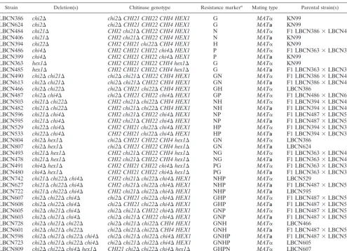
Real-time PCR.Total RNA was extracted from lyophilized KN99␣cultures that had been grown 24 h in either YPD or YPGlcNAc by using Agilent total RNA isolation kit (Agilent Technologies, Wilmington, DE) according to manu-TABLE 1. Deletion strains created in this study
Strain Deletion(s) Chitinase genotype Resistance markera Mating type Parental strain(s)
LBCN386 chi2⌬ chi2⌬CHI21 CHI22 CHI4 HEX1 G MAT␣ KN99
LBCN624 chi2⌬ chi2⌬CHI21 CHI22 CHI4 HEX1 G MATa KN99
LBCN484 chi21⌬ CHI2 chi21⌬CHI22 CHI4 HEX1 N MAT␣ F1 LBCN386⫻LBCN406
LBCN406 chi21⌬ CHI2 chi21⌬CHI22 CHI4 HEX1 N MATa KN99
LBCN394 chi22⌬ CHI2 CHI21 chi22⌬CHI4 HEX1 H MAT␣ KN99
LBCN486 chi4⌬ CHI2 CHI21 CHI22 chi4⌬HEX1 P MAT␣ F1 LBCN363⫻LBCN399
LBCN399 chi4⌬ CHI2 CHI21 CHI22 chi4⌬HEX1 P MATa KN99
LBCN363 hex1⌬ CHI2 CHI21 CHI22 CHI4 hex1⌬ G MAT␣ KN99
LBCN485 hex1⌬ CHI2 CHI21 CHI22 CHI4 hex1⌬ G MATa F1 LBCN363⫻LBCN399
LBCN490 chi2⌬chi21⌬ chi2⌬chi21⌬CHI22 CHI4 HEX1 GN MAT␣ F1 LBCN386⫻LBCN406 LBCN613 chi2⌬chi21⌬ chi2⌬chi21⌬CHI22 CHI4 HEX1 GN MATa F1 LBCN386⫻LBCN406
LBCN466 chi2⌬chi22⌬ chi2⌬CHI21 chi22⌬CHI4 HEX1 GH MAT␣ LBCN386
LBCN487 chi2⌬chi4⌬ chi2⌬CHI21 CHI22 chi4⌬HEX1 GP MAT␣ F1 LBCN486⫻LBCN624 LBCN503 chi21⌬chi22⌬ CHI2 chi21⌬chi22⌬CHI4 HEX1 NH MAT␣ F1 LBCN394⫻LBCN406 LBCN482 chi21⌬chi22⌬ CHI2 chi21⌬chi22⌬CHI4 HEX1 NH MATa F1 LBCN394⫻LBCN406 LBCN596 chi21⌬chi4⌬ CHI2 chi21⌬CHI22 chi4⌬HEX1 NP MAT␣ F1 LBCN487⫻LBCN503 LBCN595 chi21⌬chi4⌬ CHI2 chi21⌬CHI22 chi4⌬HEX1 NP MATa F1 LBCN487⫻LBCN503 LBCN529 chi22⌬chi4⌬ CHI2 CHI21 chi22⌬chi4⌬HEX1 HP MAT␣ F1 LBCN394⫻LBCN399 LBCN533 chi22⌬chi4⌬ CHI2 CHI21 chi22⌬chi4⌬HEX1 HP MATa F1 LBCN394⫻LBCN399
LBCN804 chi2⌬hex1⌬ chi2⌬CHI21 CHI22 CHI4 hex1⌬ GN MAT␣ LBCN386
LBCN807 chi2⌬hex1⌬ chi2⌬CHI21 CHI22 CHI4 hex1⌬ GN MATa LBCN624
LBCN493 chi21⌬hex1⌬ CHI2 chi21⌬CHI22 CHI4 hex1⌬ NG MAT␣ F1 LBCN363⫻LBCN406 LBCN478 chi21⌬hex1⌬ CHI2 chi21⌬CHI22 CHI4 hex1⌬ NG MATa F1 LBCN363⫻LBCN406 LBCN491 chi4⌬hex1⌬ CHI2 CHI21 CHI22 chi4⌬hex1⌬ PG MAT␣ F1 LBCN363⫻LBCN399 LBCN480 chi4⌬hex1⌬ CHI2 CHI21 CHI22 chi4⌬hex1⌬ PG MATa F1 LBCN363⫻LBCN399 LBCN742 chi21⌬chi22⌬chi4⌬ CHI2 chi21⌬chi22⌬chi4⌬HEX1 NHP MAT␣ LBCN529
LBCN627 chi21⌬chi22⌬chi4⌬ CHI2 chi21⌬chi22⌬chi4⌬HEX1 NHP MATa F1 LBCN487⫻LBCN503 LBCN722 chi21⌬chi22⌬chi4⌬ CHI2 chi21⌬chi22⌬chi4⌬HEX1 NHP MATa LBCN595
LBCN607 chi2⌬chi22⌬chi4⌬ chi2⌬CHI21 chi22⌬chi4⌬HEX1 GHP MAT␣ F1 LBCN487⫻LBCN503 LBCN608 chi2⌬chi22⌬chi4⌬ chi2⌬CHI21 chi22⌬chi4⌬HEX1 GHP MATa F1 LBCN487⫻LBCN503 LBCN605 chi2⌬chi21⌬chi4⌬ chi2⌬chi21⌬CHI22 chi4⌬HEX1 GNP MAT␣ F1 LBCN487⫻LBCN503 LBCN603 chi2⌬chi21⌬chi4⌬ chi2⌬chi21⌬CHI22 chi4⌬HEX1 GNP MATa F1 LBCN487⫻LBCN503 LBCN728 chi2⌬chi21⌬chi22⌬ chi2⌬chi21⌬chi22⌬CHI4 HEX1 GNH MAT␣ LBCN466
LBCN601 chi2⌬chi21⌬chi22⌬ chi2⌬chi21⌬chi22⌬CHI4 HEX1 GNH MATa F1 LBCN487⫻LBCN503 LBCN598 chi2⌬chi21⌬chi22⌬chi4⌬ chi2⌬chi21⌬chi22⌬chi4⌬HEX1 GNHP MATa F1 LBCN487⫻LBCN503 LBCN723 chi2⌬chi21⌬chi22⌬chi4⌬ chi2⌬chi21⌬chi22⌬chi4⌬HEX1 GNHP MAT␣ LBCN605
LBCN809 chi2⌬chi22⌬chi4⌬hex1⌬ CHI21 chi2⌬chi22⌬chi4⌬hex1⌬ GHPN MAT␣ LBCN607
a
Resistance markers: G, Geneticin; N, nourseothricin; H, hygromycin; P, phleomycin. Marker order corresponds to gene deletion order left to right.
on September 8, 2020 by guest
http://ec.asm.org/
facturer’s instructions for yeast cell cultures. First-strand cDNA was made using 1g of total RNA using the First-Strand cDNA synthesis kit for reverse tran-scription-PCR (AMV; Roche, Indianapolis, IN). This cDNA was used as a template in a real-time PCR by using Roche LightCycler 480 SYBR green I according to the manufacturer’s recommendations. A Bio-Rad CFX96 thermal cycler was programmed with the following two-step PCR cycles: initial denatur-ation for 5 min at 95°C and then 10 s at 95°C and 10 s at 60°C, with a plate read repeated in the second step for a total of 44 cycles. A melting-curve analysis was performed at the end of the reaction to confirm a single product. Standard curves were determined for each primer set, and the efficiencies were calculated. The data were normalized to glucose-6-phosphate dehydrogenase andN -myristoyl-transferase cDNA expression included with each experiment.
Chitinase activity assay.Chitinase activities were measured using (GlcNAc)1-3
conjugated to 4-methylumbelliferyl (4MU) as substrates (Sigma). Assays were at 100M substrate in MacIlvaine’s citrate-phosphate buffer at various pHs rang-ing from 3.0 to 7.0. A sample, typically 10l, was added to give a final volume of 100l in each well of a black, 96-well microplate. The initial rates of 4MU cleavage from substrate, which is a fluorogenic reaction, were determined at 30°C by using a Genios fluorescent microplate reader (Tecan, Mannedorf, Switzer-land) with Ex340/Em465 filters. Standard curves for 4MU (Sigma, St. Louis, MO) in MacIlvaine’s buffer were generated for conversion of relative fluorescent units (RFU) to nmoles of product released. Samples were crude extracts pre-pared from strains grown in liquid YPD medium at 30°C for 2 days. Cells were separated from medium by centrifugation in microfuge tubes at 14,000 rpm for 2 min. The cell pellets were suspended in PBS at their original culture volume. Homogenates were made from 3 ml of culture with the cells washed three times with PBS using centrifugation at 800⫻gfor 10 min to collect cell pellets. The cells were finally suspended in 1 ml of cold PBS and transferred to 2-ml mi-crofuge tubes with 2.2 g of 0.7-mm-diameter zirconium beads prechilled on ice (Biospec Products, Bartlesville, OK). Homogenization was performed at 4°C for 6 min by using a Disruptor Genie (Scientific Products, Bohemia, NY).
Eosin Y staining.Cells were grown in 5 ml of YPD, pelleted, and washed three times with 1 ml of MacIlvaine’s buffer (0.2 M Na2HPO4, 0.1 M citric acid [pH
6.0]). Pellet was resuspended in 500l of MacIlvaine’s buffer and stained with 30 l of Eosin Y (stock 5 mg/ml) (Sigma). Cells were incubated at room tempera-ture in the dark for 10 min. Excess dye was removed by two washes with 1 ml of MacIlvaine’s buffer and resuspended in 500l of MacIlvaine’s buffer. Cells were examined with an Olympus Vanox AHBT3 microscope by using a fluorescein isothiocyanate filter set.
CFW staining.Cells were grown in 5 ml of YPD, washed three times with 1⫻
PBS, fixed, stained with 0.5 mg of calcofluor white (CFW; Sigma)/ml, and mounted following the protocol of Pringle et al. (43). Cells were examined with an Olympus Vanox AHBT3 microscope with DAPI (4⬘,6⬘-diamidino-2-phenyl-indole) filters.
Cellular chitin and chitosan content assay.To measure the chitin and chitosan content of cells, samples were divided into two aliquots. One aliquot was treated with acetic anhydride to measure chitin plus chitosan, and the second aliquot remained untreated to measure chitin. The difference between the two measure-ments was estimated to be the amount of chitosan. Cultures were initially grown for 24 h in liquid YPD medium then diluted to an optical density at 650 nm of 0.05 in fresh medium and incubated at 30°C with shaking at 225 rpm for 68 to 72 h. The two 0.5- to 1.0-ml aliquots of each culture were transferred to tared 2-ml microfuge tubes. Cells were collected by centrifugation at 14,000 rpm for 2 min, the medium was removed, and the tubes were spun again at 14,000 rpm for 1 min so as to remove any residual medium. The weight of the cell pellet of each sample was determined and defined as the wet weight, typically 30 to 40 mg. Dry weights were measured after 2 to 3 days of evaporation at 37°C. One aliquot of pelleted cells was resuspended in 1.0 ml of 1 M sodium bicarbonate, followed by the addition of 50l of acetic anhydride. The acetylation reaction proceeded for 20 min at room temperature with occasional mixing, followed by 5 min at 100°C. Cells were pelleted as described above. Both aliquots of cells were subsequently extracted with 1 ml of 6% KOH at 80°C for 90 min. Samples were centrifuged at 14,000 rpm for 20 min, and the supernatants were discarded. Each pellet was suspended in 1 ml of PBS and spun again, and the buffer was discarded. Finally, each pellet was suspended in 0.2 ml of McIlvaine’s buffer (0.2 M Na2HPO4, 0.1
M citric acid [pH 6.0]) and frozen at⫺20°C. Upon thawing, 5l of purified
Streptomyces plicatuschitinase-63 (5 mg/ml in PBS) was added to hydrolyze the chitin to GlcNAc; samples were incubated for 2 to 3 days at 37°C and then stored at⫺20°C. For colorimetric determination of the GlcNAc, the Morgan-Elson method was adapted for microplate readers essentially as previously described (6). Chitinase-treated samples were spun at 14,000 rpm for 1 min, and each 10l of sample supernatant was combined with 10l of 0.27 M sodium borate (pH 9.0) in 0.2-ml PCR strip tubes. The samples were heated in a thermocycler
(Techne, Inc., Princeton, NJ) to 99.9°C for⬃60 s, mixed gently, and incubated further at 99.9°C for 10 min. Immediately upon cooling to room temperature, 100 l of freshly diluted DMAB solution (Ehrlich’s reagent: 10 g ofp- dimethylamino-benzaldehyde in 12.5 ml of concentrated HCl and 87.5 ml of glacial acetic acid and diluted 1:10 with glacial acetic acid) was added, and this was followed by incubation at 37°C for 20 min. Then, 100-l portions of each sample were transferred to 96-well low-evaporation microtiter plates, and the absorbance at 585 nm was recorded. Standard curves were prepared from stocks of 0.075 to 2.0 mM GlcNAc (Sigma).
Plate assays of cell wall stress.Solid YPD medium was made with 0.01, 0.03, or 0.06% SDS; 1.5 M NaCl; 0.2, 0.5, or 1.0 mg of caffeine/ml; and 0.5% Congo red (Congo red stock made in 50% ethanol) or 0.5, 1.0, or 1.5 mg of CFW/ml.C. neoformansstrains were grown to mid-log phase in YPD and diluted to 107
cells/ml, and 10-fold serial dilutions were made. For each strain, 5l of each dilution was spotted onto the solid medium and grown at 25, 30, or 37°C.
Mating assays.A cell suspension of strains with opposite mating types (␣ora) was mixed together in 500l of 1⫻PBS. Approximately 100l of this suspension was applied to each microscope slide covered in V8 agar (49) and maintained in a mating chamber (100-by-15-mm petri dish), and then 4 ml of V8 medium (minus agar) was added to the chamber to maintain nutrients and chamber humidity. The mating chambers were placed in the dark at 25°C for 4 to 5 days. Bright-field images were taken on an Olympus Vanox AHBT3 microscope at designated magnifications and are representative of three or more independent experiments.
Analysis of melanin production.Cells of each strain were taken from solid YPD medium and spread onto glucose-free asparagine medium containing 2% Bacto agar (1 g ofL-asparagine, 0.5 g of MgSO4, 3 g of KH2PO4, and 1 mg of
thiamine/liter) plus 1 mML-3,4-dihydroxyphenylalanine (L-DOPA). Incubation was at 30°C for 3 to 5 days in the dark. The amount of melanin and its retention was determined visually.
Analysis of capsule formation.Strains were streaked onto Dulbecco modified Eagle medium plates (13.4 g of Dulbecco modified Eagle medium [Sigma]/liter, 25 mM morpholinepropanesulfonic acid [pH 7.0], 1.8% agar) and incubated for 2 or 5 days at 30°C. Individual isolates were resuspended in 1:4 India ink-H2O
solution. The cells were observed by using an Olympus AHBT3 microscope.
RESULTS
Identification of chitinases inC. neoformans.The genome of
C. neoformansvar.grubiistrain H99 has been jointly sequenced by Duke University (http://cgt.genetics.duke.edu) and the Broad Institute (http://www.broad.mit.edu/annotation/genome /cryptococcus_neoformans/MultiHome.html). Using multiple BLASTp searches of the H99 genome revealed four potential endochitinase genes,CHI2,CHI21,CHI22, andCHI4, and one exochitinase gene, HEX1 (Fig. 1 and Table 2). The initial BLASTp searches used as queries the two knownS. cerevisiae
chitinases, Cts1p and Cts2p. Cts1p is the main chitinase in-volved in budding (27), and a homolog to Cts1p was not found in the genome of C. neoformans. This result was surprising sinceC. neoformansreplicates preferentially by budding. Cts2p is involved in the formation of the ascospore cell wall inS. cerevisiae (Saccharomyces Genome Database entry by E. Bogengruber et al. in 2001). The BLASTp search with the Cts2p protein sequence from S. cerevisiae yielded three ho-mologs, Chi2, Chi21, and Chi22 with E values of⬍e⫺9. Chi4
was identified using the predicted sequence forC. neoformans
Chi22 and had an E value of⬍e⫺5. C. neoformansHex1 was
found by using the Hex1 sequence fromCandida albicans(65) and had an E value of ⬍e⫺69. We further queried the H99
genome using the conserved chitinase catalytic domains found in both the endo- and exochitinases. This search method yielded no additional hits for putative endo- or exochitinases encoded byC. neoformans, indicating that we had likely iden-tified all of the chitinases encoded by C. neoformans. Each chitinase gene was also found in the genome of strain JEC21,
on September 8, 2020 by guest
http://ec.asm.org/
a C. neoformans var. neoformans strain that has also been sequenced and annotated (32).
BLASTp searches of theS. cerevisiaegenome using the five predictedC. neoformans chitinase proteins indicated that S. cerevisiaedoes not contain a homolog for either Chi4 or Hex1. Therefore, we determined their representation in other se-quenced fungal genomes. A query of the NCBI nonredundant fungal protein databases withC. neoformans’ Chi4 sequence yielded only two other basidiomycete homologs:Ustilago may-dis(E value⫽e⫺60) andMoniliophthora pernicio(E value⫽
e⫺43). Interestingly, the closest nonfungal homologs are
puta-tive chitinases of the amoebozoans,Tetrahymena and Physa-rum(E value⫽e⫺30). In contrast, queries withC. neoformans
Hex1 found several ascomycete and basidiomycete species. A SMART analysis of each of the C. neoformansputative chitinase proteins revealed that Chi2, Chi21, Chi22, and Chi4 are endochitinases (EC 3.2.1.14), with each containing a con-served Glyco_Hydo_18 domain (Pfam00704), and Hex1 as a
N-acetylhexosaminidase (EC 3.2.1.52) that contains a con-served Glyco_Hydro_20 domain, Pfam00728 (http://smart .embl-heidelberg.de) (29, 30, 51) (Fig. 1). Based on sequence similarities to endochitinases from bacteria, the four endochi-tinases ofC. neoformans belong to family 18 of the glycosyl hydrolase superfamily and group into the fungal/bacteria class reviewed by Dou-Chuan (12). Endochitinases randomly cleave chitin polymers at internal sites to generate low-molecular-mass multimers of GlcNAc (12). Chi2, Chi22, and Chi4 have two highly conserved regions within the Glyco_hydro_18 do-main: a putative substrate-binding site, SXGG, and a putative catalytic domain, DXXDXDXE (21). Chi21 also contains a putative catalytic domain overlapping the Glyco_hydro_18 do-main; however, the predicted putative substrate-binding site is located 24 amino acids outside the Glyco_hydro_18 domain toward the N terminus. The one exochitinase inC. neoformans, Hex1, is predicted to be a -(1,4)-N-acetylhexosaminidase. Hexosaminidases belong to the glycosyl hydrolase family 20 that are found in bacteria, fungi, and humans. Their main function is to complete the chitin degradation begun by the chitinases and generate GlcNAc for uptake by cells (12).
In other organisms chitinases have been reported to also contain a chitin-binding domain that is separate from the cat-alytic domain. We did not find a chitin-binding domain for any of the predicted chitinases inC. neoformans. Lack of this do-main does not appear to hamper the ability of chitinases to hydrolyze chitin (12).
N-terminal signals were predicted in four of the chitinases Chi21, Chi22, Chi4, and Hex1 using SignalIP V3 (http://www .cbs.dtu.dk/services/SignalP); these sequences are indicative of secreted proteins. The TMHMM V2 server (http://www.cbs .dtu.dk/services/TMHMM-2.0) predicted Chi2 to have an ␣-helix transmembrane-spanning domain at the N terminus that comprised amino acids 21 to 43. This indicated that Chi2 might be localized to the plasma membrane, where it could function in cell morphology or cell wall remodeling of chitin. Only one of the chitinases was predicted to contain a puta-tive glycosylphosphatidylinositol (GPI) anchor. An analysis of the predicted chitinase with the big-PI Fungal Predictor pro-gram (http://mendel.imp.ac.at/gpi/fungi_server.html) indicated that Chi22 has signature characteristics of GPI-anchored pro-teins: an N-terminal signal sequence and an omega () site located between hydrophilic and hydrophobic domains at the carboxyl terminus (Fig. 1 and Table 2). The attachment of a GPI moiety to the carboxyl terminus at the omega site of the polypeptide occurs after cleavage of the C-terminal hydropho-bic domain (13); therefore, we predicted this endochitinase FIG. 1. Predicted protein structure ofC. neoformanschitinases. The
name of each protein is indicated at the top of each protein structure. Domains were identified by BLAST searches of the NCBI database (www .ncbi.nlm.nih.gov/Structure/cdd/wrpsb.cgi) and SMART analysis tool (http://smart.embl-heidelberg.de/). Key: solid black box, predicted N-ter-minal secretion signal sequence; open rectangle, predicted transmem-brane domain on Chi2; solid triangles, predicted Glyco_hydro_18 domain of endochitinases; open rectangle, predicted Glyco_hydro_20 domain of exochitinase;, predicted GPI cleavage site, big-PI Fungal Predictor (http://mendel.imp.ac.at/gpi/fungi_server.html).
TABLE 2. C. neoformanspredicted endochitinases and hexosaminidase
Gene JEC21 NCBI serotype D
Broad Institute serotype A
Chromo-some no.
No. of predicted
introns
Predicted protein
(aa)a
N-terminal signal sequence
Predicted GPI anchorsite
(aa)
C-terminal hydrophobic domain
CHI2 XP_571896 CNAG_03412 7 12 453 None None None
CHI21 XP_567647 CNAG_02598 11 5 531 MHFVGSTTLFVILTALAVRSA None None
CHI22 XP_572898 CNAG_04245 9 3 830 MFLSTPAVLSFVLLLASQSSAQ 794 WPLTDAVRSGLGLPAV
CHI4 XP_570840 CNAG_02351 5 7 635 MYCTLATLSLLALAEA None None
HEX1 XP_571630 CNAG_06659 6 10 587 MLFNGLLEAVSLSLPFFASPSPLSA None None
a
aa, amino acid(s).
on September 8, 2020 by guest
http://ec.asm.org/
protein may be GPI anchored in the plasma membrane and/or cross-linked to-(1-6)-glucan in the cell wall ofC. neoformans. C. neoformans does not encode known chitosanases. The majority of the chitin in the vegetative cell wall ofC. neofor-mans is converted to the deacetylated form, chitosan (4, 5). Thus, we considered thatC. neoformans might make an en-zyme specifically for the degradation of chitosan. Chitosanases have been identified in other fungal species, including Aspergil-lusandGongronella spp. (8, 60, 68). However, an exhaustive search for chitosanases encoded byC. neoformansusing sev-eral of the fungal and bacterial chitosanases deposited in the NCBI database yielded no significant hits. This may indicate that C. neoformans does not have enzymes that specifically digest chitosan.
Chitinase activity in C. neoformans. We first established
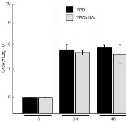
measurable chitinase activity of wild-type KN99␣cultures. Be-cause chitinase gene expression in other fungi has been re-ported to be controlled by a repressor (glucose) and inducer (products of chitin degradation) system (12), we compared activities from cells grown using glucose or GlcNAc as the principal carbon source. First, we examined whether the sub-stitution of GlcNAc for the glucose component in the growth medium would affect the replication of vegetative cells. The ability to grow on either carbon source was not adversely af-fected for the wild type (Fig. 2) or any mutant strain (data not shown). The data indicated that we could grow the cells in either liquid YPD or YPGlcNAc medium for the determina-tion of chitinase activity during vegetative growth. In addidetermina-tion, at the 24-h time point we used quantitative PCR to determine whether growth in GlcNAc induced transcription of the chi-tinase or hexosaminidase genes. Interestingly, the data indi-cated that vegetative growth in YPGlcNAc did not increase the transcription level of any of the chitinases above that of
cul-tures grown in YPD but did cause a significant increase in the transcription level ofHEX1compared to growth in YPD (P⫽
0.01) (Table 3).
Enzyme activities of wild-type cells were measured for whole cultures, culture supernatants, cell pellets, and cell homoge-nates. GlcNAc and chitin oligosaccharides, including di- and tri-GlcNAc conjugated to 4MU, were used as substrates. Ac-tivity was measured as cleavage between GlcNAc and 4MU, which is a fluorogenic reaction commonly used for chitinase activity assays (47). Activity against GlcNAc-4MU was de-tected in the whole culture, cell pellet, and cell homogenate fractions, but there was at least a 20-fold increase in activity when the cells were grown in GlcNAc compared to glucose (Fig. 3). Activity in YPD was almost undetectable in the whole culture and the cell pellet. Activity in YPD was absent in the culture supernatant and almost undetectable when grown in YPGlcNAc (Fig. 3). The data indicated activity on the mono-GlcNAc substrate is inducible by carbon source, is not se-creted, and is associated with the cell.
Total culture activity was detected with (GlcNAc)2-4MU and (GlcNAc)3-4MU in both growth media, but growth in YPGlcNAc more than doubled the activity in whole culture on these substrates (Fig. 3 and data not shown). The activity in the supernatant was similar when cells were grown on the two carbon sources, but cell-associated activity increased when grown on GlcNAc, suggesting that secreted chitinase activity is unaffected by carbon source. Together, the data of enzymatic activities of cultures suggested that chitin degradation products can regulate some activities of chitin catabolism inC. neofor-mans(Fig. 3).
Activity of Hex1. The only predicted N
-acetylhexosamini-dase ofC. neoformansis encoded byHEX1. To determine the substrate specificity of it, a strain deleted for the four-predicted endochitinase genes, but retaining the wild-typeHEX1gene,
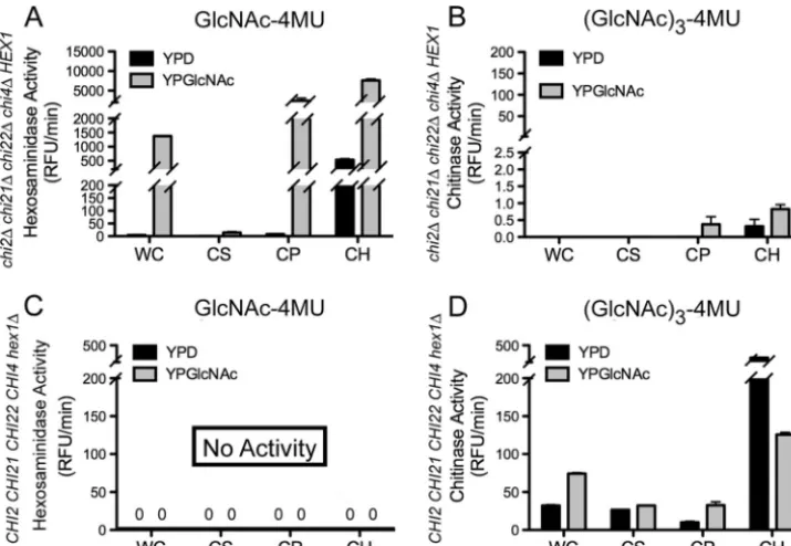
chi2⌬chi21⌬chi22⌬chi4⌬HEX1, was generated by sequential deletion and sexual crosses (see Materials and Methods and Table 3). The cell homogenate of this strain was tested using GlcNAc-4MU and (GlcNAc)3-4MU as substrates and showed abundant activity against the GlcNAc-4MU substrate but was barely detectible with the (GlcNAc)3-4MU substrate at ⬍1
RFU/min, (Fig. 4). Even after overnight incubation of the reaction, activity against the (GlcNAc)3-4MU substrate did not
increase (data not shown). It is possible that the chitinase activity on the (GlcNAc)3-4MU substrate observed in the wild
type when grown on GlcNAc as a carbon source was due to induction of hexosaminidase activity (Fig. 3); however, when the quadruple endochitinase deletion strain was grown in
TABLE 3. Induction of chitinase gene expression by growth in YPGlcNAc
Gene Fold induction of chitinase
gene expression⫾SEM
CHI2...0.34⫾0.18
CHI21...1.13⫾0.3
CHI22...0.26⫾0.29
CHI4...0.35⫾0.22
HEX1...7.11⫾2.1a
a
Statistically different from growth in YPD (P⫽0.011关Student’sttest two-tailed distribution, assuming equal variance兴).
FIG. 2. Growth assay of wild-type KN99␣in YPD and YPGlcNAc. Liquid cultures were started at 106cell/ml in YPD or YPGlcNAc as the sole source of carbon and incubated at 30°C with shaking. At time points 0, 24, and 48 h, aliquots were removed and plated. After 3 days at 30°C the CFU were determined. Each time point and carbon source includes three independent biological replicas having two technical replicates.
on September 8, 2020 by guest
http://ec.asm.org/
GlcNAc as the carbon source we still observed⬍1 RFU/min activity against (GlcNAc)3-4MU in the cell homogenate frac-tion (Fig. 4). The inability of thechi2⌬chi21⌬chi22⌬chi4⌬ HEX1 strain to release 4MU from (GlcNAc)3-4MU but to cleave GlcNAc-4MU suggested that the hexosaminidase asso-ciated with this fraction could not remove more than one GlcNAc subunit from the substrates. This is consistent with Hex1 cleaving one GlcNAc subunit from the (GlcNAc)2-4MU and higher substrates and not proceeding further; thus, Hex1 activity is not processive.
The GlcNAc-4MU substrate was cleaved as efficiently by the strain that lacks the four endochitinases as a wild-type strain, which indicates that Hex1 is the enzyme responsible for cleav-age of this substrate. Furthermore, the activity was found to be cell bound with⬍1% of the total activity released into the culture medium (Fig. 5). This activity correlates with the ac-tivity measured on this substrate in wild-type cell homogenate fractions (Fig. 3). In the absence of the four chitinases a small
amount of activity (⬍0.5 RFU/min) on the (GlcNAc)3-4MU
was observed in the cell pellet fraction (Fig. 5). This minimal activity may indicate that Hex1 has a residual ability to act processively or the (GlcNAc)3-4MU substrate contains small amounts of GlcNAc-4MU.
To confirm that the activity on the GlcNAc-4MU substrate was due to Hex1, ahex1⌬strain was generated and tested for substrate specificity. No activity against the GlcNAc-4MU sub-strate was observed (Fig. 5), although activity against the (GlcNAc)3-4MU substrate was similar to the wild type in this deletion strain (Fig. 4 and 5). This indicated that Hex1 is solely responsible for the hexosaminidase activity inC. neoformans
and that the specificity and activity of each of the four endo-chitinases could be measured by using (GlcNAc)3-4MU as a substrate in the presence of Hex1.
Endochitinase activity of Chi2 and Chi22. Deletion of all
four endochitinases virtually eliminated activity on the trimeric substrate. To deduce the activities attributable to each of the FIG. 3. Hexosaminidase and chitinase activities of wild-type KN99␣in YPD and YPGlcNAc. (A) Relative chitinase activity as detected with the fluorogenic release of 4MU from GlcNAc-4MU. Cells were assayed after 2 days of growth with shaking in liquid medium at 30°C. Measurements were performed on WC (chitinase activity of total cultures containing whole cells and supernatant), CS (culture supernatant), CP (cell pellet), and CH (cell homogenate) fractions. (B) Relative chitinase activity as detected with GlcNAc3-4MU as a substrate with measurements on the same fractions as in panel A.
FIG. 4. Cell-associated hexosaminidase activity of Hex1 isolated from cultures grown in YPD and YPGlcNAc. The genotype of each strain tested is indicated below thexaxis of graphs; a plus symbol (⫹) designates a wild-type gene and a delta symbol (⌬) designates a gene deletion. (A) Measurement of relative chitinase activity of cell homogenates for indicated strains grown for 2 days with shaking in liquid YPGlcNAc medium at 30°C with GlcNAc-4MU as a substrate. (B) Relative chitinase activity as detected with (GlcNAc)3-4MU as a substrate for the indicated strains.
on September 8, 2020 by guest
http://ec.asm.org/
four putative endochitinases, we generated a series of triple endochitinase deletion strains (Table 1), grew them in YPD or YPGlcNAc, and then measured the activity of total cultures using (GlcNAc)3-4MU as a substrate. The triple-deletion strains each retain a single endochitinase gene; thus, the activ-ity remaining in that strain is attributable to the remaining endochitinase. These triple mutants still containedHEX1, but because Hex1 activity on (GlcNAc)3-4MU is either not de-tectible or barely dede-tectible depending upon the culture frac-tion, it suggests that the specificity and activity of each of the four endochitinases could be measured using (GlcNAc)3-4MU as a substrate in the presence of Hex1. Strains containing only
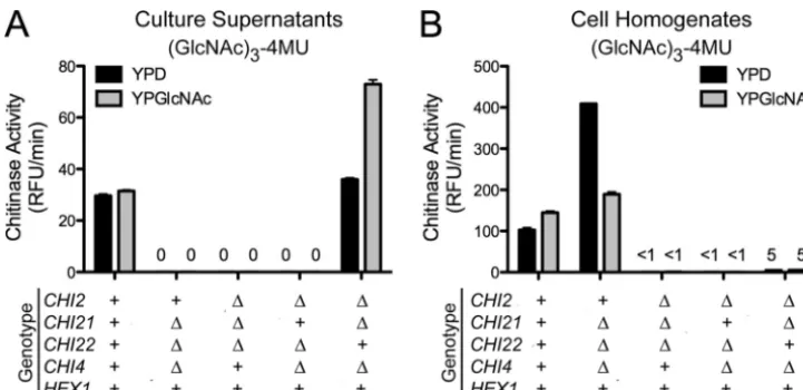
CHI4 (chi2⌬ chi21⌬ chi22⌬ CHI4 HEX1) or CHI21 (chi2⌬ CHI21 chi22⌬chi4⌬HEX1) showed no activity on (GlcNAc)3 -4MU, and further analysis with other substrates at various pHs did not reveal any chitinase activity for either (Fig. 6 and data not shown). These data suggested that Chi4 and Chi21 do not contribute to the chitinase activity observed in the wild type during vegetative growth (Fig. 3). The strain containing the endochitinaseCHI22(chi2⌬chi21⌬CHI22 chi4⌬HEX1) had measurable activity against (GlcNAc)3-4MU when the cells were grown on either GlcNAc or glucose as a carbon source, although less on the glucose carbon source (Fig. 6). The activ-ity of this strain retaining onlyCHI22was associated with the supernatant fraction and ca. 80% was secreted (Fig. 6 and see Fig. S1 in the supplemental material). These data correlated with the predicted protein structure of Chi22, which indicated an N-terminal signal sequence for secretion outside of the cell, but not with its retention in the plasma membrane or cell wall through addition of a GPI anchor (Fig. 1). The strain
contain-ing onlyCHI2(CHI2 chi21⌬chi22⌬chi4⌬HEX1) had mea-surable activity when grown on both glucose and GlcNAc, and the activity was found only in the cell homogenate fraction (Fig. 6). Again, this correlated well with Chi2’s predicted pro-tein structure containing a transmembrane-spanning domain (Fig. 1). From these studies it became evident that Chi2 activity could be distinguished from Chi22 by assaying cell homoge-nates and supernatants and suggested that Chi2 is mostly cell associated. Taken together, these data indicated Chi2 and Chi22 were responsible for the observed chitinase activity in the wild-type strain and that their activities can be somewhat influenced by carbon source.
Endochitinases are not necessary for normal vegetative
growth.In other fungi, endochitinases are required for
sepa-ration of mother and daughter cells. For example,S. cerevisiae
endochitinase Cts1p is active during vegetative growth and functions to cleave the chitin in the bud neck during cell sep-aration (27). Deletion of CTS1 in S. cerevisiae disrupts the ability of the daughter cell to separate from the mother cell, culminating in a multi-budding/pseudohyphal phenotype (25, 27). Although C. neoformans grows vegetatively by budding and is predicted to have four distinct endochitinases, only Chi22 had weak similarity (E value of 0.001) to Cts1p ofS. cerevisiae. Therefore, we sought to determine whether one or more of the endochitinases inC. neoformanswere needed to cleave the bud neck chitin during separation of the mother from the daughter cell. Surprisingly, the morphology or ability of the chitinase deletion strains to bud during growth in either rich or minimal medium was not affected in any of the deletion strains, including the quadruple endochitinase mutant strain FIG. 5. Hexosaminidase and chitinase activities of Hex1 isolated from cultures grown in YPD and YPGlcNAc. The strains tested are indicated on they axis. Strains were grown either in YPD or in YPGlcNAc, and GlcNAc-4MU or GlcNAc3-4MU was used as a substrate. Activity measurements were done as described for Fig. 3. (A) Relative chitinase activities for thechi2⌬chi21⌬chi22⌬chi4⌬HEX1strain on GlcNAc-4MU as a substrate. (B) Relative chitinase activities for thechi2⌬chi21⌬chi22⌬chi4⌬HEX1strain on (GlcNAc)3-4MU as a substrate. (C) Relative chitinase activities for theCHI2 CHI21 CHI22 CHI4 hex1⌬strain on GlcNAc-4MU as a substrate. (D) Relative chitinase activities for theCHI2 CHI21 CHI22 CHI4 hex1⌬strain on (GlcNAc)3-4MU as a substrate.
on September 8, 2020 by guest
http://ec.asm.org/
(chi2⌬chi21⌬chi22⌬chi4⌬HEX1) (Fig. 7). These data indi-cated that the endochitinases are not necessary for vegetative growth.
Chitinases do not affect chitin or chitosan localization or
content in the vegetative cell wall.Because chitinases remodel
chitin to allow for lateral cell wall expansion in other fungi reviewed by Duo-Chuan (12) and the cell wall ofC. neofor-manscontains both chitin and chitosan (4, 5), we hypothesized that one or more of the chitinases may hydrolyze the cellular chitin that remains unconverted to chitosan during cell growth inC. neoformans. If the chitinases were remodeling the lateral cell wall, deletion of these might result in altered cell size and/or an accumulation of chitin in the cell wall. We visualized the cell wall of the quadruple chitinase deletion strain using stains that are reported to bind to chitin or chitosan; CFW binds to both chitin and chitosan, and Eosin Y is specific to chitosan. The size of the cells and the staining patterns of both dyes did not differ from that of the wild type (Fig. 7). Because staining is a qualitative and not quantitative measurement, we considered that the differences in the chitin or chitosan content might be too subtle to distinguish with the staining method, so a quantitative method was used to measure the amount of cellular chitin and chitosan. Deletion of the four endochi-tinases did not have a significant effect on these measurements (data not shown). The data suggestedC. neoformans might have an alternative mechanism for controlling the cell wall chitin content and localization other than by the endochi-tinases or a different approach was needed to detect their impact.
CHI2,CHI21, andCHI4are necessary for growth on SDS.
Even though gross morphological differences in the various chitinase deletion strains were not observed, we considered that the deletions might compromise the cell wall architecture, which could become more pronounced during conditions of stress. Therefore, we tested the ability of the chitinase deletion strains to grow on a variety of chemical agents that are com-monly used to assess different aspects of cell wall integrity. SDS
FIG. 6. Chi2 and Chi22 endochitinase activity isolated from cultures grown in YPD and YPGlcNAc. All measurements were performed on triple endochitinase deletion strains containing one endochitinase (CHI2,CHI21,CHI22, orCHI4) andHEX1. Strains were grown either in YPD or in YPGlcNAc, and GlcNAc3-4MU was used as a substrate. The genotype of each strain tested is indicated below thexaxis of graphs; a plus symbol (⫹) designates a wild-type gene and a delta symbol (⌬) designates a gene deletion. (A) Relative chitinase activities from culture supernatant; (B) relative chitinase activities from cell homogenate.
FIG. 7. Analysis of KN99␣and quadruple endochitinase deletion strains for in vitro growth morphology and chitin and chitosan local-ization. (A and B) KN99␣(top panels) andchi2⌬chi21⌬chi22⌬chi4⌬ HEX1(chi-quad⌬HEX1, bottom panels) strains were grown in YPD medium to mid-log phase and stained with CFW (A) or Eosin Y (B). Images were taken at⫻100 magnification with bright-field imaging (left panels) corresponding to the CFW/Eosin Y-stained cells (right panels). The images are representative of at least three biological replications.
on September 8, 2020 by guest
http://ec.asm.org/
is used extensively to test cell wall integrity. It can disrupt the plasma membrane in a strain whose cell wall has been com-promised. Caffeine is used to test both signal transduction and cell integrity (33), while Congo red tests cell wall integrity by inhibiting the microfibril assembly of a compromised cell (48, 58). In addition, the fungal cell wall maintains turgor pressure; hence, NaCl is often used to test for osmotic stability. Finally, CFW is often used to test cell wall integrity by disrupting the assembly of chitin microfibrils (48). Thus, when the wall is compromised any or all of these compounds may lead to lack of growth or cell death.
After 5 days on YPD medium supplemented individually with these inhibitors at a variety of concentrations (see Mate-rials and Methods), the only compound that substantially in-hibited growth of the deletion strains was SDS (Fig. 8). The single-deletionchi2⌬andchi21⌬strains grew 3 to 4 orders of magnitude less than the wild type on 0.06% SDS. In addition, the chi22⌬ and chi4⌬strains were slightly more sensitive to 0.06% SDS than the wild type. The deletion of hexosaminidase (hex1⌬) had no affect under the conditions tested (Fig. 8).
Chi2 can rescue growth on SDS.Because each of the
indi-vidual deletions of the endochitinases affected the ability ofC. neoformans to survive on 0.06% SDS, we anticipated that strains carrying multiple deletions of these genes would display an additive growth defect on this medium. Three of the triple endochitinase deletion combinations CHI21 HEX1 (chi2⌬ chi22⌬chi4⌬),CHI22 HEX1(chi2⌬chi21⌬chi4⌬), andCHI4 HEX1(chi2⌬chi21⌬chi22⌬) displayed sensitivities similar to that observed with the chi2⌬ strain, being 3 to 4 orders of magnitude more sensitive than the wild type on 0.06% SDS. We found that a strain with only CHI2 HEX1 remaining (chi21⌬ chi22⌬ chi4⌬) retained the ability to grow on this medium, which had very similar growth compared to the wild type (Fig. 8). Although the single deletion ofCHI21 had an adverse affect on growth under these conditions, a strain with only CHI21 HEX1remaining did not rescue growth to wild-type levels like that of the CHI2 HEX1 strain as described above. Interestingly, the deletion of all of the endochitinases
(chi2⌬chi21⌬chi22⌬chi4⌬) resembled the phenotype of the singleCHI2deletion (Fig. 8). Combined, these data indicated that Chi2 is the main endochitinase used for cell wall mainte-nance during vegetative growth ofC. neoformans, and it alone might compensate for the other endochitinases during this growth phase.
Chitinase deletions do not adversely affect known virulence
factors.The deletion of several genes inC. neoformans
asso-ciated with chitin synthesis affects several of C. neoformans’ virulence factors. The deletion ofCHS3andCSR2cause tem-perature sensitivity at 37°C and thus a presumed inability to grow within the host (5). In addition, these deletions, as well as the triple deletion of the chitin deacetylases (CDA1, CDA2, andCDA3), share a defect in capsule formation and melanin production/retention (4, 5). These effects may be caused by the overall cell wall architecture being changed in these deletion strains. Because chitinases are known to play a role in cell morphology (1, 12), we hypothesized that their deletion might indirectly affect these virulence factors by changing the chitin or chitosan, thus the cell wall ofC. neoformans. We tested the panel of deletion strains for the ability to grow at 37°C, to produce capsule, and to produce and retain melanin. We found that the deletion of the chitinases did not affect any of these virulence factors (data not shown).
Endochitinases are necessary for mating and sporulation.
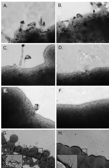
In other fungi, chitinases have been reported to be involved in the formation of spores and/or hyphal septa (1, 12). To test whether theC. neoformansendochitinases are needed for spe-cific morphological stages during mating, reactions between compatible isogenic triple and quadruple endochitinase dele-tion strains were assayed for the presence of filaments and basidiospores. After 5 days on V8 agar slides and 21 days on V8 agar plates, crosses between wild-type KN99␣and KN99a
produced several hyphae that extended distally from the yeast mat. Most hyphae had well formed basidium with long chains of basidiospores (Fig. 9). With the exception of the CHI22 HEX1 MAT␣ ⫻CHI22 HEX1 MATa(chi2⌬chi21⌬chi4⌬ ␣ ⫻ chi2⌬chi21⌬chi4⌬MATa) mating pair, all other triple endo-chitinase deletion strains produced mating structures similar to wild type in form and number. Rarely, abnormal stunted fila-ment structures were observed for theCHI22 HEX1 MAT␣ ⫻ CHI22 HEX1 MATacrosses (Fig. 9). In addition, even with the longer incubation of 21 days on V8 agar plates no mating structures were ever observed with thechi2⌬ chi21⌬chi22⌬ chi4⌬HEX1 MAT␣ ⫻chi2⌬chi21⌬chi22⌬chi4⌬HEX1 MATa
pair, suggesting that Hex1 alone could not compensate for the loss of all endochitinases during mating (Fig. 9). These data indicate Chi2, Chi21, and Chi4 are each sufficient for mating and spore production inC. neoformans.
InC. neoformanstheMATamating partner produces pher-omone in response to environmental cues such as nitrogen starvation. In response to this signal theMAT␣mating partner sends out a conjugation tube (35). Interestingly, mating reac-tions between wild-type KN99␣ and CHI22 HEX1 MATa
strains produced mating structures similar to those observed in the wild-type KN99␣ and KN99a crosses (data not shown). These data indicated that pheromone production and release by the CHI22 HEX1 MATa strain was not affected by the deletion ofCHI2,CHI21, andCHI4. In contrast, mating reac-tions between CHI22 HEX1 MAT␣ and wild-type KN99a
FIG. 8. Analysis of KN99␣and deletion strains for in vitro sensi-tivity to cell wall stressors. Strains were grown in YPD medium to mid-log phase, serially diluted 10-fold (starting with 105cells in the left column of each panel), plated on YPD solid medium or YPD solid medium supplemented with 0.06% SDS, and incubated at 30°C for 5 days. Strains are indicated at the left of the panels.
on September 8, 2020 by guest
http://ec.asm.org/
strains rarely produced any mating structures, more resem-bling theCHI22 HEX1 MAT␣ ⫻CHI22 HEX1 MATacrosses having rare abnormal stunted filament structures with no sporulation (data not shown). These data suggested the
dele-tion of CHI2, CHI21, and CHI4 in the MAT␣ background severely affected the ability of this mating partner to produce a conjugation tube in response to the MATa pheromone and indicated their importance in the initiation of mating. FIG. 9. Compatible isogenic mating crosses on V8 slides. Equal amounts of compatible mating partners␣anda, incubated in the dark at 25°C for 4 days. In the top panels, the images are⫻40 magnification. (A) KN99␣ ⫻KN99a; (B)chi21⌬chi22⌬chi4⌬␣ ⫻chi21⌬chi22⌬chi4⌬a; (C)chi2⌬chi22⌬chi4⌬␣ ⫻chi2⌬chi22⌬chi4⌬a; (D)chi2⌬chi21⌬chi4⌬␣ ⫻chi2⌬chi21⌬chi4⌬a; (E)chi2⌬chi21⌬chi22⌬␣ ⫻chi2⌬chi21⌬ chi22⌬a; (F)chi2⌬chi21⌬chi22⌬chi4⌬␣ ⫻chi2⌬chi21⌬chi22⌬chi4⌬a. In the lower panels, the images are⫻10 magnification (insets,⫻100). (G) KN99␣ ⫻KN99a; (H)chi2⌬chi21⌬chi4⌬␣ ⫻chi2⌬chi21⌬chi4⌬a. All images are representative of at least three independent experiments.
on September 8, 2020 by guest
http://ec.asm.org/
DISCUSSION
C. neoformanshas a complex life cycle. Predominantly, it is maintained in the haploid yeast form, but it can also produce hyphae and basidiospores during both monokaryotic fruiting and sexual reproduction (7, 31). Chitin is an essential compo-nent of the fungal cell wall, and in other fungi chitinases have been reported to be important for cell separation, hyphal growth, and spore production or germination (1, 12). Chitinase activity has been reported in the yeast and hyphal forms of many fungi. InC. albicans, Cht2 and Cht3 activities were found in cultures of both yeast and hypha (36).Aspergillusspp. en-code several different chitinases in their genomes; two exam-ples are ChiA, which is a GPI-anchored chitinase that func-tions in cell wall remodeling and/or cell wall maturation, and ChiB, which functions in autolysis, indicating that it has an important role in the autolytic process inA. nidulans. (45, 55, 66, 67) In addition, S. cerevisiae has two chitinases. Cts1p functions specifically during vegetative growth and aids in sep-aration of the daughter from the mother cell (27), where Cts2p functions specifically in the ascospore stage (Saccharomyces
Genome Database entry by Bogengruber et al. in 2001). Our overall goal was to gain a better understanding of the putative chitin-remodeling proteins in C. neoformans. In par-ticular, our focus was to determine how or whether the chi-tinases inC. neoformansare necessary for asexual growth and their involvement in gross sexual reproduction. Surprisingly, unlike other fungi,C. neoformanswas able to reproduce asex-ually without any of the predicted endochitinases. This may indicate that either the endochitinases are not the exclusive enzymes used for lateral cell wall expansion and bud separa-tion during vegetative growth or thatC. neoformans has an-other mechanism for chitin remodeling during this type of growth. Cts1p and Cht3 inS. cerevisiaeandC. albicans, respec-tively, are required for the separation of the daughter from the mother cell during vegetative growth (11, 27). In addition, the chitin-rich bud scars, the remnant of a cell division event that can be observed in these two fungi, appear to correlate with a requirement for a chitinase to mediate cell separation.C. neo-formanslacks chitin-rich bud scars (5), and this absence could indicate cell separation mechanisms other than chitinases. Hence, several alternative methods might be used to change the cellular chitin/chitosan amount or type to allow budding. (i) The first approach is regulation of the amount of cellular chitin at the level of chitin metabolism by reducing or increas-ing the transcription or translation of the chitin synthases and/or chitin synthase regulators, thus effectively turning on and off cellular chitin or chitosan levels. (ii) A second method would be to control the localization or activity of chitin syn-thases and chitin synthase regulators, thus turning on/off chitin synthesis in localized regions. This could allow bud formation to occur in specific areas where chitin has not been deposited. (iii) A third method would be to change the degree of chitin deacetylation of the chitin/chitosan, thus making the cellular chitin or chitosan more flexible and water soluble. This may allow a bud to form and be separated from the mother cell. Based on CFW and Eosin Y staining patterns of the vegeta-tively growing deletion strains, the chitin/chitosan localization in the cell wall and bud neck was not affected (Fig. 7 and data not shown), and the chitin/chitosan levels did not differ
signif-icantly from the wild type (data not shown). Although the transcription of the chitin synthases, chitin synthase regulators, and chitin deacetylases was not determined, these two lines of data indicate that, overall, the chitin/chitosan metabolism was not affected by the deletion of the chitinases. This indicates that the chitin synthases, chitin synthase regulators, and chitin deacetylases, as well as the localization of these chitin-produc-ing enzymes, were not grossly affected. InC. albicansand S. cerevisiaethe deletion of chitinase genes did not affect chitin synthase activity or vice versa the deletion of single chitin synthase genes had little effect on in vitro specific chitinase activity in either fungal system (52). The total vegetative chitin and chitosan content did not differ between the wild-typeC. neoformansand endochitinase deletion strains indicating that, likeC. albicansandS. cerevisiae, regulation of the chitin syn-thases and activation of the chitin synthase regulators more than likely was not affected. Perhaps the remaining Hex1 in the
chi2⌬chi21⌬chi22⌬chi4⌬HEX1strain was able to compen-sate in some small manner to allow budding. However, because Hex1 was found not to be processive, its ability to degrade cell wall chitin should be very limited. Therefore, of the options presented, a change in the type of deacetylation of the chitosan polymer (an increase in the percent deacetylation) and thus a change in the flexibility and solubility of the chitosan polymer is the most plausible. A change in flexibility of the chitin/ chitosan polymers may allow expansion of the lateral cell wall, budding through the cell wall, and the subsequent release of the daughter cell from the bud neck.
Alternatively, because the vegetative cellular chitin in the cell wall ofC. neoformansis mostly deacetylated into chitosan (4, 5), perhaps it possesses enzymes that specifically break-down chitosan. Other fungi such asAspergillusspp, Schizosac-charomyces pombe, and Gongronella spp. encode functional chitosanases (8, 53, 60); therefore, we considered thatC. neo-formansmight utilize chitosanases during vegetative growth. However, exhaustive BLAST searches of the genome of C. neoformanswith these fungal chitosanases, as well as bacterial chitosanases, yielded no significant hits. This suggested thatC. neoformans might not encode for chitosanases or has some other mechanism for the degradation of chitosan. We were unable to find any vegetative chitinase activity for either Chi21 or Chi4. Some fungal chitinases have the ability to cleave chitosan as well as chitin (40, 54, 64). Approximately 40% of the vegetative chitin in C. neoformans remains unacetylated (4), which suggests stretches of GlcNAc should still occur within the chitosan polymers inC. neoformans. Therefore, in-stead of acting upon chitin linkages (GlcNAc-GlcNAc), Chi21 and Chi4 may act specifically to degrade the linkages in cellular chitosan (GlcN-GlcN or GlcNAc-GlcN), thus degrading the “chitosan” substrate as if it were chitin.
The endochitinases do affect the more complex process of mating. Before and during mating the yeast cells ofC. neofor-mans must go through several morphological changes. Two distinct stages are cell fusion and dikaryotic filament forma-tion, both of which are prerequisites for mating inC. neofor-mans(14). A successful mating event inC. neoformansrequires multiple steps. First, haploid yeast cells of opposite mating type (aand␣) use pheromones to sense one another. Second, the two cells fuse their cytoplasm and eventually form dikaryotic filaments. The final is step is the production of basidia where