*For correspondence. Email [email protected].
© 2004 Caister Academic Press
Signal Transduction in T Helper Cells: CD4 Coreceptors
Exert Complex Regulatory Effects on T Cell Activation
and Function
Rolf König and Wenhong Zhou
Department of Microbiology and Immunology and the Sealy Center for Molecular Science; University of Texas Medical Branch, 301 University Boulevard, Galveston, TX 77555-1070, U.S.A.
Abstract
The immune system provides a highly sophisticated surveillance mechanism to detect diverse antigens and to protect the host organism from invading pathogens and altered cells (e.g., virus-infected and tumor cells). Adaptive immune responses depend on the recognition of antigen by specific antigen receptors that are expressed on the surface of T and B lymphocytes. Helper T cells provide regulatory functions and direct the adaptive immune system to respond appropriately to a particular antigen (i.e., cytotoxic T cell responses against viral infections and tumor cells, humoral responses against extracellular bacteria and parasitic worms). Helper T cells express CD4 coreceptors, which recognize conserved domains on proteins expressed by the class II major histocompatibility complex, the same proteins that present antigen to the T cell receptor. Recent progress in T cell biology has identified multiple regulatory functions of CD4 during thymocyte development and antigen stimulation of mature T helper cells. Signaling pathways induced by engagement of CD4 independently of T cell receptor signaling mediate these regulatory functions. In this review, we discuss the regulation of T cell signaling and emphasize the functional consequences of proper and improper CD4 coreceptor signaling.
Introduction
The immune system provides a highly sophisticated surveillance mechanism to detect diverse antigens and protect the host organism from invading pathogens and altered cells (e.g., virus-infected and tumor cells). Adaptive immune responses depend on the recognition of antigens by specific antigen receptors expressed on the surface of T and B lymphocytes. To initiate effector mechanisms, antigen recognition must induce intracellular signaling cascades that activate the lymphocyte and promote the differentiation to an effector cell appropriate for the
particular antigenic challenge. Importantly, regulatory mechanisms must also be present to safeguard against inadvertent self-reactivity, which could lead to autoimmunity, and to terminate immune responses, thus avoiding overexposure of the organism to toxic effectors (e.g., cytotoxic T cells, cytokines).
T lymphocytes are derived from the lymphoid lineage of hematopoietic stem cells. T cell progenitors enter the thymus where they develop into mature T lymphocytes (Res and Spits, 1999). During thymic development, the immature thymocytes undergo rearrangement of first the
β and then the α T cell receptor (TCR) genes (Khor and
Sleckman, 2002; Raulet et al., 1985). This process increases the diversity of available TCRs and assures that each T cell expresses only a single type of TCR. Only those thymocytes that have successfully completed TCR gene rearrangement will be allowed to survive. Unsuccessful rearrangement leads to programmed cell death by apoptosis.
Following rearrangement, the functionality of the maturing thymocytes is tested by interactions with thymic antigen-presenting cells (APCs). In order to be functional, the thymocytes’ TCR must be able to engage epitopes formed by short peptides bound to molecules encoded by the major histocompatibility complex (MHC). Because of the large isotypic and allelic variability of MHC molecules within each species, not all recombination events of TCR genes lead to matches with a peptide/MHC complex potentially present in the individual organism. Therefore, a positive selection event is required. Two classes of MHC molecules select T cells with different functions. MHC class I molecules bind peptides derived from proteins synthesized by the presenting cell and proteolytically processed by the cell’s proteasome (Niedermann, 2002). These peptides can combine with nascent MHC class I molecules in the endoplasmic reticulum (Norbury et al., 2001; Yewdell, 2001). MHC class II molecules bind peptides derived from extracellular, endocytosed proteins that are processed in lysosomes. Nascent and recycling MHC class II molecules bind these peptides while trafficking through lysosomes (Germain, 1994).
that will only be encountered later in life. The mechanisms and models of positive selection have been recently reviewed (Germain, 2002; Singer, 2002).
Maturing thymocytes express both CD4 and CD8 coreceptors. Following positive selection, one coreceptor gene is silenced (Leung et al., 2001; Taniuchi et al., 2002; Zou et al., 2001). Recent experimental results suggest that the duration of the selection signal determines lineage commitment with a short duration leading to differentiation to CD8+ and a long duration favoring differentiation to CD4+ thymocytes (Brugnera et al., 2000; Yasutomo et al., 2000). A negative selection step eliminates thymocytes that overtly respond to self-antigens. Estimates suggest that of all thymocytes that mature up to the double-positive (CD4+CD8+) stage, only about 3% complete maturation and emigrate from the thymus to peripheral lymphoid tissues (e.g., blood, lymph nodes, and spleen) (Surh and Sprent, 1994). Negative selection is mediated by thymic stromal cells or APCs (mainly dendritic cells) that migrate from peripheral organs into the thymus (Brocker, 1999). These cells provide a large battery, but not a complete complement, of different peptides derived from self and foreign proteins. Thymocytes that vigorously respond to the stimuli presented during negative selection will undergo apoptosis (Bommhardt et al., 2000; Robey and Fowlkes, 1994; Williams et al., 1999).
In this review, we will discuss signal transduction in CD4+ T helper (Th) cells, which regulate adaptive immune responses by promoting and directing the differentiation and activation of B cells and cytotoxic CD8+ T cells (Schoenberger et al., 1998; Snow et al., 1994). Activation of Th cells is critical for the clearance of pathogenic infections (Diepolder et al., 1998; Taylor-Robinson and Phillips, 1992) and for the destruction of tumor cells (Kahn
et al., 1991). Th cells also participate in the pathogenesis of many autoimmune disorders (Datta et al., 1997; Rizzo
et al., 1996) and in transplant rejection (Bushell et al., 1995). Th cells recognize antigen in the context of MHC class II molecules and express the CD4 coreceptor on their cell surface, which interacts with non-polymorphic regions of MHC class II (König et al., 1992; König et al., 1995). The binding of the antigen-specific TCR to antigen/MHC class II complexes initiates Th cell activation, but the engagement of CD4 by MHC class II regulates Th cell signaling, promoting progression in the cell cycle and expression of immune response genes.
T helper cell activation
The antigen-specific T cell receptor
Following thymic selection, peripheral T lymphocytes express a TCR that is composed of two transmembrane proteins, the α and the β chains, linked by a disulfide bridge.
This αβ TCR is the antigen-recognition unit of the T cell,
but it does not have intrinsic signaling capability. The ability to transduce intracellular signals is conveyed to the TCR via its mandatory and constitutive association with a multi-protein structure, termed the CD3-ζ complex. None of the
individual components of the TCR/CD3-ζ signaling machine
can be transported to the cell surface without full assembly of the complex (Sun et al., 2001).
The CD3 complex is composed of four transmembrane polypeptide chains, a γεand a δε heterodimer. These
proteins have very short extracellular domains and each intracellular domain contains a conserved protein tyrosine kinase (PTK) recognition motif, termed “Immunoreceptor Tyrosine-based Activation Motif” (ITAM). The associated, disulfide-linked ζ-dimer, sometimes substituted by a ζ-η
heterodimer, contains three ITAMs per protein chain (De Aos et al., 1997). ITAMs are substrates for src family PTKs (Iwashima et al., 1994; Weil et al., 1995), and their phosphorylation is a determining initiation event for T cell signaling (Qian et al., 1993).
The binding of ligands to the TCR triggers the activation of receptor-associated src family PTKs, such as p56lck and
p59fyn, leading to the rapid tyrosine phosphorylation of
numerous proteins. The phosphorylation of ITAMs located in the cytoplasmic tails of CD3 and ζ2 generates binding
sites for proteins bearing Src homology 2 (SH2) domains such as the cytosolic syk family PTK “ζ-associated protein
of 70 kDa” (ZAP-70) (Chan et al., 1991). Recruitment of ZAP-70 allows enhanced activation of that kinase (Lograsso et al., 1996). ZAP-70 in turn phosphorylates components of distinct downstream signaling pathways (Elder et al., 2001; Gong et al., 2001; Magnan et al., 2001). Thus, T cell activation depends on the activation of both
src family kinases and ZAP-70 (Figure 1).
The formation of signalosomes
The engagement of TCRs by antigen/MHC ligands induces a complex temporal and spatial arrangement of signaling complexes and networks. Slightly different compositions of these signaling machines – also termed “signalosomes” (Werlen et al., 2000) - using essentially the same components can induce different second messenger signals and lead to drastically diverse cellular responses. In addition, the dynamic assembly and disassembly of signalosomes is likely a major factor in regulating signal transduction networks. TCR signalosomes consist of transmembrane receptors, protein kinases, phosphatases and their substrates, all of which are organized into signaling machines by anchoring, adapter, and scaffolding proteins. Signalosomes connect events on the plasma membrane to distal signaling cascades, which ultimately modulate T cell biology. Several protein adapters, in particular “linker of activated T cells” (LAT), act as central switches that translate the quality, quantity, and duration of signals into the correct activation of specific downstream pathways (Zhang et al., 1998). Although the precise parameters that regulate the formation of signalosomes still await further clarification, recent work in this area has identified some characteristics of the signalosomes.
The relocalization of signalosomes to receptor-associated scaffolds is crucial for effective signal transduction (Delgado et al., 2000; Harder and Kuhn, 2000; Werlen et al., 2000). Adapter proteins with SH2 domains bind to the phosphorylated ζ chain. Among these proteins
is the “Src homology 2 protein of beta-cells” (Shb), which recruits LAT (Welsh et al., 1998; Zhang et al., 1998), a substrate for ZAP-70 (Figure1). Tyrosine phosphorylation of LAT leads to the recruitment of additional signaling molecules with SH2 motifs (Figures 1 and 2), including the adapter “growth factor receptor-bound protein 2” (Grb2), the phospholipase Cγ1 (PLCγ1), and the p85 subunit of
phosphatidylinositol 3-kinase (PI 3-kinase) (Paz et al., 2001; Sommers et al., 2001; Yablonski and Weiss, 2001; Zhang et al., 1998).
Active PLCγ1 hydrolyzes phosphotidylinositol
biphosphate (PIP2), producing diacylglycerol (DAG) and 1,4,5-inositol triphosphate (IP3). DAG in turn activates the serine/threonine kinase family of protein kinase C (PKC), while IP3 induces calcium ion (Ca2+) mobilization in the
cytosol (Corado et al., 1990; May et al., 1986). Thus, ZAP-70 amplifies the TCR signal by specifically phosphorylating downstream components such as LAT and PLCγ1 (Figures
1 and 2).
In this way, the signalosome expands in molecular complexity and amplifies the TCR initiated signal. Importantly, LAT can also bind proteins that negatively regulate TCR signaling. The “SH2 domain-containing hematopoietic phosphotyrosine phosphatase”, SHP-1, associates with LAT upon TCR stimulation (Kosugi et al., 2001; Su et al., 2001) and prevents further phosphorylation of the adapter by ZAP-70, suggesting a potential conversion from an “activating” to an “inhibiting” signalosome. Similarly, the “C-terminal src kinase” (Csk) relocalizes to rafts by docking to the transmembrane adapter, Csk-binding protein (Cbp), also known as “phosphoprotein associated with glycosphingolipid-enriched microdomains” (PAG) (Brdicka
et al., 2000). In rafts, Csk inhibits src family PTKs by phosphorylating their regulatory tyrosines, and thus blocks TCR-mediated signal transduction (Vang et al., 2001). Figure 1. The TCR signaling machinery and activation of PLCγ1. The initial signalosome consists of the TCR, the associated CD3/ζ2 complex, the co-receptor CD4, the phosphatase CD45, and the srk kinases p56lckand p59fyn. The syk kinase ZAP-70 is recruited following phosphorylation of tyrosines located in
ITAMs of CD3 and ζ2 by srk kinases. See text for details.
SH2
SH2
SH1
Calcineurin PLCγ1
NF-ATn
NF-ATc
IL-2 Gene
PIP2
IP3
[Ca
2+]
NF-ATc
NFATc
PKC
ZAP-70
DG
CD45
SH3
SH2
SH1
Tyr505
SH3
SH2
SH1
CD4
CD3
TCR
ζ
2p56
lckp59
fynP P
P
P
P
P P
P
The CD4 coreceptor
The best-characterized coreceptors, and most important for the discussions presented in this review, are the membrane glycoproteins CD4 and CD8. In mature T cells, the expression of CD4 and CD8 is mutually exclusive. In general, CD4+ T cells respond to antigen presented by MHC class II molecules, and CD8+ T cells respond to MHC class I-presented antigens (Bierer et al., 1989; Swain, 1983). Therefore, CD4+ and CD8+ T cells are considered to be restricted by MHC class II and MHC class I, respectively. Importantly, MHC class restriction is independent of the Th or cytotoxic functions displayed by mature T cells (Krensky et al., 1982; Meuer et al., 1982; Swain, 1981). The correlation between CD4 or CD8 expression and MHC recognition results from direct interactions between the coreceptors and relatively monomorphic regions of the membrane-proximal, immunoglobulin-like domains of the MHC molecules (Connolly et al., 1990; Gao et al., 1997; Kern et al., 1998; König et al., 1992; König et al., 1995; Salter et al., 1990). Coreceptors are associated with the TCR/CD3-ζ
complex upon T cell activation. Their presence in the TCR multi-component signaling machine amplifies or modulates the activation signal. Often, their presence is absolutely
required, but not sufficient, for productive signaling, i.e., signaling that results in cell cycle progression and effector functions. Interestingly, three-dimensional live cell imaging of fluorescence resonance energy transfer between CD3-ζ
and CD4 demonstrates recruitment of CD4 to the CD3-ζ
complex dependent on antigen-stimulation of the TCR (Zal
et al., 2002). Importantly, the antigen threshold for T cell activation is affected by the presence of CD4 in the TCR/ CD3 complex. In the presence of CD4, a single MHC class II molecule presenting antigen suffices to induce Ca2+ flux, and 10 class II MHC/antigen complexes cause the formation of an immunological synapse (Irvine et al., 2002). Blocking of CD4 reduces the sensitivity to antigen stimulation by more than three-fold (Irvine et al., 2002).
The structural basis of interactions between CD4 and MHC class II molecules
The proteins encoded by the class II MHC consist of two non-covalently associated integral membrane polypeptide chains, the α and the β chains (König et al., 1996). Each
polypeptide chain forms two protein domains (α1, α2, β1,and β2). The α1 and β1 domains are highly polymorphic
and combine to form an antigen peptide-binding groove (Brown et al., 1988; Brown et al., 1993). Using a combination of mutational and functional analysis, we identified amino acid residues on MHC class II molecules that mediate the interaction with CD4 (König et al., 1992; König et al., 1995). A major CD4 binding site, forming a short loop exposed on the surface of the class II β2 domain,
consists of amino acids 134-148 (Cammarota et al., 1992; König et al., 1992). Specifically, glutamic acid 137 is mandatory for mediating the interactions with CD4, and an alanine substitution at this position alone will disrupt all T cell functions mediated by engagement of CD4 (König
et al., 1992). A second interaction site is located within the
α2 domain of MHC class II (Gaubin et al., 1999; König et al., 1995). Importantly, both sites are required for mediating interactions with CD4. We have recently reviewed the literature on interactions between MHC molecules and coreceptors of the TCR (König, 2002).
Functional consequences of CD4 engagement by monoclonal antibodies
The crystal structure of the exodomains of human CD4 suggests that the membrane proximal domains of CD4 may promote dimerization (Wu et al., 1997). Extraction of CD4 oligomers from freshly isolated T lymphocytes and lymphoid cell lines also indicates that oligomerization may be an intrinsic property of CD4 molecules (Lynch et al., 1999). Many cell surface receptors transduce signals following oligomerization (Papoff et al., 1999; Rodriguez-Frade et al., 1999; White and Tartaglia, 1999). Therefore, a common procedure to test whether CD4 can activate intracellular signaling pathways utilizes crosslinking anti-CD4 antibodies alone or in conjunction with anti-TCR antibodies (Luo and Sefton, 1990; Pallier et al., 1998; Prasad et al., 1993; Ravichandran et al., 1993; Veillette et al., 1989). However, antibody-mediated crosslinking prevents co-localization of CD4 with TCR molecules (Ratcliffe et al., 1992), and does not adequately reflect CD4-mediated signaling induced by engaging MHC class II during antigen activation. Figure 2. LAT and the MAP kinase signaling pathway. Phosphorylation of
LAT by ZAP-70 induces recruitment of multiple adapter proteins and the bifunctional guanine nucleotide exchange factor, Son of sevenless (Sos-1), leading to the activation of the MAP kinase signaling pathway. See text for details.
Raf-1
Mek
MAP Kinase
Ras
GTP
GDP
Ras
Grb2
Shc
Sos-1
LAT
Ligation of CD4 by anti-CD4 antibodies or the envelope glycoprotein of the human immunodeficiency virus (HIV), gp120, inhibits antigen-dependent and -independent T cell activation (Jabado et al., 1994; Jauliac et al., 1998; Marschner et al., 2002). The inhibition of T cell activation induced by CD4 ligation depends on the cytoplasmic domain of CD4 (Marschner et al., 2002). Binding of these cross-linking ligands decreases the DNA-binding activity of the nuclear transcription factors NF-AT, NF-κB, and
AP-1, thus preventing IL-2 synthesis (Jabado et al., 1994). These results indicate that ligation of CD4 apart from the TCR/CD3 complex induces negative, regulatory signals that prevent the activation of nuclear factors necessary for IL-2 gene transcription and cell proliferation. Interestingly, memory CD4+ T cells appear to be more susceptible to negative signals transduced via CD4 as their activation can be prevented by both antibody-mediated CD4 ligation and MHC class II molecules on APCs in the absence of antigen (Farber et al., 1995).
Experiments using various monoclonal anti-CD4 antibodies recognizing different epitopes suggested the existence of specific signaling epitopes on CD4 (Milia et al., 1997). Depending on the epitope recognized by a specific monoclonal antibody, different signaling pathways are activated. Also, the simultaneous engagement of non-overlapping CD4 epitopes can modify the signals from individual epitopes (Milia et al., 1997). These experiments and structure-function analysis of the CD4-MHC class II interaction suggest that MHC class II molecules bind to a
broad region on the membrane-distal domains of CD4 (König et al., 1996), and induce signals via CD4 that differ from those induced by other natural CD4 ligands (e.g., gp120 and IL-16) or monoclonal antibodies. Therefore, we used a dual approach to identify CD4-mediated signals induced by MHC class II engagement. First, we separated TCR-mediated and CD4-mediated signals by restricting the ability of MHC class II to interact with CD4 during antigen stimulation (Zhou and König, 2003). Second, we avoided antibody-mediated crosslinking, but employed a peptide mimetic to engage the CD4 epitope recognized by MHC class II (Zhou and König, 2003). This peptide binds to CD4+ T cells (Shen et al., 1996) and soluble CD4 (Cammarota et al., 1992).
The formation of the immunological synapse
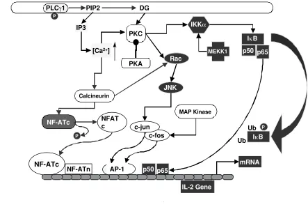
TCR-antigen/MHC interactions initiate the formation of a specialized junction between T cells and APCs, the immunological synapse (Monks et al., 1998). Stimulation-and cytoskeleton-dependent processes cluster TCR/CD3 complexes in the center of the synapse, also termed the “central zone of the supramolecular activation cluster” (cSMAC), whereas adhesion molecules such as LFA-1 form a ring surrounding the central area called the peripheral SMAC (pSMAC) (Delon and Germain, 2000; Grakoui et al., 1999). After stimulation of the T cell, CD4 coreceptors are rapidly recruited into the cSMAC, but migrate towards the periphery within a few minutes, while TCR/CD3 complexes stabilize within the central area Figure 3. The signaling pathways induced by stimulation of the TCR/CD3/ζ2 complex mediate translocation of NF-AT, AP-1, and NF-κB transcription factors to the nucleus, leading to IL-2 gene expression. See text for details.
p50 IκB
p65
p50 p65
c-fos c-jun
JNK
AP-1 Calcineurin
P
PLCγ1
mRNA
NF-ATn
NF-ATc
IL-2 Gene
MAP Kinase PIP2
IP3
[Ca2+]
NF-ATc NFATc
PKC DG
Rac
IκB
Ub Ub
MEKK1
IKKα
PP
PP
(Krummel et al., 2000). Both CD4 and CD8 associate with the PTK p56lck, and the efficient transport of p56lck into the
cSMAC is a major function of these coreceptors (Holdorf
et al., 2002).
T cell signaling pathways
One of the earliest antigen-induced signaling events is the mobilization of Ca2+, which is essential for Th cell activation (Wülfing et al., 1997). This second messenger activates several cytosolic enzymes, initiating downstream signaling cascades (Berridge, 1993a). However, antigen stimulation also generates signals through the TCR that antagonize Th cell activation. For example, TCR-mediated signals activate the 3',5'-cyclic adenosine monophosphate (cAMP)-dependent protein kinase, PKA (Laxminarayana and Kammer, 1996; Skalhegg et al., 1992). PKA initiates a signaling pathway that inhibits antigen-induced T cell proliferation and cytokine production (Paliogianni et al., 1993; Vang et al., 2001). Therefore, to achieve full activation of Th cells, TCR-mediated signals must be modified. We have recently identified signaling pathways induced by MHC class II engagement of CD4. These TCR-independent signals participate in the regulation of intracellular concentrations of both Ca2+ and cAMP (Zhou and König, 2003).
Calcium mobilization
The TCR-induced signal transduction leads to the activation of PLCγ1 (Figure 1). Binding of IP3 to its receptor in the
membrane of the endoplasmic reticulum induces the release of Ca2+ into the cytosol (Corado et al., 1990). The subsequent increase in intracellular free Ca2+ opens Ca2+ -regulated Ca2+ channels in the plasma membrane, inducing additional Ca2+ influx (Berridge, 1993b; Tsien et al., 1982). Intracellular free Ca2+ acts as an essential second messenger for T cell activation (Weiss et al., 1984). Its regulatory effects on T cell activation are mediated via calmodulin, a Ca2+-binding protein expressed in all eukaryotic cells (Zhang et al., 1998). Effective T cell activation leading to IL-2 secretion requires that intracellular Ca2+ levels be elevated for a period of 1-2 h (Karttunen and Shastri, 1991; Negulescu et al., 1994). Sustained Ca2+ signaling is required for maintaining the transcription factor “Nuclear Factor of Activated T cells” (NFAT) in the nucleus in an active form (Loh et al., 1996; Timmerman et al., 1996). NFAT is a key transcriptional regulator of the IL-2 gene (Rooney et al., 1995) (Figure 3).
Ca2+ signaling is required for various lymphocyte activities, for example cell motility is impeded by increases in Ca2+ (Donnadieu et al., 1994; Negulescu et al., 1996). Increases in Ca2+ cause changes in the cytoskeletal structure (Wülfing and Davis, 1998), induce cell death in immature thymocytes (Andjelic et al., 1993), differentiation (Sloan-Lancaster et al., 1997), and activation (Gearing et al., 1985). Thus, a single second messenger can elicit multiple cellular responses. The type of response induced may depend on the amplitude, duration, and temporal fluctuations of Ca2+ mobilization. For example, activation of NF-κB is induced by high levels of Ca2+, because of this
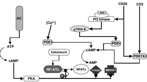
Figure 4. Cyclic AMP exerts multiple regulatory effects on T cell activation and is itself regulated by multiple signaling pathways that activate or block adenylyl cyclase and activate phosphodiesterases. Stimulatory effects are indicated by a line ending in an arrow, whereas inhibitory effects are depicted by a line ending in a filled circle. See also effects of PKA on Raf-1 (Figure 2) and PKC (Figure 3). The overall effect of PKA I activation is inhibition of IL-2 gene expression.
PI3 kinase
SH2
[Ca
2+]
PDE4
cAMP
AMP
PDE1
cAMP
PDE7A1
PKA
ATP
p70S6 K
CD3
Calcineurin
NF-ATc
NFATc c-myc MEKK1P
CD28
transcription factor’s low Ca2+ sensitivity. In contrast, even low elevations of Ca2+ levels, if maintained over a prolonged period of time, selectively activate NFAT, because NFAT is highly sensitive to Ca2+ but is rapidly inactivated after Ca2+ removal (Dolmetsch et al., 1997).
Cyclic AMP, adenylyl cyclases, and cyclic nucleotide phosphodiesterases
Cyclic AMP has been defined as an intracellular second messenger to a wide variety of hormones and neurotransmitters. In T cells, numerous studies have shown that elevated cAMP levels antagonize T cell activation. In addition to inhibiting T cell proliferation (Lingk et al., 1990), cAMP suppresses the production of IL-2 and IFN-γ in
activated Th cells (Zidek, 1999). Opposite effects on IL-5 production from activated Th cells have been observed in different studies (Staples et al., 2000). TCR signaling alone elevates adenylyl cyclase (AC) activity and intracellular cAMP (Feuerstein et al., 1996). Importantly, TCR-independent signals induced by CD4 engagement regulate
cAMP levels (Zhou and König, 2003).
In T cells, the cAMP level is controlled by two types of enzymes: ACs and cyclic nucleotide phosphodiesterases (PDEs). ACs catalyze the production of cAMP from ATP, whereas PDEs control the rate of cAMP degradation to AMP. At least eleven families of PDEs have been classified by their primary sequences, substrate specificities, susceptibility to selective inhibitors, and tissue location (Manganiello et al., 1995; Soderling and Beavo, 2000). Among them, PDE families 1,3,4, and 7 have been found in T cells (Ekholm et al., 1997; Giembycz et al., 1996; Hurwitz et al., 1990). The activity of each of these four families is controlled by distinct mechanisms. The PDE1 family is stimulated by Ca2+/calmodulin (Charbonneau et
al., 1991). The PDE3 family is inhibited by cyclic GMP (Tang
et al., 1997). Activation of the PDE4 family has been linked to different pathways. In 3T3-F442A fibroblasts, PDE4 activity is induced by stimulation of p70S6 kinase (Mackenzie et al., 1998), whereas in FDCP2 myeloid cells, PDE4 is activated via MAP kinase-dependent pathways
[Ca
2+]
AC
cAMP ATP
PDE1
PDE4
AMP
cAMPB.
[Ca
2+]
AC
cAMP ATP
PDE1 PDE4
AMP cAMP
C.
Th cell
MHCII•Ag
TCR•CD3
[Ca2+]
AC
cAMP
ATPA.
CD4
(Ahmad et al., 1999). In T cells, CD3 and CD28-mediated signals activate the PDE7 family (Li et al., 1999).
Elevation of cAMP has long been regarded as inhibitory to T cell activation (Figures 3 and 4). However, inhibition of basal PKA I activity leads to decreased TCR-triggered IL-2 production (Sugiyama et al., 1997; Zhou and König, 2003). Also, purified human T cells stimulated by mitogens transiently up-regulate AC and PDE activities with different kinetics for different PDE isozymes (Kanda and Watanabe, 2001). Both observations suggest a requirement of cAMP for T cell activation. We have recently demonstrated that cAMP is required for cell cycle progression and cytokine gene expression following antigenic activation of Th cells (Zhou and König, 2003). Thus, a precise kinetic regulation of the intracellular cAMP concentration is required for Th cell activation.
Cyclic AMP-dependent kinase
The second messenger, cAMP, activates a class of cyclic nucleotide-gated ion channels. It also directly activates the guanine-nucleotide-exchange factors Epac1 and Epac2, which are able to activate Rap1 by promoting its release of GDP and binding to GTP (De Rooij et al., 1998; Kawasaki
et al., 1998). Rap is a small Ras-like GTPase that can suppress the oncogenic transformation of cells by Ras. It is also involved in other cellular activities, including cell differentiation, T cell anergy, and platelet activation (Boussiotis et al., 1997; Franke et al., 1997; York et al., 1998).
The cAMP-dependent protein kinase, PKA, is the principal intracellular cAMP receptor (Beebe, 1994; Walsh and Van Patten, 1994). In the absence of cAMP, PKA is an enzymatically inactive, tetrameric holoenzyme, consisting of two catalytic subunits and two regulatory subunits. The cooperative binding of four cAMP molecules to two sites on each regulatory subunit drastically decreases the binding affinity between regulatory and catalytic subunits, and induces dissociation into dimeric regulatory and two monomers of catalytic subunits (Doskeland et al., 1993). Once freed from the regulatory subunits, the catalytic subunits display serine/threonine kinase activity (Houge
et al., 1990).
PKA I, but not PKA II, mediates the inhibitory role of cAMP on T cell proliferation induced by TCR signaling (Aukrust et al., 1999; Skalhegg et al., 1992). PKA I antagonizes T cell activation at multiple levels (Chen and Rothenberg, 1994; Ramstad et al., 2000; Van Oirschot et al., 2001; Vang et al., 2001), one of which is to activate the protein tyrosine kinase, Csk (Vang et al., 2001). Csk inhibits Lck activity(Abraham and Veillette, 1990). By inhibiting Lck, PKA I can diminish T cell activation at the initiation stage. This suggests that at the early stage of T cell activation, PKA I activity needs to be restricted. PKA I also phosphorylates Raf-1 to block the MAP kinase pathway (Ramstad et al., 2000) (Figures 2 and 3). In the nucleus, activation of PKA prevents stable protein-DNA interactions at the NF-κB, NFAT, and AP1 binding sites of the IL-2
enhancer (Chen and Rothenberg, 1994) (Figures 3 and 4). In addition, PKA I activity also inhibits cyclin D3 expression and induces the cyclin-dependent kinase inhibitor p27kip1 (Van Oirschot et al., 2001). For T cells to
enter the S phase of the cell cycle, D-type cylins, including cyclin D3, are synthesized during the G1 phase (Boonen
et al., 1999). These cyclins can bind to cyclin-dependent kinase (Cdk) and form an active kinase complex that phosphorylates and inactivates retinoblastoma protein, pRb (Weinberg, 1995). Inactivation of pRb then allows cells to pass through the late G1 phase restriction point and enter the S phase. However, cyclin D/Cdk complexes can associate with the Cdk inhibitor p27kip1, thus rendered
inactive (Firpo et al., 1994; Nourse et al., 1994). Therefore, in addition to induction of cyclin D, downregulation of p27kip1
is also required for the initiation of T cell proliferation (Boonen et al., 1999; Firpo et al., 1994; Nourse et al., 1994). Hence, inhibition of cyclin D3 expression and induction of p27kip1 by PKA I both block T cell cycle progression. Little
is known about the distribution of PKA I in activated T cells. The only study reported so far demonstrated co-localization of the PKA I holoenzyme with the TCR/CD3 complex in human peripheral blood T cells after crosslinking with anti-CD3 mAb for 30 min (Skalhegg et al., 1994).
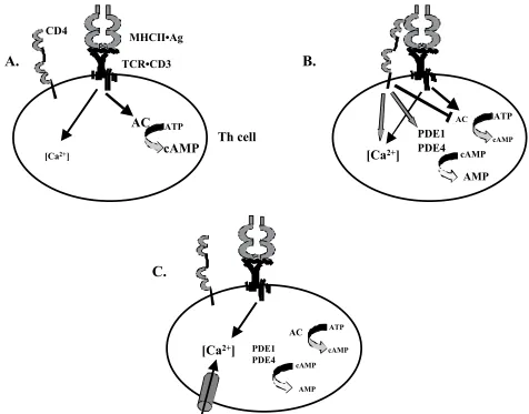
Functional consequences of CD4 engagement by MHC class II
Interactions between CD4 and MHC class II increase antigen-induced Th cell proliferation and cytokine production. Initially, it was thought that CD4 functioned as an adhesion molecule, enhancing contact between Th cells and APCs (Gay et al., 1987). However, CD4 and MHC class II interact with extremely low affinity (Weber and Karjalainen, 1993; Xiong et al., 2001), and soluble CD4 does not affect the binding of soluble antigen/MHC class II complexes to immobilized TCR (Xiong et al., 2001). Furthermore, CD4-MHC class II interactions do not increase the binding avidity between Th cells and APCs (Hamad et al., 1998; Metz et al., 1997; Zhou and König, 2003). Thus, an adhesive effect of CD4 is unlikely, and the major function of CD4 may be its active participation in Th cell signaling (Sleckman et al., 1991; Zhou and König, 2003).
Role of CD4 in modulating T cell signal transduction
of CD4 are critical for successful Th cell activation. An important characteristic for effective regulation of intracellular cAMP by CD4 signaling is the temporal and spatial sequence of CD4 localization in the signaling synapse. Recruitment of CD4 depends on binding of the TCR to its specific antigen/MHC class II ligand (Krummel
et al., 2000). Thus, an initial TCR-mediated increase in AC activity may be countered by CD4-mediated AC inhibition and PDE activation to promote early T cell activation events. Exclusion of CD4 from the central TCR cluster to the periphery of the signaling synapse within a few min after the initial Ca2+ signal (Krummel et al., 2000) may then remove the block on cAMP increases to promote proliferation and cytokine production.
The activities of ACs are controlled by hormones, neurotransmitters, chemotactic transducers, and other molecules. A common mechanism to inhibit AC activity is through the activation of Gi proteins (e.g., Gi1, Gi2, and Gi3), which are coupled to receptors for effector molecules. Members of the Gi family of proteins positively regulate T cell activation (Lippert et al., 2000). A 32-kDa GTP-binding protein associates with CD4 and CD8 in human T cell lines (Telfer and Rudd, 1991). Therefore, inhibition of AC by CD4-MHC class II engagement may be mediated via Gi proteins. CD4 may serve as a docking protein, carrying Gi proteins to the signaling synapse in activated Th cells, similar to the mechanism by which CD4 promotes p56lck function
(Holdorf et al., 2002).
Therefore, we propose that transduction of costimulatory signals induced by MHC class II engagement represent a major function of the CD4 coreceptor and that CD4-mediated signals participate in the precise regulation of intracellular levels of cAMP following T cell stimulation.
Role of CD4 in regulating T cell homeostasis
We have recently reported that coreceptor interactions with MHC molecules also regulate peripheral T cell homeostasis and the survival of naïve T cells in the absence of antigenic stimulation (König, 2002; König et al., 2002). We discovered this function of CD4 in a transgenic mouse strain that we generated for the purpose of elucidating the role of CD4 engagement by MHC class II in thymic selection and peripheral activation of Th cells. These transgenic mice express mutant MHC class II molecules defective in their ability to engage CD4 (Gilfillan et al., 1998). Despite the inability of the mutant MHC class II expressed on thymic epithelial cells to interact with CD4, positive thymic selection into the CD4 lineage proceeds, and CD4+ thymocytes mature. However, levels of single-positive CD4+ thymocytes in these mice are less than one third of those in normal mice. Furthermore, peripheral, naïve CD4+ T cells undergo apoptosis at an approximately three-fold higher rate in mutant MHC class II transgenic mice than do CD4 lineage cells in normal mice (Maroto et al., 1999). The increased frequency of apoptotic cells is due to a lack of CD4-MHC class II interactions in peripheral lymphoid organs, because naïve CD4+ T cells from normal mice adoptively transferred into mutant MHC class II transgenic hosts meet the same fate as do endogenous Th cells (Shen and König, 2001). Similarly, CD4-deficient T helper lineage cells are apoptosis-prone, and fail to survive after adoptive
transfer into irradiated hosts (Strong et al., 2001). Thus, CD4 lineage T cells require CD4-MHC class II interactions for efficient positive selection in the thymus and for maintenance of homeostasis in the periphery. Following the selection in the thymus for cells with TCRs that appropriately recognize self-MHC/peptide ligands, a peripheral recertification process enhances the stringency for proper matching between TCR restriction and coreceptor recognition of MHC (König et al., 2002). This process depends on signals through CD4 (Shen and König, 2001; Strong et al., 2001).
At the present time, we do not know whether the defect in peripheral Th cell homeostasis in the absence of CD4 engagement by MHC class II is related to an impaired regulation of intracellular cAMP levels. However, this is an attractive hypothesis and future research is targeted towards answering this question.
Outlook
The receptors that induce T cell signaling and the intracellular proteins mediating specific signal transduction pathways have been mostly identified. It is evident that signalosomes in T cells differ in composition depending on the extracellular signal received and the requirements of the stimulated cell. Depending on the level of participation of coreceptors in the induction and modulation of signaling pathways, the composition of established signalosomes may change over time in order to permit the fine-tuning of effector signaling pathways. Future research will focus on determining the kinetics of assembly, the dynamics of composition, and the compartmentalization of components of signalosomes induced during T cell activation. An important aspect will also be to define the interactions between different signaling pathways.
A clear understanding of Th cell signaling will provide a blueprint for the rationale intervention in multiple immune effector functions. Th cells promote adaptive immune responses against infective microorganisms and tumor cells, but also hamper tissue and organ transplantation. In addition, improper activation of Th cells can cause autoimmune diseases. Thus, targeting coreceptor-induced signaling pathways could improve vaccine development and therapeutic approaches to infectious diseases and cancer.
Acknowledgements
This work was supported in part by grants from the American Heart Association and the National Science Foundation. W.Z. acknowledges support from the McLaughlin Fellowship Fund, the American Foundation for Aging Research, the Christina Fleischmann Foundation, and the Association for Women in Science.
References
Abraham, N. and Veillette, A. 1990. Activation of p56lck
through mutation of a regulatory carboxy-terminal tyrosine residue requires intact sites of autophosphorylation and myristylation. Mol Cell Biol 10: 5197-5206.
Degerman, E., Pierce, J.H., and Manganiello, V.C. 1999. IL-3 and IL-4 activate cyclic nucleotide phosphodiesterases 3 (PDE3) and 4 (PDE4) by different mechanisms in FDCP2 myeloid cells. J. Immunol. 162: 4864-4875.
Andjelic, S., Jain, N., and Nikolic-Zugic, J. 1993. Immature thymocytes become sensitive to calcium-mediated apoptosis with the onset of CD8, CD4, and the T cell receptor expression: a role for bcl-2? J Exp Med 178: 1745-1751.
Aukrust, P., Aandahl, E.M., Skalhegg, B.S., Nordoy, I., Hansson, V., Tasken, K., Froland, S.S., and Muller, F. 1999. Increased activation of protein kinase A type I contributes to the T cell deficiency in common variable immunodeficiency. J Immunol 162: 1178-1185.
Beebe, S.J. 1994. The cAMP-dependent protein kinases and cAMP signal transduction. Semin Cancer Biol 5: 285-294.
Berridge, M.J. 1993a. Cell signalling. A tale of two messengers. Nature 365: 388-389.
Berridge, M.J. 1993b. Inositol trisphosphate and calcium signalling. Nature 361: 315-325.
Bierer, B.E., Sleckman, B.P., Ratnofsky, S.E., and Burakoff, S.J. 1989. The biologic roles of CD2, CD4, and CD8 in T-cell activation. Annu Rev Immunol 7: 579-599.
Bommhardt, U., Scheuring, Y., Bickel, C., Zamoyska, R., and Hunig, T. 2000. MEK activity regulates negative selection of immature CD4+CD8+ thymocytes. J Immunol 164: 2326-2337.
Boonen, G.J., Van Dijk, A.M., Verdonck, L.F., Van Lier, R.A., Rijksen, G., and Medema, R.H. 1999. CD28 induces cell cycle progression by IL-2-independent down-regulation of p27kip1 expression in human peripheral T lymphocytes.
Eur J Immunol 29: 789-798.
Boussiotis, V.A., Freeman, G.J., Berezovskaya, A., Barber, D.L., and Nadler, L.M. 1997. Maintenance of human T cell anergy: blocking of IL-2 gene transcription by activated Rap1. Science 278: 124-128.
Brdicka, T., Pavlistova, D., Leo, A., Bruyns, E., Korinek, V., Angelisova, P., Scherer, J., Shevchenko, A., Hilgert, I., Cerny, J., Drbal, K., Kuramitsu, Y., Kornacker, B., Horejsi, V., and Schraven, B. 2000. Phosphoprotein associated with glycosphingolipid-enriched microdomains (PAG), a novel ubiquitously expressed transmembrane adaptor protein, binds the protein tyrosine kinase csk and is involved in regulation of T cell activation. J Exp Med 191: 1591-1604.
Brocker, T. 1999. The role of dendritic cells in T cell selection and survival. Journal of Leukocyte Biology. 66: 331-335. Brown, J.H., Jardetzky, T., Saper, M.A., Samraoui, B., Bjorkman, P.J., and Wiley, D.C. 1988. A hypothetical model of the foreign antigen binding site of class II histocompatibility molecules. Nature 332: 845-850. Brown, J.H., Jardetzky, T.S., Gorga, J.C., Stern, L.J., Urban,
R.G., Strominger, J.L., and Wiley, D.C. 1993. Three-dimensional structure of the human class II histocompatibility antigen HLA-DR1. Nature 364: 33-39. Brugnera, E., Bhandoola, A., Cibotti, R., Yu, Q., Guinter, T.I., Yamashita, Y., Sharrow, S.O., and Singer, A. 2000. Coreceptor reversal in the thymus: signaled CD4+8+ thymocytes initially terminate CD8 transcription even
when differentiating into CD8+ T cells. Immunity 13: 59-71.
Bushell, A., Morris, P.J., and Wood, K.J. 1995. Transplantation tolerance induced by antigen pretreatment and depleting anti-CD4 antibody depends on CD4+ T cell regulation during the induction phase of the response. Eur. J. Immunol. 25: 2643-2649.
Cammarota, G., Scheirle, A., Takacs, B., Doran, D.M., Knorr, R., Bannwarth, W., Guardiola, J., and Sinigaglia, F. 1992. Identification of a CD4 binding site on the beta 2 domain of HLA-DR molecules. Nature 356: 799-801. Capone, M., Romagnoli, P., Beermann, F., Macdonald,
H.R., and Van Meerwijk, J.P. 2001. Dissociation of thymic positive and negative selection in transgenic mice expressing major histocompatibility complex class I molecules exclusively on thymic cortical epithelial cells. Blood 97: 1336-1342.
Chan, A.C., Irving, B.A., Fraser, J.D., and Weiss, A. 1991. The zeta chain is associated with a tyrosine kinase and upon T-cell antigen receptor stimulation associates with ZAP-70, a 70-kDa tyrosine phosphoprotein. Proc Natl Acad Sci U S A 88: 9166-9170.
Charbonneau, H., Kumar, S., Novack, J.P., Blumenthal, D.K., Griffin, P.R., Shabanowitz, J., Hunt, D.F., Beavo, J.A., and Walsh, K.A. 1991. Evidence for domain organization within the 61-kDa calmodulin-dependent cyclic nucleotide phosphodiesterase from bovine brain. Biochemistry 30: 7931-7940.
Chen, D. and Rothenberg, E.V. 1994. Interleukin 2 transcription factors as molecular targets of cAMP inhibition: delayed inhibition kinetics and combinatorial transcription roles. J Exp Med 179: 931-942.
Chidgey, A.P. and Boyd, R.L. 2001. Thymic stromal cells and positive selection. APMIS 109: 481-492.
Connolly, J.M., Hansen, T.H., Ingold, A.L., and Potter, T.A. 1990. Recognition by CD8 on cytotoxic T lymphocytes is ablated by several substitutions in the class I alpha 3 domain: CD8 and the T-cell receptor recognize the same class I molecule. Proc Natl Acad Sci USA 87: 2137-2141. Corado, J., Le Deist, F., Griscelli, C., and Fischer, A. 1990. Inositol 1,4,5-trisphosphate- and arachidonic acid-induced calcium mobilization in T and B lymphocytes. Cell Immunol 126: 245-254.
Datta, S.K., Kaliyaperumal, A., Mohan, C., and Desai-Mehta, A. 1997. T helper cells driving pathogenic anti-DNA autoantibody production in lupus: nucleosomal epitopes and CD40 ligand signals. Lupus 6: 333-336. De Aos, I., Metzger, M.H., Exley, M., Dahl, C.E., Misra, S.,
Zheng, D., Varticovski, L., Terhorst, C., and Sancho, J. 1997. Tyrosine phosphorylation of the CD3-epsilon subunit of the T cell antigen receptor mediates enhanced association with phosphatidylinositol 3-kinase in Jurkat T cells. J Biol Chem 272: 25310-25318.
De Rooij, J., Zwartkruis, F.J., Verheijen, M.H., Cool, R.H., Nijman, S.M., Wittinghofer, A., and Bos, J.L. 1998. Epac is a Rap1 guanine-nucleotide-exchange factor directly activated by cyclic AMP. Nature 396: 474-477.
Delgado, P., Fernandez, E., Dave, V., Kappes, D., and Alarcon, B. 2000. CD3delta couples T-cell receptor signalling to ERK activation and thymocyte positive selection. Nature 406: 426-430.
the immunological synapse. Curr Biol 10: R923-933. Diepolder, H.M., Jung, M.C., Keller, E., Schraut, W.,
Gerlach, J.T., Gruner, N., Zachoval, R., Hoffmann, R.M., Schirren, C.A., Scholz, S., and Pape, G.R. 1998. A vigorous virus-specific CD4+ T cell response may contribute to the association of HLA-DR13 with viral clearance in hepatitis B. Clin. Exp. Immunol. 113: 244-251.
Dolmetsch, R.E., Lewis, R.S., Goodnow, C.C., and Healy, J.I. 1997. Differential activation of transcription factors induced by Ca2+ response amplitude and duration. Nature 386: 855-858.
Donnadieu, E., Bismuth, G., and Trautmann, A. 1994. Antigen recognition by helper T cells elicits a sequence of distinct changes of their shape and intracellular calcium. Curr Biol 4: 584-595.
Doskeland, S.O., Maronde, E., and Gjertsen, B.T. 1993. The genetic subtypes of cAMP-dependent protein kinase—functionally different or redundant? Biochim Biophys Acta 1178: 249-258.
Ekholm, D., Hemmer, B., Gao, G., Vergelli, M., Martin, R., and Manganiello, V. 1997. Differential expression of cyclic nucleotide phosphodiesterase 3 and 4 activities in human T cell clones specific for myelin basic protein. J. Immunol. 159: 1520-1529.
Elder, M.E., Skoda-Smith, S., Kadlecek, T.A., Wang, F., Wu, J., and Weiss, A. 2001. Distinct T cell developmental consequences in humans and mice expressing identical mutations in the DLAARN motif of ZAP-70. J Immunol 166: 656-661.
Farber, D.L., Luqman, M., Acuto, O., and Bottomly, K. 1995. Control of memory CD4 T cell activation: MHC class II molecules on APCs and CD4 ligation inhibit memory but not naive CD4 T cells. Immunity 2: 249-259.
Feuerstein, N., Firestein, R., Aiyar, N., He, X., Murasko, D., and Cristofalo, V. 1996. Late induction of CREB/ATF binding and a concomitant increase in cAMP levels in T and B lymphocytes stimulated via the antigen receptor. J. Immunol. 156: 4582-4593.
Firpo, E.J., Koff, A., Solomon, M.J., and Roberts, J.M. 1994. Inactivation of a Cdk2 inhibitor during interleukin 2-induced proliferation of human T lymphocytes. Mol Cell Biol 14: 4889-4901.
Franke, B., Akkerman, J.W., and Bos, J.L. 1997. Rapid Ca2+-mediated activation of Rap1 in human platelets. Embo J 16: 252-259.
Gao, G.F., Tormo, J., Gerth, U.C., Wyer, J.R., Mcmichael, A.J., Stuart, D.I., Bell, J.I., Jones, E.Y., and Jakobsen, B.K. 1997. Crystal structure of the complex between human CD8 alpha alpha and HLA-A2. Nature 387: 630-634.
Gaubin, M., Houlgatte, R., Dettin, M., Scarinci, C., Martin, M., Guardiola, J., Di Bello, C., and Piatier-Tonneau, D. 1999. Definition of the alpha 2 region of HLA-DR molecules involved in CD4 binding. Hum Immunol 60: 273-281.
Gay, D., Maddon, P., Sekaly, R., Talle, M.A., Godfrey, M., Long, E., Goldstein, G., Chess, L., Axel, R., Kappler, J., and Marrack, P. 1987. Functional interaction between human T-cell protein CD4 and the major histocompatibility
complex HLA-DR antigen. Nature 328: 626-629. Gearing, A.J., Wadhwa, M., and Perris, A.D. 1985.
Interleukin 2 stimulates T cell proliferation using a calcium flux. Immunol Lett 10: 297-302.
Germain, R.N. 1994. MHC-dependent antigen processing and peptide presentation: providing ligands for T lymphocyte activation. Cell 76: 287-299.
Germain, R.N. 2002. T-cell development and the CD4-CD8 lineage decision. Nature Rev Immunol 2: 309-322. Giembycz, M.A., Corrigan, C.J., Seybold, J., Newton, R.,
and Barnes, P.J. 1996. Identification of cyclic AMP phosphodiesterases 3, 4 and 7 in human CD4+ and CD8+ T-lymphocytes: role in regulating proliferation and the biosynthesis of interleukin-2. Br. J. Pharmacol. 118: 1945-1958.
Gilfillan, S., Shen, X., and König, R. 1998. Selection and function of CD4+ T lymphocytes in transgenic mice expressing mutant MHC class II molecules deficient in their interaction with CD4. J Immunol 161: 6629-6637. Gong, Q., Jin, X., Akk, A.M., Foger, N., White, M., Gong,
G., Wardenburg, J.B., and Chan, A.C. 2001. Requirement for tyrosine residues 315 and 319 within zeta chain-associated protein 70 for T cell development. J Exp Med 194: 507-518.
Grakoui, A., Bromley, S.K., Sumen, C., Davis, M.M., Shaw, A.S., Allen, P.M., and Dustin, M.L. 1999. The immunological synapse: a molecular machine controlling T cell activation. Science 285: 221-227.
Hamad, A.R., O’herrin, S.M., Lebowitz, M.S., Srikrishnan, A., Bieler, J., Schneck, J., and Pardoll, D. 1998. Potent T cell activation with dimeric peptide-major histocompatibility complex class II ligand: the role of CD4 coreceptor. J. Exp. Med. 188: 1633-1640.
Harder, T. and Kuhn, M. 2000. Selective accumulation of raft-associated membrane protein LAT in T cell receptor signaling assemblies. J Cell Biol 151: 199-208.
Harder, T. and Kuhn, M. 2001. Immunoisolation of TCR signaling complexes from Jurkat T leukemic cells. Sci STKE 2001: PL1.
Hogquist, K.A. 2001. Signal strength in thymic selection and lineage commitment. Curr Opin Immunol 13: 225-231.
Holdorf, A.D., Lee, K.H., Burack, W.R., Allen, P.M., and Shaw, A.S. 2002. Regulation of Lck activity by CD4 and CD28 in the immunological synapse. Nature Immunol. 3: 259-264.
Houge, G., Steinberg, R.A., Ogreid, D., and Doskeland, S.O. 1990. The rate of recombination of the subunits (RI and C) of cAMP-dependent protein kinase depends on whether one or two cAMP molecules are bound per RI monomer. J Biol Chem 265: 19507-19516.
Hurwitz, R.L., Hirsch, K.M., Clark, D.J., Holcombe, V.N., and Hurwitz, M.Y. 1990. Induction of a calcium/ calmodulin-dependent phosphodiesterase during phytohemagglutinin-stimulated lymphocyte mitogenesis. J. Biol. Chem. 265: 8901-8907.
Irvine, D.J., Purbhoo, M.A., Krogsgaard, M., and Davis, M.M. 2002. Direct observation of ligand recognition by T cells. Nature 419: 845-849.
and Weiss, A. 1994. Sequential interactions of the TCR with two distinct cytoplasmic tyrosine kinases. Science 263: 1136-1139.
Jabado, N., Le Deist, F., Fisher, A., and Hivroz, C. 1994. Interaction of HIV gp120 and anti-CD4 antibodies with the CD4 molecule on human CD4+ T cells inhibits the binding activity of NF-AT, NF-kappa B and AP-1, three nuclear factors regulating interleukin-2 gene enhancer activity. Eur J Immunol 24: 2646-2652.
Jauliac, S., Mazerolles, F., Jabado, N., Pallier, A., Bernard, F., Peake, J., Fischer, A., and Hivroz, C. 1998. Ligands of CD4 inhibit the association of phospholipase Cgamma1 with phosphoinositide 3 kinase in T cells: regulation of this association by the phosphoinositide 3 kinase activity. Eur J Immunol 28: 3183-3191.
Kahn, M., Sugawara, H., Mcgowan, P., Okuno, K., Nagoya, S., Hellstrom, K.E., Hellstrom, I., and Greenberg, P. 1991. CD4+ T cell clones specific for the human p97 melanoma-associated antigen can eradicate pulmonary metastases from a murine tumor expressing the p97 antigen. J. Immunol. 146: 3235-3241.
Kanda, N. and Watanabe, S. 2001. Regulatory roles of adenylate cyclase and cyclic nucleotide phosphodiesterases 1 and 4 in interleukin-13 production by activated human T cells. Biochem. Pharmacol. 62: 495-507.
Karttunen, J. and Shastri, N. 1991. Measurement of ligand-induced activation in single viable T cells using the lacZ reporter gene. Proc Natl Acad Sci U S A 88: 3972-3976. Kawasaki, H., Springett, G.M., Mochizuki, N., Toki, S., Nakaya, M., Matsuda, M., Housman, D.E., and Graybiel, A.M. 1998. A family of cAMP-binding proteins that directly activate Rap1. Science 282: 2275-2279.
Kern, P.S., Teng, M.K., Smolyar, A., Liu, J.H., Liu, J., Hussey, R.E., Spoerl, R., Chang, H.C., Reinherz, E.L., and Wang, J.H. 1998. Structural basis of CD8 coreceptor function revealed by crystallographic analysis of a murine CD8 alpha alpha ectodomain fragment in complex with H-2K. Immunity 9: 519-530.
Khor, B. and Sleckman, B.P. 2002. Allelic exclusion at the TCRbeta locus. Curr Opin Immunol 14: 230-234. König, R. 2002. Interactions between MHC molecules and
co-receptors of the TCR. Curr Opin Immunol 14: 75-83. König, R., Fleury, S., and Germain, R.N. 1996. The structural basis of CD4-MHC class II interactions: coreceptor contributions to T cell receptor antigen recognition and oligomerization-dependent signal transduction. Curr. Top. Microbiol. Immunol. 205: 19-46. König, R., Huang, L.Y., and Germain, R.N. 1992. MHC class II interaction with CD4 mediated by a region analogous to the MHC class I binding site for CD8. Nature 356: 796-798.
König, R., Shen, X., and Germain, R.N. 1995. Involvement of both major histocompatibility complex class II alpha and beta chains in CD4 function indicates a role for ordered oligomerization in T cell activation. J. Exp. Med. 182: 779-787.
König, R., Shen, X., Maroto, R., and Denning, T.L. 2002. The role of CD4 in regulating homeostasis of T helper cells. Immunol Res 25: 115-130.
Kosugi, A., Sakakura, J., Yasuda, K., Ogata, M., and
Hamaoka, T. 2001. Involvement of SHP-1 tyrosine phosphatase in TCR-mediated signaling pathways in lipid rafts. Immunity 14: 669-680.
Krensky, A.M., Clayberger, C., Reiss, C.S., Strominger, J.L., and Burakoff, S.J. 1982. Specificity of OKT4+ cytotoxic T lymphocyte clones. J Immunol 129: 2001-2003. Krummel, M.F., Sjaastad, M.D., Wulfing, C., and Davis,
M.M. 2000. Differential clustering of CD4 and CD3zeta during T cell recognition. Science 289: 1349-1352. Laxminarayana, D. and Kammer, G.M. 1996. Activation of
type I protein kinase A during receptor-mediated human T lymphocyte activation. J. Immunol. 156: 497-506. Leitenberg, D., Balamuth, F., and Bottomly, K. 2001.
Changes in the T cell receptor macromolecular signaling complex and membrane microdomains during T cell development and activation. Semin Immunol 13: 129-138. Leung, R.K., Thomson, K., Gallimore, A., Jones, E., Van Den Broek, M., Sierro, S., Alsheikhly, A.R., Mcmichael, A., and Rahemtulla, A. 2001. Deletion of the CD4 silencer element supports a stochastic mechanism of thymocyte lineage commitment. Nat Immunol 2: 1167-1173. Li, L., Yee, C., and Beavo, J.A. 1999. CD3- and
CD28-dependent induction of PDE7 required for T cell activation. Science 283: 848-851.
Lingk, D.S., Chan, M.A., and Gelfand, E.W. 1990. Increased cyclic adenosine monophosphate levels block progression but not initiation of human T cell proliferation. J Immunol 145: 449-455.
Lippert, E., Jacques, Y., and Hermouet, S. 2000. Positive regulation of human T cell activation by Gi2 proteins and interleukin-8. J. Leukoc. Biol. 67: 742-748.
Lograsso, P.V., Hawkins, J., Frank, L.J., Wisniewski, D., and Marcy, A. 1996. Mechanism of activation for Zap-70 catalytic activity. Proc Natl Acad Sci U S A 93: 12165-12170.
Loh, C., Carew, J.A., Kim, J., Hogan, P.G., and Rao, A. 1996. T-cell receptor stimulation elicits an early phase of activation and a later phase of deactivation of the transcription factor NFAT1. Mol Cell Biol 16: 3945-3954. Luo, K.X. and Sefton, B.M. 1990. Cross-linking of T-cell surface molecules CD4 and CD8 stimulates phosphorylation of the lck tyrosine protein kinase at the autophosphorylation site. Mol. Cell. Biol. 10: 5305-5313. Lynch, G.W., Sloane, A.J., Raso, V., Lai, A., and Cunningham, A.L. 1999. Direct evidence for native CD4 oligomers in lymphoid and monocytoid cells. Eur. J. Immunol. 29: 2590-2602.
Mackenzie, S.J., Yarwood, S.J., Peden, A.H., Bolger, G.B., Vernon, R.G., and Houslay, M.D. 1998. Stimulation of p70S6 kinase via a growth hormone-controlled phosphatidylinositol 3-kinase pathway leads to the activation of a PDE4A cyclic AMP-specific phosphodiesterase in 3T3-F442A preadipocytes. Proc Natl Acad Sci U S A 95: 3549-3554.
Magnan, A., Di Bartolo, V., Mura, A.M., Boyer, C., Richelme, M., Lin, Y.L., Roure, A., Gillet, A., Arrieumerlou, C., Acuto, O., Malissen, B., and Malissen, M. 2001. T cell development and T cell responses in mice with mutations affecting tyrosines 292 or 315 of the ZAP-70 protein tyrosine kinase. J Exp Med 194: 491-505.
Degerman, E. 1995. Diversity in cyclic nucleotide phosphodiesterase isoenzyme families. Arch. Biochem. Biophys. 322: 1-13.
Maroto, R., Shen, X., and König, R. 1999. Requirement for efficient interactions between CD4 and MHC class II molecules for survival of resting CD4+ T lymphocytes in vivo and for activation-induced cell death. J Immunol 162: 5973-5980.
Marschner, S., Hunig, T., Cambier, J.C., and Finkel, T.H. 2002. Ligation of human CD4 interferes with antigen-induced activation of primary T cells. Immunol Lett 82: 131-139.
May, W.S., Lapetina, E.G., and Cuatrecasas, P. 1986. Intracellular activation of protein kinase C and regulation of the surface transferrin receptor by diacylglycerol is a spontaneously reversible process that is associated with rapid formation of phosphatidic acid. Proc Natl Acad Sci U S A 83: 1281-1284.
Metz, D.P., Farber, D.L., König, R., and Bottomly, K. 1997. Regulation of memory CD4 T cell adhesion by CD4-MHC class II interaction. J. Immunol. 159: 2567-2573. Meuer, S.C., Hussey, R.E., Hodgdon, J.C., Hercend, T.,
Schlossman, S.F., and Reinherz, E.L. 1982. Surface structures involved in target recognition by human cytotoxic T lymphocytes. Science 218: 471-473. Milia, E., Di Somma, M.M., Majolini, M.B., Ulivieri, C.,
Somma, F., Piccolella, E., Telford, J.L., and Baldari, C.T. 1997. Gene activating and proapoptotic potential are independent properties of different CD4 epitopes. Mol. Immunol. 34: 287-296.
Monks, C.R., Freiberg, B.A., Kupfer, H., Sciaky, N., and Kupfer, A. 1998. Three-dimensional segregation of supramolecular activation clusters in T cells. Nature 395: 82-86.
Negulescu, P.A., Krasieva, T.B., Khan, A., Kerschbaum, H.H., and Cahalan, M.D. 1996. Polarity Of T Cell Shape, Motility, and Sensitivity to Antigen. Immunity 4: 421-430. Negulescu, P.A., Shastri, N., and Cahalan, M.D. 1994. Intracellular calcium dependence of gene expression in single T lymphocytes. Proc Natl Acad Sci U S A 91: 2873-2877.
Niedermann, G. 2002. Immunological functions of the proteasome. Curr Top Microbiol Immunol 268: 91-136. Norbury, C.C., Princiotta, M.F., Bacik, I., Brutkiewicz, R.R.,
Wood, P., Elliott, T., Bennink, J.R., and Yewdell, J.W. 2001. Multiple antigen-specific processing pathways for activating naive CD8+ T cells in vivo. J Immunol 166: 4355-4362.
Nourse, J., Firpo, E., Flanagan, W.M., Coats, S., Polyak, K., Lee, M.H., Massague, J., Crabtree, G.R., and Roberts, J.M. 1994. Interleukin-2-mediated elimination of the p27Kip1 cyclin-dependent kinase inhibitor prevented by
rapamycin. Nature 372: 570-573.
Paliogianni, F., Kincaid, R.L., and Boumpas, D.T. 1993. Prostaglandin E2 and other cyclic AMP elevating agents inhibit interleukin 2 gene transcription by counteracting calcineurin-dependent pathways. J. Exp. Med. 178: 1813-1817.
Pallier, A., Jauliac, S., Jabado, N., Fischer, A., and Hivroz, C. 1998. Differential CD4-dependent inhibition of JNK but not Erk-2 activities in human naive and memory CD4+ T
cell populations. Int. Immunol. 10: 869-876.
Papoff, G., Hausler, P., Eramo, A., Pagano, M.G., Di Leve, G., Signore, A., and Ruberti, G. 1999. Identification and characterization of a ligand-independent oligomerization domain in the extracellular region of the CD95 death receptor. J. Biol. Chem. 274: 38241-38250.
Paz, P.E., Wang, S.J., Clarke, H., Lu, X.B., Stokoe, D., and Abo, A. 2001. Mapping the Zap-70 phosphorylation sites on LAT (linker for activation of T cells) required for recruitment and activation of signalling proteins in T cells. Biochemical Journal 356: 461-471.
Prasad, K.V., Kapeller, R., Janssen, O., Repke, H., Duke-Cohan, J.S., Cantley, L.C., and Rudd, C.E. 1993. Phosphatidylinositol (PI) 3-kinase and PI 4-kinase binding to the CD4-p56lck complex: the p56lck SH3 domain binds
to PI 3-kinase but not PI 4-kinase. Mol. Cell. Biol. 13: 7708-7717.
Qian, D., Griswold-Prenner, I., Rosner, M.R., and Fitch, F.W. 1993. Multiple components of the T cell antigen receptor complex become tyrosine-phosphorylated upon activation. J Biol Chem 268: 4488-4493.
Rabinowitz, J.D., Beeson, C., Wulfing, C., Tate, K., Allen, P.M., Davis, M.M., and Mcconnell, H.M. 1996. Altered T cell receptor ligands trigger a subset of early T cell signals. Immunity 5: 125-135.
Ramstad, C., Sundvold, V., Johansen, H.K., and Lea, T. 2000. cAMP-dependent protein kinase (PKA) inhibits T cell activation by phosphorylating ser-43 of raf-1 in the MAPK/ERK pathway. Cell Signal 12: 557-563.
Ratcliffe, M.J., Coggeshall, K.M., Newell, M.K., and Julius, M.H. 1992. T cell receptor aggregation, but not dimerization, induces increased cytosolic calcium concentrations and reveals a lack of stable association between CD4 and the T cell receptor. J. Immunol. 148: 1643-1651.
Raulet, D.H., Garman, R.D., Saito, H., and Tonegawa, S. 1985. Developmental regulation of T-cell receptor gene expression. Nature 314: 103-107.
Ravichandran, K.S., Lee, K.K., Songyang, Z., Cantley, L.C., Burn, P., and Burakoff, S.J. 1993. Interaction of Shc with the zeta chain of the T cell receptor upon T cell activation. Science 262: 902-905.
Res, P. and Spits, H. 1999. Developmental stages in the human thymus. Seminars in Immunology 11: 39-46. Rizzo, L.V., Silver, P., Wiggert, B., Hakim, F., Gazzinelli,
R.T., Chan, C.C., and Caspi, R.R. 1996. Establishment and characterization of a murine CD4+ T cell line and clone that induce experimental autoimmune uveoretinitis in B10.A mice. J. Immunol. 156: 1654-1660.
Robey, E. and Fowlkes, B.J. 1994. Selective events in T cell development. Annu Rev Immunol 12: 675-705. Rodriguez-Frade, J.M., Vila-Coro, A.J., De Ana, A.M., Albar,
J.P., Martinez, A.C., and Mellado, M. 1999. The chemokine monocyte chemoattractant protein-1 induces functional responses through dimerization of its receptor CCR2. Proc. Natl. Acad. Sci. U. S. A. 96: 3628-3633. Rooney, J.W., Sun, Y.L., Glimcher, L.H., and Hoey, T. 1995.
Novel NFAT sites that mediate activation of the interleukin-2 promoter in response to T-cell receptor stimulation. Mol. Cell. Biol. 15: 6299-6310.
Garrett, T.P., Clayberger, C., Krensky, A.M., Norment, A.M., Littman, D.R., and Parham, P. 1990. A binding site for the T-cell co-receptor CD8 on the alpha 3 domain of HLA-A2. Nature 345: 41-46.
Schoenberger, S.P., Toes, R.E., Van Der Voort, E.I., Offringa, R., and Melief, C.J. 1998. T-cell help for cytotoxic T lymphocytes is mediated by CD40-CD40L interactions. Nature 393: 480-483.
Shen, X., Hu, B., Mcphie, P., Wu, X., Fox, A., Germain, R.N., and König, R. 1996. Peptides corresponding to CD4-interacting regions of murine MHC class II molecules modulate immune responses of CD4+ T lymphocytes in
vitro and in vivo. J. Immunol. 157: 87-100.
Shen, X. and König, R. 2001. Post-thymic selection of peripheral CD4+ T-lymphocytes on class II major histocompatibility antigen-bearing cells. Cell. Mol. Biol. 47: 87-96.
Singer, A. 2002. New perspectives on a developmental dilemma: the kinetic signaling model and the importance of signal duration for the CD4/CD8 lineage decision. Curr Opin Immunol 14: 207-215.
Skalhegg, B.S., Landmark, B.F., Doskeland, S.O., Hansson, V., Lea, T., and Jahnsen, T. 1992. Cyclic AMP-dependent protein kinase type I mediates the inhibitory effects of 3',5'-cyclic adenosine monophosphate on cell replication in human T lymphocytes. J. Biol. Chem. 267: 15707-15714.
Skalhegg, B.S., Tasken, K., Hansson, V., Huitfeldt, H.S., Jahnsen, T., and Lea, T. 1994. Location of cAMP-dependent protein kinase type I with the TCR-CD3 complex. Science 263: 84-87.
Sleckman, B.P., Rosenstein, Y., Igras, V.E., Greenstein, J.L., and Burakoff, S.J. 1991. Glycolipid-anchored form of CD4 increases intercellular adhesion but is unable to enhance T cell activation. J. Immunol. 147: 428-431. Sloan-Lancaster, J., Steinberg, T.H., and Allen, P.M. 1997.
Selective loss of the calcium ion signaling pathway in T cells maturing toward a T helper 2 phenotype. J Immunol 159: 1160-1168.
Snow, E.C., Pittner, B., and Reid, S. 1994. T helper cell regulation of normal and neoplastic B cell growth. Semin. Immunol. 6: 311-326.
Soderling, S.H. and Beavo, J.A. 2000. Regulation of cAMP and cGMP signaling: new phosphodiesterases and new functions. Curr. Op. Cell Biol. 12: 174-179.
Sommers, C.L., Menon, R.K., Grinberg, A., Zhang, W., Samelson, L.E., and Love, P.E. 2001. Knock-in mutation of the distal four tyrosines of linker for activation of T cells blocks murine T cell development. J Exp Med 194: 135-142.
Staples, K.J., Bergmann, M., Barnes, P.J., and Newton, R. 2000. Stimulus-specific inhibition of IL-5 by cAMP-elevating agents and IL-10 reveals differential mechanisms of action. Biochem Biophys Res Commun 273: 811-815.
Strong, J., Wang, Q., and Killeen, N. 2001. Impaired survival of T helper cells in the absence of CD4. Proc Natl Acad Sci U S A 98: 2566-2571.
Su, M.W., Yu, C.L., Burakoff, S.J., and Jin, Y.J. 2001. Targeting Src homology 2 domain-containing tyrosine phosphatase (SHP-1) into lipid rafts inhibits CD3-induced T cell activation. J Immunol 166: 3975-3982.
Sugiyama, H., Chen, P., Hunter, M.G., and Sitkovsky, M.V.
1997. Perturbation of the expression of the catalytic subunit C alpha of cyclic AMP-dependent protein kinase inhibits TCR-triggered secretion of IL-2 by T helper hybridoma cells. J Immunol 158: 171-179.
Sun, Z.Y.J., Kim, H.S., Wagner, G., and Reinherz, E.L. 2001. Mechanisms contributing to T cell receptor signaling and assembly revealed by the solution structure of an ectodomain fragment of the CD3 epsilon gamma heterodimer. Cell 105: 913-923.
Surh, C.D. and Sprent, J. 1994. T-cell apoptosis detected in situ during positive and negative selection in the thymus. Nature 372: 100-103.
Swain, S.L. 1981. Significance of Lyt phenotypes: Lyt2 antibodies block activities of T cells that recognize class 1 major histocompatibility complex antigens regardless of their function. Proc Natl Acad Sci U S A 78: 7101-7105. Swain, S.L. 1983. T cell subsets and the recognition of
MHC class. Immunol Rev 74: 129-142.
Tang, K.M., Jang, E.K., and Haslam, R.J. 1997. Expression and mutagenesis of the catalytic domain of cGMP-inhibited phosphodiesterase (PDE3) cloned from human platelets. Biochem J 323 ( Pt 1): 217-224.
Taniuchi, I., Sunshine, M.J., Festenstein, R., and Littman, D.R. 2002. Evidence for distinct CD4 silencer functions at different stages of thymocyte differentiation. Mol Cell 10: 1083-1096.
Taylor-Robinson, A.W. and Phillips, R.S. 1992. Functional characterization of protective CD4+ T-cell clones reactive to the murine malaria parasite Plasmodium chabaudi. Immunology 77: 99-105.
Telfer, J.C. and Rudd, C.E. 1991. A 32-kD GTP-binding protein associated with the CD4-p56lck and CD8-p56lck T
cell receptor complexes. Science 254: 439-441. Timmerman, L.A., Clipstone, N.A., Ho, S.N., Northrop, J.P.,
and Crabtree, G.R. 1996. Rapid shuttling of NF-AT in discrimination of Ca2+ signals and immunosuppression. Nature 383: 837-840.
Tsien, R.Y., Pozzan, T., and Rink, T.J. 1982. T-cell mitogens cause early changes in cytoplasmic free Ca2+ and membrane potential in lymphocytes. Nature 295: 68-71. Van Oirschot, B.A., Stahl, M., Lens, S.M., and Medema, R.H. 2001. Protein kinase A regulates expression of p27(kip1) and cyclin D3 to suppress proliferation of leukemic T cell lines. J Biol Chem 276: 33854-33860. Vang, T., Torgersen, K.M., Sundvold, V., Saxena, M., Levy,
F.O., Skalhegg, B.S., Hansson, V., Mustelin, T., and Tasken, K. 2001. Activation of the COOH-terminal Src kinase (Csk) by cAMP-dependent protein kinase inhibits signaling through the T cell receptor. J Exp Med 193: 497-507.
Veillette, A., Bookman, M.A., Horak, E.M., Samelson, L.E., and Bolen, J.B. 1989. Signal transduction through the CD4 receptor involves the activation of the internal membrane tyrosine-protein kinase p56lck. Nature 338:
257-259.
Walsh, D.A. and Van Patten, S.M. 1994. Multiple pathway signal transduction by the cAMP-dependent protein kinase. Faseb J 8: 1227-1236.
Weber, S. and Karjalainen, K. 1993. Mouse CD4 binds MHC class II with extremely low affinity. Int. Immunol. 5: 695-698.
Regulation of Zap-70 by Src family tyrosine protein kinases in an antigen-specific T-cell line. J Biol Chem 270: 2791-2799.
Weinberg, R.A. 1995. The retinoblastoma protein and cell cycle control. Cell 81: 323-330.
Weiss, M.J., Daley, J.F., Hodgdon, J.C., and Reinherz, E.L. 1984. Calcium dependency of antigen-specific (T3-Ti) and alternative (T11) pathways of human T-cell activation. Proc Natl Acad Sci U S A 81: 6836-6840.
Welsh, M., Songyang, Z., Frantz, J.D., Trub, T., Reedquist, K.A., Karlsson, T., Miyazaki, M., Cantley, L.C., Band, H., and Shoelson, S.E. 1998. Stimulation through the T cell receptor leads to interactions between SHB and several signaling proteins. Oncogene 16: 891-901.
Werlen, G., Hausmann, B., and Palmer, E. 2000. A motif in the alphabeta T-cell receptor controls positive selection by modulating ERK activity. Nature 406: 422-426. White, D.W. and Tartaglia, L.A. 1999. Evidence for
ligand-independent homo-oligomerization of leptin receptor (OB-R) isoforms: a proposed mechanism permitting productive long-form signaling in the presence of excess short-form expression. J. Cell. Biochem. 73: 278-288.
Williams, C.B., Engle, D.L., Kersh, G.J., Michael White, J., and Allen, P.M. 1999. A kinetic threshold between negative and positive selection based on the longevity of the T cell receptor-ligand complex. J Exp Med 189: 1531-1544. Wu, H., Kwong, P.D., and Hendrickson, W.A. 1997. Dimeric
association and segmental variability in the structure of human CD4. Nature 387: 527-530.
Wülfing, C. and Davis, M.M. 1998. A receptor/cytoskeletal movement triggered by costimulation during T cell activation. Science 282: 2266-2269.
Wülfing, C., Rabinowitz, J.D., Beeson, C., Sjaastad, M.D., Mcconnell, H.M., and Davis, M.M. 1997. Kinetics and extent of T cell activation as measured with the calcium signal. J. Exp. Med. 185: 1815-1825.
Xiong, Y., Kern, P., Chang, H.C., and Reinherz, E.L. 2001