Lower Segment Caesarean Section: Evidence-Based Practice
Mohd Rushdan MN
Department Obstetrics & Gynaecology, Hospital Sultanah Bahiyah, Alor Setar, Kedah, Malaysia.
Introduction
Caesarean section has become the most common major surgical procedure which is performed worldwide. Caesarean section is considered as a lifesaving procedure for both mother and baby. It is estimated that 18.5 million caesarean sections are performed yearly, worldwide. In the United States, more than one million caesarean sections are being performed, annually. Overall rates of caesarean section have increased in the last 30 years without significant improvement in perinatal or maternal outcomes. Reasons that may account for the increase in caesarean section rate are: (a) increase in repeat caesarean sections, (b) delay in childbirth and reduced parity, (c) decrease in vaginal breech deliveries, (d) decreased perinatal mortality with caesarean delivery, (e) non-reassuring fetal heart rate traces and (f) fear of malpractice litigation. Caesarean deliveries for failure to progress in labour and abnormal fetal heart rate tracings represent the majority of indications for emergency caesarean section. The caesarean delivery rate ranges from 22 % to 40% in developed countries, far exceeding the WHO’s recommended upper limit of 15%, a theoretical estimation which is supported by several observational studies (1). These studies assessed the association between caesarean section rates and mortality and morbidity in mothers and neonates, and found no reduction in those indicators when the caesarean section rate was more than 15% (2,3). Regarding the lower limit, it has been argued that caesarean section rates of 5% could achieve major improvement on maternal outcomes. However, for neonatal health, rates between 5% and 10% have been reported to attain better outcomes. According to the WHO Health Report 2010, based on the data obtained
from 137 countries around the world, 50.4% of these countries had a caesarean section rate of more than 15% (4). Caesarean section is much safer now than in the past, due to improvement in surgical techniques, use of antibiotic prophylaxis, thromboprophylaxis, use of regional anaesthesia and overall improvement in the perioperative care. However, it is still associated with an increase in the risk of maternal morbidity and mortality compared with vaginal delivery. It is also known that caesarean section performed in labour carries a higher morbidity as compared to planned surgery. Infection is the most common postoperative complication attributable to caesarean deliveries ranging from 6-18%. With the use of routine prophylactic antibiotics, the rate of postoperative infection has been reduced to less than 2% in some centres (5). The other potential complications of caesarean delivery are bleeding, thromboembolism (especially with emergency caesarean section), anaesthetic complications, urinary tract injuries and risk to the subsequent pregnancy such as placenta accreta, abruptio placentae, placenta praevia and repeat caesarean section (6). The WHO Global Survey had reported that increased caesarean section rates are associated with a higher risk of postpartum antibiotic use and increased maternal morbidity and mortality as well as an increase in fetal mortality rates, with a higher number of babies admitted to intensive care units for seven days compared to babies born vaginally (3). In order to achieve a better maternal and neonatal outcomes, caesarean section rate should be kept within the recommended rate and the techniques of performing this procedure must be based on evidences. The surgeons who performed this procedure should be competent and should be able to handle unexpected complications related to this procedure.
Dato’ Dr. Mohd Rushdan Md Nor, Department Obstetrics & Gynaecology, Hospital Sultanah Bahiyah, Alor Setar, Kedah, Malaysia. Email: [email protected]
Correspondence:
Indications for Caesarean Delivery
Maternal Indications
The indications of caesarean delivery can be divided into maternal indications, foetal indications and both. The majority of caesarean deliveries are performed for previous caesarean deliveries, breech presentation, abnormal labour progress and foetal distress; these factors account for 85% of all caesarean deliveries. Maternal indications include: (a) previous caesarean delivery or failed trial of scar, (b) cephalo-pelvic disproportion, (c) obstruction to the birth canal e.g. ovarian or uterine tumour, (d) lesions at the lower genital tracts such as obstructive vaginal septum, severe vulvo-vaginal condylomata obstructing the birth canal, active herpes, cervical cancer and others, (e) maternal contraindication to Valsalva manoeuvers (leading to increase intrathorasic pressure) such as left heart valvular stenosis, dilated aortic valve root, arterio-venous malformations, recent retinal detachment, (f) maternal conditions in which the stress of normal labour significantly increase the risk of cardiac failure and cardiopulmonary collapse such as in severe primary pulmonary hypertension, Eisenmenger’s syndrome and others, (g) woman who have previously undergo vaginal reconstructive surgery or fistula repair, (h) maternal severe pre-eclampsia or pre-eclampsia with unfavourable cervix, (i) placenta praevia major and (h) maternal request caesarean delivery despite extensive counseling.
Patients who requested to undergo caesarean delivery without medical indications must be counselled by attending Obstetrician who should provide the best possible evidence-based counseling, and at the same time respect her autonomy in the decision making. The following are information that should be given to such patient: (a) caesarean delivery on maternal request should be avoided if possible by women who have not completed their family, (b) caesarean should not be performed before 39th week pregnancy or without verifying fetal lung maturity, (c) caesarean delivery has a potential risk of respiratory problems for the baby and (d) caesarean section on maternal request is associated with a longer maternal hospital stay and increased risk of future pregnancy complications pertaining to placenta praevia, placenta accreta, repeat caesarean delivery etc.
Fetal Indications
The most common fetal indications for caesarean delivery are fetal distress and breech presentation. The fetal indications of caesarean delivery are: (a)
malpresentation such as transverse and oblique lie at term, (b) breech presentation, if the mother refuses or fails external cephalic version, (c) brow presentation in labour, (d) prolonged acidaemia such as in severe intrauterine growth restriction, (e) macrosomic baby with previous history of shoulder dystocia, (f) non-vertex presentation of leading twin or higher order of multiple pregnancy, (g) fetal anomalies that preclude normal delivery, such as neural tube defects with large defects that may rupture during normal vaginal delivery, fetal hydrocephalus, skeletal dysplasia such as osteogenesis imperfecta, fetuses with huge abdominal mass, (h) mother with active or symptomatic genital herpes infection, (i) maternal HIV infection with viral count > 1,000 copies/ml and (j) vasa praevia.
Fetal and Maternal Indications
Some of the indications listed in the previous sections are overlapping such as placenta praevia and occasionally the actual causes cannot be confirmed, for example in cases of cephalo-pelvic disproportion. Patients with failed instrumental deliveries should be delivered by caesarean section for maternal and fetal reasons. Expeditious delivery is also crucial for the safety of both mother and the baby in severe abruptio placenta. Caesarean section may also indicate for both maternal and fetal reasons in maternal sepsis secondary to prolong absent of membranes but early delivery is not anticipated. In this condition, allowing longer duration of labour may impose significant risk of maternal and fetal septicaemia.
Caesarean Section: Techniques
Preparation
Patients should be given complete information about caesarean section such as the indications, the procedure involved, potential complications and the implications for future pregnancies. Informed consent is obtained and if the caesarean section is a planned procedure, she should be admitted at least a day before. According to NICE guidelines (2011), the risk of respiratory morbidity is increased in babies born by caesarean section before labour, but this risk decreases significantly after 39 weeks (7). Therefore, planned caesarean section should be routinely be carried out by 39 weeks. Haemoglobin assessment should be done prior to surgery and anaemia corrected if necessary. Routine cross-matching, coagulation profile and preoperative ultrasound for localization of the placenta should not be offered to pregnant women who are healthy with otherwise uncomplicated pregnancies (7). Regional anaesthesia is the preferred method because it is safer and results in less maternal and neonatal morbidity as compared to general anaesthesia. To reduce the risk of hypotension during surgery, volume pre-loading with intravenous crystalloid or colloid is recommended. To reduce the risk of aspiration pneumonitis, patient should be given antacids and drugs to reduce gastric volumes and acidity before caesarean section. If general anaesthesia is employed, the patient should be preoxygenated, cricoid pressure applied and rapid sequence induction carried out to reduce the risk of aspiration. Healthcare providers should practice universal precautions such as wearing goggles and double-gloving while performing or assisting caesarean section.
Begin by ensuring that checklists for preoperative preparations (partly mentioned above) are complete. Before starting the operation, fetal heart activity should be verified and the indication for caesarean sections is still valid. The presentation and position of the fetal head should be assessed and consideration given to whether the woman is for tubal ligation. Since general anaesthesia carries significant risks to pregnant woman such as difficult intubation and aspiration of the stomach contents, spinal anaesthesia is recommended. The main risk of spinal anaesthesia is hypotension, which can be prevented by preloading the woman with intravenous fluid, elevation of her lower limbs and ensuring left lateral tilt. Routine administration of prophylactic antibiotics, given before the skin incision reduces the risk of maternal infection (7). Cases that are more complicated such as placenta praevia, transverse lie and obstructed labour require additional pre-planning to avoid potential complications. For example, in the case of placenta praevia major, the preparation includes : (a)
optimization of the haemoglobin levels to about 11 g% prior to surgery, (b) ensuring availability of cross matched blood in theatre, (c) presence of senior or experienced obstetrician and anaesthetist, (d) availability of uterotonic agents and (e) availability of balloon for uterine temponade.
Abdominal Entry, Skin and Uterine Incision
The patient is placed in a slight left lateral tilt position to minimize supine hypotension which can reduce placental perfusion secondary to aorto-caval compression. Preoperative skin cleansing with chlorhexidine-alcohol is superior to cleansing with povidone iodine for surgical site preparation with overall infection rate of 9.5% and 16.1%, respectively. Many Obstetricians prefer low transverse incision for routine caesarean sections for its effectiveness and good cosmetic result. In certain circumstances when complications are anticipated, or when the surgeon is less experienced, or previous scar is a midline, vertical sub umbilical incision is a preferred technique. Vertical incision has an advantage of simplicity, shorter duration, lesser blood loss and allows extension to improve the exposure if needed. Slightly longer time is taken to enter the peritoneal cavity with transverse skin incision, however they are usually less painful, associated with a smaller risk of an incisional hernia and can provide excellent visualization of the pelvis. This incision is also cosmetically more acceptable. There are several types of lower transverse incision such as Pfannenstiel incision, Maylard’s incision and Joel Cohen incision. Pfannenstiel incision is the most commonly performed skin incision for Cesarean section. In Pfannenstiel incision, the incision is curved slightly cephalad at the level of the pubic hairline or about 2 fingerbreadth above the symphysis pubis. The incision extends slightly beyond the lateral borders of the rectus muscle bilaterally. A Maylard’s incision is made approximately 2-3 cm above the symphysis pubis and it involves the transverse division of the rectus muscle, ligation of the inferior epigastric vessels and transverse opening of the peritoneum. The third type of transverse incision is Joel Cohen incision and this technique is described below (Fig. 1.0).
skin incision to birth of the baby (7). The main feature of Joel-Cohen-based techniques is blunt dissection and separation of tissues along natural tissue planes, using a minimum of sharp dissection.
Before an incision is made, rotation of the uterus should be noted and, if possible, corrected, so that the incision will not be asymmetrical, with risk of extension to the opposite site. Before the incision is made, check whether the regional anaesthesia is effective. Intravenous prophylactic antibiotics shouldbe given before skin incision to reduce the risk of maternal infection. Foetal heart activity must be assessed immediately before the surgery. Patient is placed in supine position with slightly left lateral tilt to minimize aotocaval compression. The skin is cleaned and draped. The Joel Cohen incision is a straight transverse incision made 3cm below the level of a straight line joining the antero-posterior iliac spines (NICE guideline described Joel Cohen incision as straight skin incision made 3cm above the symphysis pubis (7). The skin incision is made and carried down to the anterior sheath of the rectus fascia. Incise the subcutaneous tissues and anterior rectus sheath in the midline. The use of separate surgical knives to incise the skin and the deeper tissues at caesarean section is not recommended because it does not decrease wound infection. Extend the incision of anterior rectus sheath laterally with slightly open scissors tip or stretching the tissues with the fingers (digital dissection). Then, separate the rectus muscle vertically and open the parietal peritoneum. Enlarge the abdominal opening laterally using fingers in opposite direction to separate
the rectus muscle and the peritoneum to expose the abdominal cavity. Randomized trials have found that compared with Pfananstiel transverse incision, the Joel Cohen incision is quicker, less risk of morbidity and subsequent adhesions. Pick up and open the loose visceral peritoneum covering the lower uterine segment, once the uterus is exposed and push bladder inferiorly, made a small transverse incision with a scalpel and enlarged the opening by fingers stretching laterally to expose the amniotic membranes. Rupture the amniotic membranes and aspirate the amniotic fluid. Extend the uterine opening either by blunt lateral extension especially if the lower segment is well formed. In one randomized trial, blunt lateral extension of the uterine incision results in less wound extension towards the lateral wall of the uterus and less bleeding. Blunt expansion of the uterus may be
Figure 1.0: Type of skin incision: A: Pfannenstiel, B: Joel-Cohen, C: Maylard, D: Midline vertical
Conventional method of caesarean section Misgav Ladach Technique Caesarean Section
(Refer to Figure 1.2)
1. Pfannenstiel incision
2. Use of instruments/sharp dissection while opening the abdomen and extending the incision on the lower uterine segment.
3. Double layer uterine closure
4. Closure of the abdomen in layers with or without closure of peritoneum.
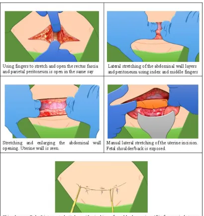
1. Joel Cohen’s skin incision: A straight incision about 3 cm below a line joining the anterior superior iliac spines. 2. Minimal use of instruments: Initial sharp incision about 3
cm then followed by stretching technique. Abdominal wall layers were separated by stretching with index and third fingers. Parietal peritoneum was also opened in the same way.
3. Digital lateral stretching of the uterine incision with exteriorization of the uterus.
4. Single or double layered uterine closure (majority double layered)
5. Non-closure of the visceral and parietal peritoneal layers. 6. Closure of the abdomen in two layers: Rectus fascia and
skin. References:
1. Xavier P, Ayres-De-Campos D, Reynolds A, et al. The modified Misgav-Ladach versus the Pfannensteil-Kerr technique for caesarean section: a randomized trial. Acta Obstet Gynecol Scand, 2005;84:878-882.
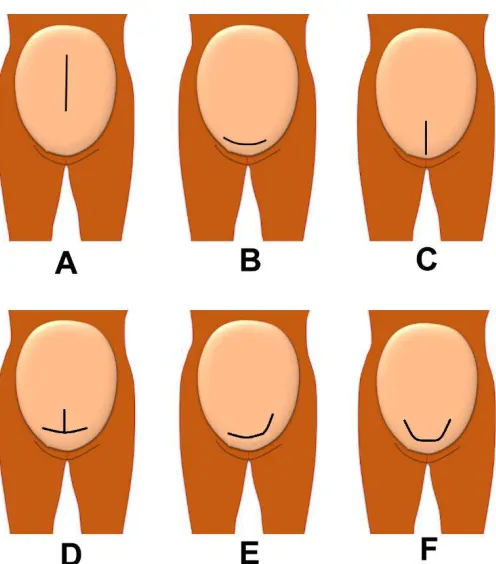
Figure 1.1: Type of uterine incision. A: Classical CS, B: Transverse Lower Segment CS, C: Vertical lower segment CS, D: Inverted-T incision, E: J-Shape incision and F: U-shape incision.
performed either in lateral direction or cephalad-caudal direction. A study by Cromi et al. shown that the later method was associated with less unintended extension and blood loss of more than 1,500 mls (8). If the foetal head is deeply engaged in the pelvis, the head must be elevated carefully above the level of uterine incision and rotated into the occipito-anterior position before it can be delivered through the incision. The active management of third stage is similar to vaginal delivery and can be implemented during caesarean delivery. Ten units of oxytocin should be given intravenously immediately after delivery of the baby. If there are concerns regarding continued loss of blood from the uterine incision, haemostatic clamps can be applied early to the cut edges and haemostatic sutures can be applied to the angles of the uterine incision.
Delivery of the Placenta
Delivering the placenta by applying steady gentle traction on the cord is performed. Randomized controlled trial comparing delivery of placenta by controlled cord traction versus manual removal by inserting the hand inside the uterus to separate the placenta showed that the traction method results in less blood loss and postoperative endometritis. There are many more trials reporting the similar outcome but the trials were conducted in generally well-resourced
settings. However, there is no reason to believe that the conclusion of this finding may not be applicable to under-resourced settings. WHO recommended that manual removal of placenta should be avoided as a routine procedure during caesarean delivery. However, in the presence of significant bleeding (e.g. from a placenta praevia or an unexpected tear in the low uterine segment), manual removal of placenta may be indicated in order to have a quicker and better access to the uterine incision or tear (9,10). Generally, uterine massage and control cord traction is a preferred technique is majority of cases.
Closing the Uterus
Figure 1.2: Main differences in Misgav-Ladach Technique of Caesarean Section compared to conventional methods.
Randomized trials have shown that no clinically important differences between single and double closure of the uterus (11,12).The uterine closure is done using absorbable suture (e.g chromic catgut, vicryl) size 1 on round body needle. In a few non-randomized comparison studies, subsequent uterine rupture was less common in two layers techniques (13,14). Therefore, at present time, there is an inadequate evidence to recommend either approach with certainty. According to NICE guideline, the effectiveness and safety of single layer closure of the uterine incision is uncertain. Therefore, except within a research context, the uterine incision should be sutured with two layers. Before closing the abdomen,
could possibly explain the higher rate of complications in these patients including higher risk of adhesion formations.
Closing the Abdomen
Once haemostasis is secured and no other abnormalities detected, the abdomen can be closed. Non-closure of both visceral and parietal peritoneum have been shown to reduce operating time, postoperative fever, wound infection, use of analgesia and length of hospital stay. There is no evidence to justify the closing either of the peritoneum. The anterior rectus sheath is usually closed with a continuous absorbable suture (preferably polyglycolic acid suture material on cutting needle). There are no clear advantages of routinely draining or suturing the subcutaneous tissues but possible benefits have been reported if the subcutaneous layer is thick (more than 2 cm) (15). Subcuticular suturing using an absorbable suture material is the recommended method of skin closure. Skin stapler is another method of skin closure and this method is recommended in some centers (16). However, a meta-analysis had reported that staple closure is associated with slightly higher risk of wound complications (17). However, stapling technique could shortened the operative time by 3-9 minutes. In some center, 2-3 mattress sutures to approximate the skin accomplish the skin closure. Combination of Joel Cohen abdominal incision, the uterus is exteriorized, single-layer closure of the uterus, non-closure of the peritoneum, 2-3 mattress sutures to approximate the skin and tissue forceps applied to the spaces between the skin sutures for five minutes have been found to reduce the blood loss, reduce the operating time and improve postoperative recovery. This method is also known and Misgav-Ladach techniques, developed by Stark et al. (18,19,20). Infiltration of local anaesthetic under the edge of the wound reduces the requirement of postoperative analgesia. Removal blood clot from vagina using swab stick and uterine massage to stimulate uterine contractions; this will facilitate the drainage of the clots and lochia from the uterine cavity.
Postoperative Care
Postoperatively the patient is monitored every 15 minutes for the first 1-2 hrs. The monitoring includes vital signs and urine output. The uterus must be assessed for its tone and vagina is inspected for any abnormal bleeding, the amount of vaginal bleeding is recorded on a pad chart. If the patient did not receive epidural or spinal anaesthesia, their pain can be controlled by administration of intramuscular or intravenous narcotics. Regarding women who have
had intrathecal opioids or patient-controlled analgesia, there should be a minimum hourly observation of respiratory rate, sedation and pain scores for at least 12 hrs for diamorphine and 24 hrs for morphine. In the absence of contraindications, non-steroidal anti-inflammatory drugs should be given as an adjunct for pain control, because they reduce the need for opioids. Adequate pain control after caesarean section is extremely important not only to facilitate recovery and ambulation, but also to ensure that the breastfeeding is initiated as soon as possible. When the vital signs are stable, the frequency of monitoring can be increased to every hour. A patient should receive approximately 3-4l of intravenous fluid for the first 24 hrs. If the surgery is uncomplicated, they can be allowed to take fluids after 12 hrs and oral intake can be advanced accordingly. The urinary catheter can be removed 12-24 hrs postoperatively once the patient is ambulatory but no sooner than 12 hrs after the last epidural “top up” dose. Breastfeeding can be initiated with assistance within a few hrs after delivery and patient can be discharged on the second postoperative day if no complications. The caesarean section wound care includes: (a) removing the dressing 24 hrs after caesarean section, (b) inspect for any signs of infection, (c) encouraging the patient to wear loose, comfortable clothes and cotton underwear, (d) gently cleaning and drying the wound daily and (e) if the skin closure is done using non-absorbable sutures or staples, removal of sutures can be done on the seventh day after surgery. Postpartum thromboprophylaxis is indicated in patient with increased risk of thromboembolism. To prevent thromboembolism, patient should be advised on early ambulation, maintaining adequate hydration and wearing graduated compression stockings. Patient with moderate risk of thromboembolism (according to RCOG guideline) should be offered subcutaneous heparin for 5-7 days.
Factors Reducing the Likelihood of Caesarean Section
Consultant obstetricians should be involved in the decision making for caesarean section, (e) judicious use of electronic fetal monitoring as it is associated with an increased likelihood of caesarean sections. When caesarean section is contemplated because of an abnormal fetal heart rate pattern, in cases of suspected fetal acidosis, fetal blood sampling should be offered if it is technically possible and there are no contraindications (7).
There are no strong evidence that walking in labour, non-supine position during the second stage of labour, immersion in water during labour, epidural analgesia during labour and the use of alternative medicines (acupuncture, aromatherapy, hypnosis, herbal products, nutritional supplements and others) will influence the caesarean section rate.
References
1. World Health Organization. Appropriate technology for birth. Lancet 1985; 2(8452): 436-7.
2. Althabe F, Sosa C, Belizán JM, Gibbons L, Jacquerioz F, Bergel E. Cesarean section rates and maternal and neonatal mortality in low, medium and high income countries: an ecological study. Birth 2006; 33(4): 270-7.
3. Villar J, Valladares E, Wojdyla D, et al. Caesarean delivery rates and pregnancy outcomes: the 2005 WHO global survey on maternal and perinatal health in Latin America. Lancet 2006; 367(9525): 1819-29.
4. Gibbons L, Belizan JM, Lauer JA, Betran AP, Merialdi M, Althabe F. The Global Numbers and Costs of Additionally Needed and Unnecessary Caesarean Sections Performed per Year: Overuse as a Barrier to Universal Coverage; World Health Report 2010 Background Paper, 30. Geneva: World Health Organization, 2010.
5. Benedetto C, Marozio L, Prandi G, Roccia A, Blefari S, Fabris C. Short-term maternal and neonatal outcomes by mode of delivery. A case-controlled study. Eur J Obstet Gynecol Reprod Biol 2007; 135(1): 35-40.
6. Lydon-Rochelle M, Holt VL, Easterling TR, Martin DP. First-birth cesarean and placental abruption or previa at second birth. Obstet Gynecol 2001; 97(5 Pt 1): 765-9.
7. National Institute of Health and Clinical Excellent (NICE). Caesarean section. NICE Clinical Guideline [CG132], 2011.
8. Cromi A, Ghezzi F, Di Naro E, Siesto G, Loverno G, Bolis P. Blunt expansion of the low transverse uterine incision at caesarean delivery: a randomized comparison of 2 techniques. Am J Obstet Gynecol 2008; 199(3): 292.e1-6.
9. Anorlu RI, Maholwana B, Hofmeyr GJ. Methods of delivering the placenta at caesarean section. Cochrane Database Syst Rev 2008 Jul 16; (3): CD004737.
10. Goonewardene M. Methods of delivering the placenta at caesarean section: RHL commentary. The WHO Reproductive Health Library; Geneva: World Health Organization, 2009.
11. Chapman SJ, Owen J, Hauth JC. One- versus two-layer closure of a low transverse cesarean: the next pregnancy. Obstet Gynecol 1997; 89(1): 16-8.
12. Durnwald C, Mercer B. Uterine rupture, perioperative and perinatal morbidity after single-layer and double-layer closure at cesarean delivery. Am J Obstet Gynecol 2003; 189(4): 925-9.
13. Bujold E, Bujold C, Hamilton EF, Harel F, Gauthier RJ. The impact of a single-layer or double-layer closure on uterine rupture. Am J Obstet Gynecol 2002; 186(6): 1326-30.
14. Gyamfi C, Juhasz G, Gyamfi P, Blumenfeld Y, Stone JL. Single-versus double-layer uterine incision closure and uterine rupture. J Matern Fetal Neonatal Med 2006; 19(10): 639-43.
15. Chelmow D, Rodriguez EJ, Sabatini MM. Suture closure of subcutaneous fat and wound disruption after cesarean delivery: a meta-analysis. Obstet Gynecol 2004; 103(5 Pt 1): 974-80.
16. Rousseau JA, Girard K, Turcot-Lemay L, Thomas N. A randomized study comparing skin closure in cesarean sections: staples vs subcuticular sutures. Am J Obstet Gynecol 2009; 200(3): 265.e1-4.
cesarean delivery: a systematic review and meta-analysis. Obstet Gynecol 2011; 117(3): 682-90.
18. Hofmeyr GJ, Mathai M, Shah A, Novikova N. Techniques for caesarean section. Cochrane Database Syst Rev 2008; (1): CD004662.
19. Abalos E. Surgical techniques for caesarean section: RHL commentary. The WHO
Reproductive Health Library; Geneva: World Health Organization, 2009.