Título artículo / Títol article:
The
link
between
resting-state
functional
connectivity and cognition in MS patients
Autores / Autors:
Cruz Gómez, Álvaro Javier ; Ventura Campos,
Noelia ; Belenguer Benavides, Antonio ;Ávila
Rivera, César ; Forn Frías, Cristina
Revista:
Mult Scler 2014 20: 338 originally published online 4
July 2013
Versión / Versió:
Post-print del autor
Cita bibliográfica / Cita
bibliogràfica (ISO 690):
CRUZ-GÓMEZ, Álvaro J., et al. The link
between resting-state functional connectivity and
cognition in MS patients.
Multiple Sclerosis
Journal
, 2013, p. 1352458513495584.
Multiple Sclerosis Journal
0(0) 1 –11
© The Author(s) 2013 Reprints and permissions:
sagepub.co.uk/journalsPermissions.nav DOI: 10.1177/1352458513495584 msj.sagepub.com MULTIPLE SCLEROSIS MSJ JOURNAL
Introduction
Multiple sclerosis (MS) is an autoimmune disease that causes demyelination that may alter the dynamic communi-cation within nodes of large-scale brain networks, then con-tributing to the characteristic deficits observed in MS patients, including neuropsychological impairment.1
Functional magnetic resonance imaging (fMRI) has been extensively used to study the cognitive status of MS patients. Many of these studies2–8 have used fMRI in
con-junction with attention or working memory tasks because these cognitive functions recruit frontal and parietal areas that seem to work together, requiring correct interconnec-tion between both cortices.3 A major finding of these
stud-ies is that, at least in early phases of MS when cognitive impairment is not yet detectable, patients activate addi-tional brain areas to compensate for potential funcaddi-tional
deficits.2,6,7 This observation has been supported and
expanded on by studies describing enhanced connectivity as a neuroplasticity mechanism that probably compensates for cognitive deficits at early stages of the disease.3,5,8,9
Despite this possible convergence among different stud-ies, interpreting brain activity patterns during cognitive
The link between resting-state functional
connectivity and cognition in MS patients
Álvaro J Cruz-Gómez
1, Noelia Ventura-Campos
1,
Antonio Belenguer
2, Cesar Ávila
1and Cristina Forn
1Abstract
Background/Objective: The objective of this paper is to explore differences in resting-state functional connectivity between cognitively impaired and preserved multiple sclerosis (MS) patients.
Methods: Sixty MS patients and 18 controls were assessed with the Brief Repeatable Battery of Neuropsychologi-cal Tests (BRB-N). A global Z score of the BRB-N was obtained and allowed us to classify MS patients as cognitively impaired and cognitively preserved (n = 30 per group). Functional connectivity was assessed by independent component analysis of resting-state networks (RSNs) related to cognition: the default mode network, left and right frontoparietal and salience network. Between-group differences were evaluated and a regression analysis was performed to describe relationships among cognitive status, functional connectivity and radiological variables.
Results: Compared to cognitively preserved patients and healthy controls, cognitively impaired patients showed a lesser degree of functional connectivity in all RSNs explored. Cognitively preserved patients presented less connectivity than the control group in the left frontoparietal network. Global Z scores were positively and negatively correlated with brain parenchymal fraction and lesion volume, respectively.
Conclusion: Decreased cognitive performance is accompanied by reduced resting state functional connectivity and directly related to brain damage. These results support the use of connectivity as a powerful tool to monitor and predict cognitive impairment in MS patients.
Keywords
Resting state functional connectivity, cognitive impairment, default network, right frontoparietal network, left frontoparietal network
Date received: 14 March 2013; revised: 3 June 2013; accepted: 5 June 2013
1 Departament de Psicología Bàsica, Clínica i Psicobiología, Universitat Jaume I, Spain.
2Servicio de Neurología, Hospital General de Castellón, Spain.
Corresponding author:
Cristina Forn, Departament Psicología Bàsica, Clínica i Psicobiología, Campus Riu Sec, Fac. Ciències de la Salut, Universitat Jaume I, E-12071 Castelló, Spain.
Email: [email protected]
task performance can present difficulties, particularly when trying to identify compensatory adaptations in neurological populations. First, to conclude that there is functional reor-ganisation secondary to a possible deficit,5–7 patients must
execute the task with similar accuracy as the control group. Second, differences in brain activation patterns may arise from using different strategies to solve a task, especially if tasks involve complex cognitive operations, thus compli-cating interpretation of results.5,10 These drawbacks can be
avoided by using fMRI while participants remain motion-less with their eyes closed, providing a window into neu-ronal processes. In this situation, RSNs consume the vast majority of the brain’s resources and may prove a richer source of disease-related signal changes in patients with a range of different cognitive profiles regardless of task exe-cution.11 The few studies describing differences in RSN
synchronisation associated with the cognitive status of MS patients had somewhat conflicting results. Some showed evidence of decreased resting-state functional connectivity (rs-FC) in MS patients compared with healthy partici-pants.12,13 Others found that MS patients with normal
cog-nitive performance and less structural damage showed greater rs-FC than patients with cognitive impairment and greater structural damage.12,14–16 These authors interpreted
enhanced rs-FC as a possible compensatory mechanism that can promote the maintenance of cognitive competence in the initial stages of MS. However, other results seem to contradict this proposal. Faivre et al.17 evidenced increased
rs-FC in MS patients compared with healthy controls (HCs) but found a negative correlation between rs-FC and execu-tion of the Paced Auditory Serial Addiexecu-tion Test assessing cognitive function. Moreover, Hawellek et al.1 described
increased rs-FC as directly related to cognitive impairment, contradicting the hypothesis of adaptive or compensatory effects.
Taking these precedents into account, the present work aims at providing new evidence on the possible use of RSN activity to characterise cognitive deficits of MS patients. For this, we concentrated not only on functional connectiv-ity in the default mode network (DMN) but also in less explored RSNs: the left and right frontoparietal networks (LFPN and RFPN, respectively) and salience network. More specifically, we assessed the functional connectivity of these three RSNs in HCs and two groups of MS patients differing in cognitive status. We sought to find possible functional connectivity alterations underlying the cognitive impairment observed in some MS patients, and to assess the possible existence of compensatory mechanisms in MS patients with preserved cognition.
Methods
Participants
A total of 60 right-handed MS patients (39 women) were recruited from the Hospital General de Castellón and
diagnosed with relapsing–remitting MS according to the revised McDonald criteria.18 Exclusion criteria included
alcohol or other drug abuse, and history of psychiatric or any other cerebral diseases. Patients were not enrolled in the study if they received treatment with corticosteroids in the two months prior to the investigation. The disease severity of all patients was measured with the Expanded Disability Status Scale (EDSS) during the week of the scanning procedure.19 A control group of 18 right-handed
HCs (eight women) with no history of medical disability were also included in this study.
Cognitive assessment
All participants were neuropsychologically assessed with the Brief Repeatable Battery of Neuropsychological Tests (BRB-N) validated for the Spanish population20 that
includes the Selective Reminding Test (SRT) and 10/36 Spatial Recall Test (SPART), which respectively assess verbal and visuospatial learning. Furthermore, the Symbol Digit Modalities Test (SDMT) and Paced Auditory Serial Addition Test-3 seconds (PASAT-3) were used as measures of information processing speed and working memory. Finally, the Word List Generation Test (WLGT) was used as an index of executive function.
Following the criteria described by Calabrese et al.,21
MS participants whose scores were two standard deviations below the corresponding normative mean on at least one test of the BRB-N were considered cognitively impaired (CI; n = 30) and the rest were considered cognitively pre-served (CP; n = 30).
Approval was received from the local ethical standards committee on human experimentation, and written informed consent was obtained from all subjects participat-ing in the study.
MRI data acquisition
FMRI resting-state data were acquired on a 1.5 T scanner (Siemens Avanto, Erlangen, Germany). A total of 270 vol-umes were recorded over nine minutes using a gradient-echo T2*-weighted gradient-echo-planar imaging sequence (repetition time (TR)/echo time (TE) = 2000/30 ms, matrix = 64 × 64 × 30, voxel size = 3.5 × 3.5 × 4.02 mm, flip angle = 90º). During the resting sequence, participants were instructed to stay motionless and relaxed with their eyes closed, to not fall asleep and to think of nothing in particular. Prior to the functional sequences, sagittal high-resolution three-dimensional (3D) MPRAGE T1 images were acquired (TR = 11 ms, TE = 4.9 ms, field of view (FOV) = 24 cm, matrix = 256 × 224 × 176, voxel size = 1 × 1 × 1 mm).
Brain and lesion volume measurements
Using high-resolution 3D images and the segmentation tool of Statistical Parametric Mapping 8 (SPM8; Wellcome
Trust Centre for Neuroimaging, London, UK), the brain parenchymal fraction (BPF) was computed for each par-ticipant according to the procedure described by Sanfilipo and colleagues.22
After converting sagittal T1 images to axial images, T1-hypointense lesions were visually identified and semi-automatically drawn with Jim software (Version 5.0, Xinapse Systems, Northants, UK; http://www.xinapse. com). We used the T1-acquired images previously described by Ceccarelli et al.23 to be more precise in detecting the
lesions because we acquired 176 images. Lesion masks for each patient were created (transforming the regions of interest into independent images) using the same Jim soft-ware and then were binarised using the ImCalc module in SPM8.
RSN analysis
Rs-FC images were preprocessed using SPM8. Preprocessing included slice-timing correction for inter-leaved acquisitions using sinc-interpolation and resampling with the middle (29th) slice in time as a reference point. Head motion correction, spatial normalisation with a resa-mpled voxel size of 3 mm3 to the Montreal Neurological
Institute (MNI) space and spatial smoothing with an iso-tropic Gaussian kernel of 4-mm full width at half maximum (FWHM).
Independent component analysis (ICA) was conducted for all participants using the Group ICA of FMRI Toolbox (http://icatb.sourceforge.net/groupica.htm)24 to find the
predefined RSN independent components (ICs). Group-level spatial ICA was applied using the minimum descrip-tion length criteria to determine the optimal number of ICs, and using the Infomax ICA algorithm25 to extract 20 ICs.
Twenty iterations of ICA were performed using ICASSO software to determine the reliability of the ICA algorithm,26
and the estimated centrotypes were used as representative ICs. The individual IC maps and time courses were puted using back-reconstruction based on aggregate com-ponents of the ICA and the results from the data reduction step.27 Finally, nine consistent ICs were extracted that are
described in the supplementary material and Supplementary Figure 1. Considering the objectives of the present study, three networks related to cognitive processes were selected for further evaluation: (a) the DMN;28 (b) the frontoparietal
network that can be dissociated into the LFPN and RFPN, which represent two lateralised components encompassing frontal, parietal and temporal areas, and the cingulate gyrus;29 and (c) the salience network that functions to
seg-regate the most relevant stimuli in order to guide behav-iour.30 An analysis of variance (ANOVA), including age
and gender as covariates, was calculated to compare rs-FC networks among groups (HC, CI patients and CP patients). Reported results survived family-wise error (FWE) correc-tion for multiple comparisons at the cluster level (p < 0.05)
determined by Monte Carlo simulations using the AlphaSim utility in REST software (http://www.restfmri.net; p < 0.005 voxel-wise threshold, cluster-size criterion of 12 voxels). Next, we conducted different regression analyses to observe the relationship between rs-FC and: (a) global cognitive Z scores computed using all BRB-N tests and according to the criteria developed by Sepulcre et al.;20 and
(b) structural damage in terms of T1-lesion load and the BPF.
Behavioural statistical analysis
Statistical analyses were performed using the Statistical Package for the Social Sciences (version 17.0, Chicago, IL, USA). Demographic, clinical, MRI and cognitive variables were compared among groups using one-way ANOVAs followed by the Scheffé post-hoc test. A regression analysis was used to assess if the rs-FC at the different RSNs (DMN, LFPN, RFPN and salience network) or the radiological variables (T1-lesion load and BPF) considered in this study could predict the cognitive status (global Z score of the BRB-N) of MS patients.
Results
Demographic, clinical, radiological and neuropsychologi-cal results for all participants are reported in Table 1. CI patients were older than HCs and presented greater physi-cal disability than CP patients. With regard to MRI param-eters, CI patients also presented lower BPFs than HCs and CP patients. The group of CP patients also differed in the measure of the BPF compared with HCs. As expected, MS patients characterised as CI showed poorer performance than CP patients and HCs on all BRB-N tests. More spe-cifically, the numbers of CI patients obtaining scores below normal boundaries were: 22 (SRT), nine (SPART), seven (SDMT), 17 (PASAT-3) and 14 (WLGT).
A linear regression-based analysis confirmed that cogni-tive performance (global Z scores) of all MS patients could be satisfactorily predicted (overall R2 = 0.357, p < 0.001)
when using all neuropathological (BPF and lesion load) and neurofunctional parameters assessed in this study as predictive variables. Examination of the standardised regression coefficients revealed that the BPF (beta = 0.597,
p < 0.001) and connectivity of the DMN (beta = 0.287, p = 0.051) were the most relevant variables to estimate the cog-nitive status of MS patients.
RSN results
Differences among groups in the RSNs are presented in Table 2 and Figure 1. Regarding the DMN, CI patients showed less rs-FC compared with HCs and CP patients, whereas both group of patients (CI and CP) showed less rs-FC at the LFPN compared with HCs. On the other hand,
less rs-FC was also observed in the RFPN and salience net-work in CI patients compared with CP patients.
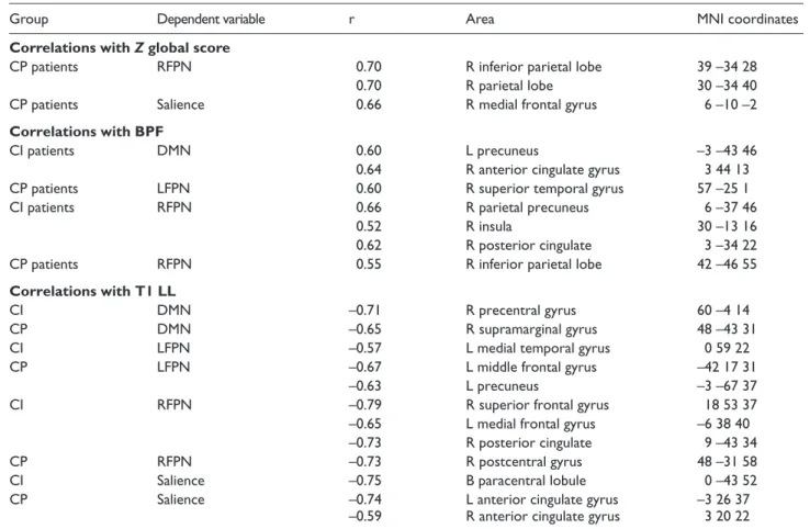
Relationships found among cognitive status, and radio-logical and neurofunctional variables of MS patients are presented in Table 3 and Figures 2–4. First, only CP patients showed positive correlations between global cognitive Z
scores and rs-FC in the RFPN and salience network, pre-dominantly focused on frontal and parietal areas. Furthermore, positive correlations between BPFs and rs-FC at the DMN, LFPN and RFPN, including parietal areas and the anterior cingulate gyrus, were also found in CI and CP patients. Finally, negative correlations among rs-FC in all explored RSNs and T1-lesion load were also found in both patient groups. These negative correlations were observed again at several frontal and parietal areas, including the anterior and posterior cingulate gyri.
Discussion
In the present study, we explored the existence of adaptive functional connectivity changes associated with cognitive function in the DMN, LFPN, RFPN and salience network of MS patients. Our results reveal that CI patients display less rs-FC among different brain areas belonging to these RSNs, thus supporting the notion that RSN alterations may play a significant role in MS cognitive disturbances. On the other hand, CP patients exhibited a degree of connectivity indistinguishable from that of the HC group but stronger in several nodes of the explored RSNs compared with CI patients. These findings might be regarded as providing further support to the previously suggested importance of preserving connectivity within the RSNs to retain normal cognitive competence.
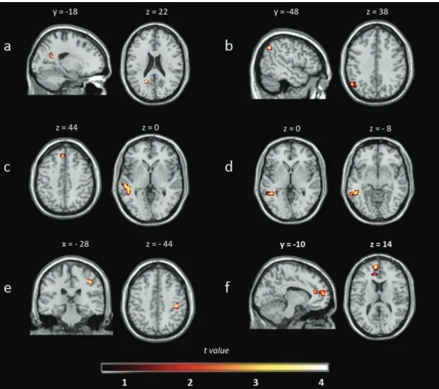
Figure 1. (a) Default mode network (DMN): decreased resting-state functional connectivity (rs-FC) in cognitively impaired (CI) patients compared with healthy controls (HCs); (b) DMN: decreased rs-FC in CI compared with cognitively preserved (CP) patients; (c) left frontoparietal network (LFPN): decreased rs-FC in CI patients compared with HCs; (d) LFPN: decreased rs-FC in CP patients compared with HCs; (e) right frontoparietal network (RFPN): decreased rs-FC in CI compared with CP patients; (f) salience network: decreased rs-FC in CI compared with CP patients. Images are presented in neurological convention and thresholded at p < 0.005 (k = 12 voxels), corrected for multiple comparisons using Monte Carlo simulations.
Tab
le 1.
Main demogra
phic
, clinical,
MRI and neur
opsychological characteristics of all par
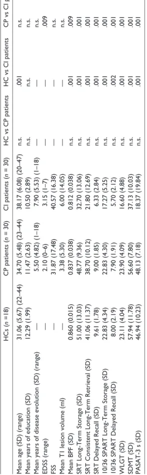
ticipants. HCs ( n =18) CP patients ( n = 30) CI patients ( n = 30) HC vs CP patients HC vs CI patients CP vs CI patients
Mean age (SD) (range)
31.06 (5.67) (22–44) 34.70 (5.48) (23–44) 38.17 (6.08) (20–47) n.s. .001 n.s.
Mean years of education (SD)
12.29 (1.99) 11.47 (2.63) 10.50 (2.89) n.s. n.s. n.s.
Mean years of disease evolution (SD) (range)
— 5.50 (4.82) (1–18) 7.90 (5.53) (1–18) — — n.s. EDSS (range) — 2.10 (0–6) 3.15 (1–7) — — .009 FSS — 31.87 (17.48) 40.57 (16.38) — — n.s.
Mean T1 lesion volume (ml)
— 3.38 (5.30) 6.00 (14.05) — — n.s. Mean BPF (SD) 0.860 (0.015) 0.837 (0.038) 0.812 (0.038) n.s. .001 .009 SRT Long-Term Storage (SD) 51.00 (13.03) 48.77 (9.36) 32.70 (13.06) n.s. .001 .001
SRT Consistent Long-Term Retrieval (SD)
41.06 (11.37) 38.70 (10.12) 21.80 (12.69) n.s. .001 .001 SRT Delayed Recall (SD) 9.61 (1.78) 9.00 (1.85) 6.33 (2.84) n.s. .001 .001
10/36 SPART Long-Term Storage (SD)
22.83 (4.34) 22.83 (4.30) 17.27 (5.25) n.s. .001 .001
10/36 SPART Delayed Recall (SD)
8.00 (2.19) 7.90 (1.91) 5.70 (2.12) n.s. .002 .001 WLGT (SD) 23.11 (4.04) 23.90 (4.09) 16.60 (4.88) n.s. .001 .001 SDMT (SD) 57.94 (11.78) 56.60 (7.80) 37.13 (10.03) n.s. .001 .001 PASAT-3 s (SD) 46.94 (10.23) 48.13 (7.18) 18.37 (19.84) n.s. .001 .001 MRI: magnetic r esonance imaging; n.s.: not significant; HC: health y contr ol; CI: cognitiv el y impair ed; CP: cognitiv el y pr eser ved; EDSS:
Expanded Disability Status Scale;
FSS: Fatigue Se verity Scale; BPF: brain pa-rench ymal fraction; SR T: Selectiv e Reminding Test; SP AR T: Spatial Recall Test; WLGT : W or d List Generation Test; SDMT
: Symbol Digit Modalities
Test; P ASA T-3 s: Paced Auditor y Serial Ad dition 3-Second Test.
More specifically, and in close similarity to previous studies,12,13 we observed that CI patients showed less
con-nectivity in the DMN and LFPN compared with HCs. Interestingly, CP patients exhibited a greater degree of rs-FC than CI patients in the DMN, RFPN and salience network. The abovementioned findings are in agreement with other studies reporting that MS patients with pre-served cognitive abilities display normal or enhanced rs-FC in RSNs,14–16,31 and these results have often been
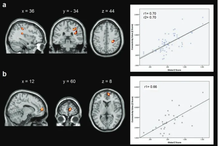
interpreted as adaptive functional changes compensating for cognitive deficits. Following this rationale, we may conclude that in our study, CP patients showed adaptive connectivity changes to compensate for potential cogni-tive deficits, although alternacogni-tive explanations must also be considered (see below). The potential association between appropriate rs-FC and the cognitive status of MS patients was further reinforced by the results of our cor-relational analyses demonstrating that global cognitive Z
scores of CP patients were positively correlated with degree of rs-FC in the medial frontal and inferior parietal areas of the salience network and RFPN, respectively. Loitfelder et al.31 recently reported that MS patients might
require high rs-FC in the inferior parietal cortex and the angular gyrus to attain correct performance in attention and working memory tasks. Therefore, our findings are in agreement with accumulating evidence that seem to con-verge towards a relationship between reduced functional connectivity in the RSNs and cognitive impairment in MS patients.
Most RSN research on MS patients has primarily focused on the DMN.12,13 However, there are other
net-works associated with resting processes that may be of special relevance when studying cognitive deficits in MS patients. For example, the LFPN and RFPN are RSNs that are highly consistent among participants and that only recently have started to receive proper atten-tion in the context of MS research.14,17 The LFPN and
RFPN engage areas distant from the frontal and parietal lobes, which may be especially prone to MS pathophys-iology. In fact, many recent fMRI studies have explored the engagement of frontoparietal networks associated with performance in attention and working memory tasks under the assumption that disconnection among distal frontoparietal areas may underlie primary cogni-tive deficits in MS.3,6,8 Therefore, the LFPN and RFPN
may be especially relevant to understanding cognitive impairment in MS patients, which seems to be sup-ported by our results demonstrating clear differences among groups in those networks. In this regard, CP patients did not show a significant increase in LFPN rs-FC but did show greater RFPN rs-FC compared with CI patients, and the magnitude of this increase was cor-related with cognitive performance (global Z scores). Combining these results, we may deduce that although CI as well as CP patients showed deficits in the LFPN,
Table 3. Correlations among resting-state networks, and cognitive and radiological variables. Thresholded at p < 0.005 (k = 12 voxels) corrected for multiple comparisons using Monte Carlo simulations
Group Dependent variable r Area MNI coordinates
Correlations with Z global score
CP patients RFPN 0.70 R inferior parietal lobe 39 –34 28
0.70 R parietal lobe 30 –34 40
CP patients Salience 0.66 R medial frontal gyrus 6 –10 –2
Correlations with BPF
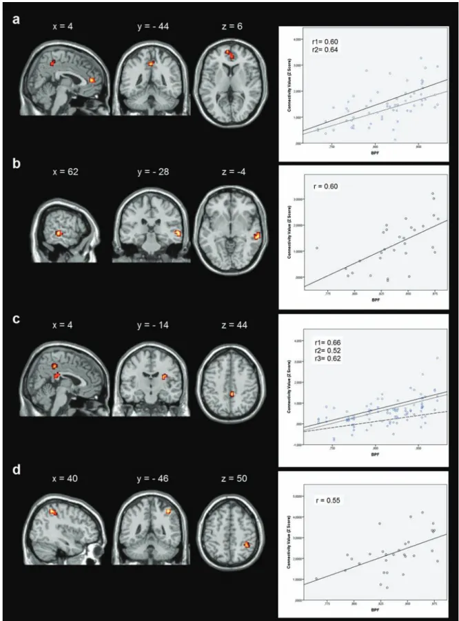
CI patients DMN 0.60 L precuneus –3 –43 46
0.64 R anterior cingulate gyrus 3 44 13
CP patients LFPN 0.60 R superior temporal gyrus 57 –25 1
CI patients RFPN 0.66 R parietal precuneus 6 –37 46
0.52 R insula 30 –13 16
0.62 R posterior cingulate 3 –34 22
CP patients RFPN 0.55 R inferior parietal lobe 42 –46 55
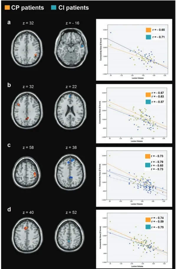
Correlations with T1 LL
CI DMN –0.71 R precentral gyrus 60 –4 14
CP DMN –0.65 R supramarginal gyrus 48 –43 31
CI LFPN –0.57 L medial temporal gyrus 0 59 22
CP LFPN –0.67 L middle frontal gyrus –42 17 31
–0.63 L precuneus –3 –67 37
CI RFPN –0.79 R superior frontal gyrus 18 53 37
–0.65 L medial frontal gyrus –6 38 40
–0.73 R posterior cingulate 9 –43 34
CP RFPN –0.73 R postcentral gyrus 48 –31 58
CI Salience –0.75 B paracentral lobule 0 –43 52
CP Salience –0.74 L anterior cingulate gyrus –3 26 37
–0.59 R anterior cingulate gyrus 3 20 22
HC: healthy control; CI: cognitively impaired; CP: cognitively preserved; MNI: Montreal Neurological Institute; L: left; R: right; B: bilateral; DMN: default mode network; LFPN: left frontoparietal network; RFPN: right frontoparietal network; BPF: brain parenchymal fraction; T1 LL: T1-lesion load.
Table 2. Mean (SD) values of z scores of resting state activity within the clusters that show significant differences among HC, CI and CP patients. Results are thresholded at p < 0.005 (k = 12 voxels) corrected for multiple comparisons using Monte Carlo simulations.
Cluster MNI space HC CP CI p
DMN
CI <HC L parietal lobe –21 –58 22 2.80 (1.15) — 1.79 (1.08) .004
CI<CP L parietal lobe –54 –61 37 — 1.98 (0.68) 1.37 (0.72) .009
LFPN
CI<HC L superior temporal lobe 63 –49 –5 1.90 (0.64) — 1.28 (0.68) .006
L medial frontal lobe –3 35 43 1.33 (0.59) — 0.79 (0.63) .017
CP<HC L temporal lobe –63 –49 –5 1.92 (0.60) 1.17 (0.72) — .000
RFPN
CI<CP R postcentral gyrus 42 –25 43 — 1.04 (0.49) 0.62 (0.40) .003
Salience
CI<CP L medial frontal lobe –6 56 13 — 3.04 (0.71) 1.99 (0.53) .000
L anterior cingulate –12 38 16 — 1.56 (0.55) 0.95 (0.48) .000
HC: healthy control; CI: cognitively impaired; CP: cognitively preserved; MNI: Montreal Neurological Institute; L: left; R: right; DMN: default mode network; LFPN: left frontoparietal network; RFPN: right frontoparietal network.
only CP patients could retain normal cognitive compe-tence by sustaining proper connectivity among RFPN areas. The notion that increased recruitment of the right
side is critical for cognitive performance was previously posed by fMRI studies using tasks that require engage-ment of the LFPN and RFPN.2,5,8
Figure 2. Correlations between resting-state functional connectivity (rs-FC) and global cognitive Z scores. (a) Correlation in the right frontoparietal network (RFPN) in cognitively preserved (CP) patients; (b) correlation in the salience network in CP patients. Images are presented in neurological convention and thresholded at p < 0.005 (k = 12 voxels), corrected for multiple comparisons using Monte Carlo simulations.
We also studied the salience network associated with behavioural control functions.30 The relationship between
rs-FC in this network and the cognitive status of MS patients had not been previously explored in MS patients, although a recent study described an association between decreased connectivity in this network in relapsing–remit-ting MS patients and their clinical disability compared with HCs.32 In our study, CI patients showed reduced rs-FC at
the anterior cingulate gyrus in the salience network com-pared with the CP group. The significance of anterior cin-gulate activation for cognition in MS is supported by the results of Rocca et al.12 describing reduced connectivity at
the anterior cingulate gyrus in CI patients compared with CP patients.
Cerebral reorganisation is secondary to structural dam-age.14,33,34 Previous data suggest that these functional
restructuration processes appear when levels of brain dam-age are low but that these processes can no longer be trig-gered when damage is more extensive.33 Following this line
of reasoning, we investigated the role of radiological vari-ables in the different connectivity networks. MS patients (either CI or CP) with higher volume of lesions showed less
rs-FC in all cognitive networks explored as compared to those patients with less lesion volume, thus suggesting that disease in white matter disrupt the pathways that mediate the transmission of information across brain networks.35 In
this regard, we also observed that BPFs were positively correlated with rs-FC networks in both groups of patients; that is, patients with more brain volume were more cogni-tively preserved than patients with less brain volume. As expected from previous studies,35 atrophy was
accompa-nied by alterations in the networks’ connectivity and both kinds of alterations probably underlie the reduction of cog-nitive performance observed in MS patients. This proposal is also supported by the results of the regression analysis performed in the present study, which revealed that the BPF was the best predictor of cognitive performance of MS patients.
In conclusion, this study extends our knowledge about functional alterations of RSNs in MS patients and their pos-sible relationship with cognitive performance. We observed that brain injury was accompanied by reduced functional connectivity at different RSNs, which may probably be responsible for the onset and progression of cognitive
Figure 3. Correlations between resting-state functional connectivity (rs-FC) and brain parenchymal fractions (BPF). (a) Correlation in the default mode network (DMN) in cognitively impaired (CI) patients; (b) correlation in the left frontoparietal network (LFPN) in cognitively preserved (CP) patients; (c) correlation in the right frontoparietal network (RFPN) in CI patients; (d) correlation in the RFPN in CP patients. Images are presented in neurological convention and thresholded at p < 0.005 (k = 12 voxels), corrected for multiple comparisons using Monte Carlo simulations.
Figure 4. Correlations between resting-state functional connectivity (rs-FC) and T1-lesion load (LL). (a) Correlation in the default mode network (DMN); (b) correlation in the left frontoparietal network (LFPN); (c) correlation in the right frontoparietal network (RFPN); (e) (d) correlation in the salience network. Images are presented in neurological convention and thresholded at p < 0.005 (k = 12 voxels), corrected for multiple comparisons using Monte Carlo simulations.
deficits in MS patients. However, and contrary to our initial hypothesis, CP patients did not exhibit a stronger degree of connectivity than HCs in any of the RSNs evaluated. In this regard, it should be noted that the discrepant size of the groups may have led to a reduction in statistical potency of our analysis resulting in false-negative findings. Future studies assessing rs-FC will help to further clarify the use of RSN activity markers to characterise and predict cognitive performance in MS patients and to determine if engaging compensatory neuroplastic mechanisms is required to retain cognitive competence despite disease progression.
Conflict of interest statement None declared.
Funding
This work was supported by the Brainglot project of the CONSOLIDER-INGENIO 2010 Programme (CSD2007-00012). The project was also supported by grants PSI2010-20168 from MINECO, P1·1B2011-09 from Universitat Jaume I and an educa-tional grant from Biogen Idec.
References
1. Hawellek DJ, Hipp JF, Lewis CM, et al. Increased functional connectivity indicates the severity of cognitive impairment in multiple sclerosis. Proc Natl Acad Sci U S A 2011; 108: 19066–19071.
2. Audoin B, Ibarrola D, Ranjeva JP, et al. Compensatory cor-tical activation observed by fMRI during a cognitive task at the earliest stage of MS. Hum Brain Mapp 2003; 20: 51–58.
3. Au Duong MV, Boulanouar K, Audoin B, et al. Modulation of effective connectivity inside the working memory network in patients at the earliest stage of multiple sclerosis. Neuroimage
2005; 24: 533–538.
4. Au Duong MV, Audoin B, Boulanouar K, et al. Altered func-tional connectivity related to white matter changes inside the working memory network at the very early stage of MS. J Cereb Blood Flow Metab 2005; 25: 1245–1253.
5. Cader S, Cifelli A, Abu-Omar Y, et al. Reduced brain func-tional reserve and altered funcfunc-tional connectivity in patients with multiple sclerosis. Brain 2006; 129: 527–537.
6. Forn C, Barros-Loscertales A, Escudero J, et al. Cortical reor-ganization during PASAT task in MS patients with preserved working memory functions. Neuroimage 2006; 31: 686–691. 7. Forn C, Barros-Loscertales A, Escudero J, et al. Compensa-tory activations in patients with multiple sclerosis during pre-served performance on the auditory N-back task. Hum Brain Mapp 2007; 28: 424–430.
8. Forn C, Rocca MA, Valsasina P, et al. Functional magnetic resonance imaging correlates of cognitive performance in patients with a clinically isolated syndrome suggestive of multiple sclerosis at presentation: An activation and connec-tivity study. Mult Scler 2012; 18: 153–163.
9. Bonnet MC, Allard M, Dilharreguy B, et al. Cognitive com-pensation failure in multiple sclerosis. Neurology 2010; 75: 1241–1248.
10. Cifelli A and Matthews PM. Cerebral plasticity in mul-tiple sclerosis: Insights from fMRI. Mult Scler 2002; 8: 193–199.
11. Fox MD and Greicius M. Clinical applications of resting state functional connectivity. Front Syst Neurosci 2010; 4: 19. 12. Rocca MA, Valsasina P, Absinta M, et al. Default-mode
net-work dysfunction and cognitive impairment in progressive MS. Neurology 2010; 74: 1252–1259.
13. Bonavita S, Gallo A, Sacco R, et al. Distributed changes in default-mode resting-state connectivity in multiple sclerosis.
Mult Scler 2011; 17: 411–422.
14. Roosendaal SD, Schoonheim MM, Hulst HE, et al. Resting state networks change in clinically isolated syndrome. Brain
2010; 133: 1612–1621.
15. Hulst HE and Geurts JJ. Gray matter imaging in multiple scle-rosis: What have we learned? BMC Neurol 2011; 11: 153. 16. Sumowski JF, Wylie GR, Leavitt VM, et al. Default network
activity is a sensitive and specific biomarker of memory in multiple sclerosis. Mult Scler 2013; 19: 199–208.
17. Faivre A, Rico A, Zaaraoui W, et al. Assessing brain connec-tivity at rest is clinically relevant in early multiple sclerosis.
Mult Scler 2012; 18: 1251–1258.
18. Polman CH, Reingold SC, Banwell B, et al. Diagnostic cri-teria for multiple sclerosis: 2010 revisions to the McDonald criteria. Ann Neurol 2011; 69: 292–302.
19. Kurtzke JF. Rating neurologic impairment in multiple scle-rosis: An expanded disability status scale (EDSS). Neurology
1983; 33: 1444–1452.
20. Sepulcre J, Vanotti S, Hernández R, et al. Cognitive impairment in patients with multiple sclerosis using the Brief Repeatable Battery-Neuropsychology test. Mult Scler 2006; 12: 187–195. 21. Calabrese M, Agosta F, Rinaldi F, et al. Cortical lesions
and atrophy associated with cognitive impairment in relapsing–remitting multiple sclerosis. Arch Neurol 2009; 66: 1144–1150.
22. Sanfilipo MP, Benedict RHB, Zivadinov R, et al. Correction for intracranial volume in analysis of whole brain atrophy in multiple sclerosis: The proportion vs. residual method. Neu-roimage 2004; 22: 1732–1743.
23. Ceccarelli A, Bakshi R and Neema M. MRI in multiple sclero-sis: A review of the current literature. Curr Opin Neurol 2012; 25: 402–409.
24. Calhoun VD, Adali T, Pearlson GD, et al. A method for mak-ing group inferences from functional MRI data usmak-ing inde-pendent component analysis. Hum Brain Mapp 2001; 14: 140–151.
25. Bell AJ and Sejnowski TJ. An information-maximization approach to blind separation and blind deconvolution. Neural Comput 1995; 7: 1129–1159.
26. Himberg J, Hyvärinen A and Esposito F. Validating the inde-pendent components of neuroimaging time series via cluster-ing and visualization. Neuroimage 2004; 22: 1214–1222. 27. Erhardt EB, Rachakonda S, Bedrick EJ, et al. Comparison of
multi-subject ICA methods for analysis of fMRI data. Hum Brain Mapp 2011; 32: 2075–2095.
28. Raichle ME, MacLeod AM, Snyder AZ, et al. A default mode of brain function. Proc Natl Acad Sci U S A 2001; 98: 676–682. 29. Damoiseaux JS, Rombouts SA, Barkhof F, et al. Consistent
resting-state networks across healthy subjects. Proc Natl Acad Sci U S A 2006; 103: 13848–13853.
30. Menon V and Uddin LQ. Saliency, switching, attention and control: A network model of insula function. Brain Struct Funct 2010; 214: 655–667.
31. Loitfelder M, Filippi M, Rocca M, et al. Abnormalities of rest-ing state functional connectivity are related to sustained atten-tion deficits in MS. PloS One 2012; 7: e42862.
32. Rocca MA, Valsasina P, Martinelli V, et al. Large-scale neu-ronal network dysfunction in relapsing–remitting multiple sclerosis. Neurology 2012; 79: 1449–1457.
33. Schoonheim MM, Geurts JJ and Barkhof F. The limits of functional reorganization in multiple sclerosis. Neurology
2010; 74: 1246–1247.
34. Rocca MA, Gatti R, Agosta F, et al. Influence of task com-plexity during coordinated hand and foot movements in MS patients with and without fatigue. A kinematic and functional MRI study. J Neurol 2009; 256: 470–482.
35. Filippi M and Rocca MA. MRI and cognition in multiple scle-rosis. Neurol Sci 2010; 3 (Suppl 2): S231–S234.