Open Research Online
The Open University’s repository of research publications
and other research outputs
Integration of Toxicity Data from Experiments and
Non-Testing Methods within a Weight of Evidence
Procedure
Thesis
How to cite:
Golbamaki Bakhtyari, Azadi (2018). Integration of Toxicity Data from Experiments and Non-Testing Methods within a Weight of Evidence Procedure. PhD thesis The Open University.
For guidance on citations see FAQs.
c
2018 The Author Version: Version of Record
Copyright and Moral Rights for the articles on this site are retained by the individual authors and/or other copyright owners. For more information on Open Research Online’s data policy on reuse of materials please consult the policies page.
Integration of Toxicity Data from Experiments and
Non-Testing Methods within a Weight of Evidence
Procedure
Thesis submitted for the Degree of Doctor of Philosophy
School of Life, Health and Chemical Sciences
Azadi Golbamaki Bakhtyari
Environmental Chemistry and Toxicology Laboratory
IRCCS - Istituto di Ricerche Farmacologiche “Mario Negri”
Milan, Italy
I
Abstract
Assessment of human health and environmental risk is based on multiple sources of
information, requiring the integration of the lines of evidence in order to reach a conclusion. There is an increasing need for data to fill the gaps and new methods for the data integration. From a regulatory point of view, risk assessors take advantage of all the available data by means of weight of evidence (WOE) and expert judgement approaches to develop conclusions about the risk posed by chemicals and also nanoparticles. The integration of the physico-chemical properties and toxicological effects shed light on relationships between the molecular properties and biological effects, leading us to non-testing methods. (Quantitative) structure-activity relationship ((Q)SAR) and read-across are examples of non-testing methods. In this dissertation, (i) two new structure-based carcinogenicity models, (ii) ToxDelta, a new read-across model for mutagenicity endpoint and (iii) a genotoxicity model for the metal oxide nanoparticles are introduced. Within the latter section, best professional judgement method is employed for the selection of reliable data from scientific publications to develop a data base of nanomaterials with their genotoxicity effect. We developed a decision tree model for the classification of these nanomaterials.
The (Q)SAR models used in qualitative WOE approaches mainly lack transparency resulting in risk estimates needing quantified uncertainties. Our two structure-based carcinogenicity models, provide transparent reasoning in their predictions. Additionally, ToxDelta provides better supported techniques in read-across terms based on the analysis of the differences of the molecules structures. We propose a basic qualitative WOE framework that couples the in silico models predictions with the inspections of the similar compounds. We demonstrate the
application of this framework to two realistic case studies, and discuss how to deal with different and sometimes conflicting data obtained from various in silico models in qualitative WOE terms to facilitate structured and transparent development of answers to scientific questions.
II
III
Acknowledgements
This research was supported by the EU-TOXRISK project. The EU-ToxRisk project has received funding from the European Union's Horizon 2020 research and innovation programme under grant agreement No 681002.
Disclaimer
This dissemination reflects only the author's view and the EU-ToxRisk Commission is not responsible for any use that may be made of the information it contains.
IV
Table of Contents
List of Tables and Figures ... 1
Publications ... 4
Abbreviations ... 5
CHAPTER 1 ... 7
Introduction ... 7
1.1 Genotoxicity, Carcinogenicity and Mutagenicity ... 7
1.1.1 Gene Mutation ... 9
1.1.2 Mutation in Cancer Cells ... 9
1.1.3 Base Alteration ... 10
1.2 Genotoxic and Carcinogenic Chemicals ... 11
1.3 The Animal Test(s) ... 12
1.3.1 Rodent Carcinogenicity Bioassay ... 12
1.3.2 Some Notes about Rodent Carcinogenicity Bioassay ... 13
1.4 Ames Test ... 14
1.5 Nongenotoxic Carcinogens ... 14
1.5.1 Modes of Action of Human Nongenotoxic Carcinogens ... 15
1.6 Genotoxic Carcinogens ... 15
1.6.1 Modes of Action of Human Genotoxic Carcinogens ... 16
1.7 Carcinogenic Categories of the Substances ... 18
1.8 Structural Alert Lists for Carcinogenicity and Mutagenicity ... 19
1.9 Current Hazard Identification Procedures and Related Considerations ... 20
1.10 (Q)SAR and REACH ... 21
V
1.12 Genotoxicity Assessments in the EU Regulations ... 24
1.13 Read-across ... 25
1.14 Classification and Labelling and Chemical Safety Assessment ... 26
1.15 Development and Optimisation of Alternative Methods ... 27
1.15.1 Importance of Mode of Action and Weight of Evidence Approach ... 27
1.15.2 Alternative Methods for Detecting Nongenotoxic Carcinogens ... 27
1.16 Quantitative Structure–Activity Relationship (QSAR) ... 28
1.17 Software Packages for Mutagenicity and Carcinogenicity Predictions ... 29
1.17.1 VEGA Platform... 29 1.17.2 DEREK Nexus ... 29 1.17.3 TOPKAT ... 30 1.17.4 MultiCASE ... 30 1.17.5 QSAR Toolbox ... 30 1.17.6 Toxtree ... 31 1.17.7 LAZAR ... 31 1.17.8 ACD/Tox Suite ... 31
1.17.9 Leadscope Model Applier ... 32
1.17.10 SARpy ... 32
1.18 Weight of Evidence ... 32
1.18.1 Weight of Evidence Method Classification ... 34
1.18.2 Weight of Evidence Application ... 36
1.18.3 Weight of Evidence Approach in Nanomaterials Risk Assessment ... 36
VI
1.19.1 Part 1- New Clues on Carcinogenicity-Related Substructures Derived From
Mining Two Large Datasets of Chemical Compounds ... 39
1.19.2 Part 2 - Toxdelta: A New Program to Assess How Dissimilarity Affects the Effect of Chemical Substances. ... 39
1.19.3 Part 3 – Genotoxicity Induced by Metal Oxide Nanoparticles: a Weight of Evidence Study and Effect of Particle Surface and Electronic Properties ... 39
CHAPTER 2 ... 41
2. Aim of the Study ... 41
2.1 Part 1 – Carcinogenicity Models ... 43
2.2 Part 2 – ToxDelta ... 45
2.3 Part 3 – Metal Oxide NMs Genotoxicity Model ... 46
CHAPTER 3 ... 48
3. Materials and Methods ... 48
3.1 Part 1 – Carcinogenicity Models ... 48
3.1.1 Carcinogenesis Data Sources ... 48
3.1.2 Comparison between the ANTARES Dataset and the ISSCAN/CGX Dataset ... 49
3.1.3 Data for Model Validation ... 49
3.1.4 Active Molecular Fragments Identification by SARpy... 50
3.1.5 Extracting Active Fragments ... 52
3.1.6 Internal Evaluation of the Models ... 53
3.1.7 External Evaluation of the Models ... 54
3.2 Part 2 – ToxDelta ... 55
3.2.1 Database of Active and Inactive Structural Alerts ... 55
VII
3.2.3 ToxDelta Implementation ... 57
3.3 Part 3 – Metal Oxide NMs Genotoxicity Model ... 61
3.3.1 Data Collection and Assessment ... 61
3.3.2 Weight of Evidence Approach in the Evaluation of the Data Set ... 63
3.3.3 Case Studies for Illustrating the Weight of Evidence Evaluation ... 64
3.3.4 Computational Analysis of nanomaterials Structure and Descriptor Generation: Quantum-chemical Descriptors... 65
3.3.5 Classification SAR Modelling Methods ... 66
3.4 Weight of Evidence Approach in the Analysis of Results of Different In Silico Methods for the Mutagenicity Assessment of Chemicals ... 71
CHAPTER 4 ... 72
4. Results and Discussions ... 72
4.1 Part 1- Carcinogenicity Models ... 72
4.1.1 R Model ... 72
4.1.2 E Model ... 72
4.1.3 Analysis of the Combination of the Prediction Results of the R and E Models .... 73
4.1.4 Fragments Analysis ... 74
4.2 Part 2- ToxDelta ... 79
4.2.1 Case Study 1: Benzodiazepine Derivatives... 79
4.2.2 Case Study 2: Androstane Derivatives ... 82
4.3 Part 3 – Metal Oxide NMs Genotoxicity Model ... 86
4.3.1 Data Quality Assessment ... 86
4.3.2 Quantum Mechanical Descriptor Calculations ... 88
VIII
4.4 Weight of Evidence Approach in In Silico Models for the Mutagenicity Assessment of
Chemicals ... 92
CHAPTER 5 ... 93
5. Conclusions ... 93
5.1 Part 1- Carcinogenicity Models ... 93
5.2 Part 2- ToxDelta ... 95
5.3 Part 3- Metal Oxide NMs Genotoxicity Model ... 97
5.4 Overall Conclusion ... 100
CHAPTER 6 ... 102
6. Example of the Use of Non-Testing Methods within a Weight of Evidence Framework 102 6.1 First Case Study: Valproic Acid ... 103
6.2 Second Case Study: Diclofenac ... 111
References ... 121
Appendices ... 131
Metal Oxide nanomaterials Genotoxicity Model Supplementary Information ... 131
New clues on carcinogenicity-related substructures derived from mining two large datasets of chemical compounds ... 163
Structural Alerts for Carcinogen Compounds (R Model) ... 182
Structural Alerts for Carcinogen Compounds (E model) ... 186
ToxDelta: A New Program to Assess How Dissimilarity Affects the Effect of Chemical Substances ... 189
1
List of Tables and Figures
Tables:
Table 1. Structural alerts belonging to certain mechanistic domains
Table 2. Test methods most commonly used for genotoxicity/mutagenicity testing Table 3. Weight of evidence methods
Table 4. Criteria for the usefulness and quality assessment of the data set for the (Q)SAR modelling: extent of comet assay conditions checklist. General parameters have been used to assess each data point and the results are reported in Table S1.A (Appendices) where all questions are answered in a yes or no fashion.
Table 5. Comet assay experimental results for all selected metal oxide nanomaterials used for (Q)SAR modelling
Table 6. Acronyms, short definitions and units of the molecular descriptors calculated by MOPAC2012.
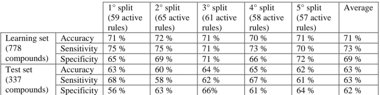
Table 7. R model internal and external validation for five different splits and the average of the model performance
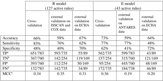
Table 8. R model and E model internal and external validation
Table 9. The combination of the predictions of the R and E models on the ECHA external validation set
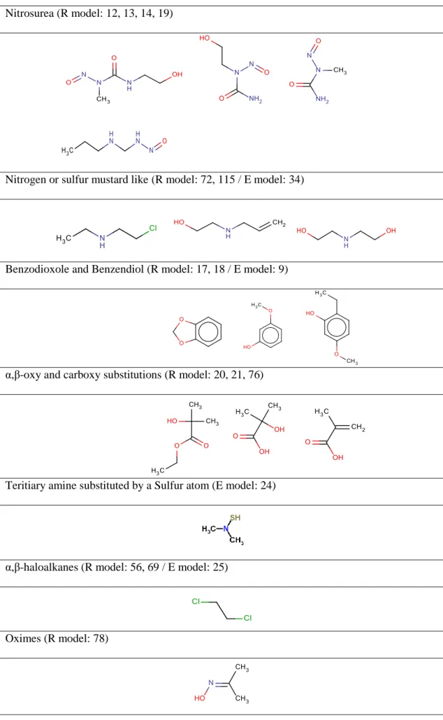
Table 10. New carcinogenic structural alerts identified by SARpy in the R and E models Table 11. Chemicals structures in the ISSCAN-CGX data set from which structural alert 24 has been extracted
Table 12. The two case studies: Case study 1) target molecule: Diazepam, source molecule: Flunitrazepam; Case study 2) target molecule: cholestan-6-one, 3-bromo-, cyclic 1,2-ethanediyl mercaptole, S,S,S',S'-tetraoxide, (3-beta,5-alpha)-, source molecule: mepitiostane, and the results of ToxDelta: maximum common substructure and dissimilar fragments.
Table 13. Assignment of the reliability of in vitro comet assays based on the criteria defined in Huk et al. The assignment questions were treated in a “yes” or “no” fashion. In a weight of
2
evidence approach, data points that presented 1st class property were used to assign the genotoxic or non-genotoxic fate to the metal oxide nanomaterials with the same chemical core composition.
Table 14. Summary of the prediction results of non-testing models for Valproic acid Table 15. Experimental and prediction values for some examples of similar chemicals to Valproic acid (CAS number: 99-66-1) in the training set and test set of T.E.S.T.
Table 16. Optional tabular format for summarizing weight of evidence assessment of Valproic acid
Table 17. Summary of the prediction results of non-testing models for Diclofenac Table 18. Experimental and prediction values for N-[(2,4,5-Trichlorophenoxy)acetyl]-L-aspartic acid (CAS 66789-80-8) and N-[4-(4-Amino-3-chlorobenzyl)-2-chlorophenyl]acetamide (CAS 91575-28-9) as examples of similar chemicals to Diclofenac in the outcome of T.E.S.T. Table 19. The mutagenicity and non-mutagenicity rules identified by ToxRead in the structure of Diclofenac
Table 20. Optional tabular format for summarizing weight of evidence assessment of Diclofenac
Figures:
Figure 1. Multistage carcinogenesis
Figure 2. Parallels between recombination and certain types of mutational repair. Figure 3. Pairing between the normal form of the bases
Figure 4. Alkylation-induced specific mispairings
Figure 5. (a) Structures of common intercalating agents and (b) their interaction with DNA Figure 6. The MCS between two molecules is shown with bold lines, and the other branches are the differences
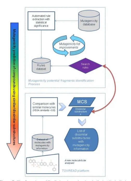
Figure 7. The flow chart of ToxDelta: the molecular similarity/dissimilarity structure analysis software for the mutagenicity endpoint
Figure 8. The ‘rpart’ classification tree model on the data set of 16 metal oxide nanomaterials.
3
Figure 10. A) ToxDelta outcome for the comparison between Valproic acid (molecule #1) and 2-[Ethyl(nitroso)amino]ethanol (CAS 13147-25-6) (molecule #2) The maximum common substructure is at the top of the panel. The dissimilar substructures are listed below their corresponding molecule. B) The identified dissimilar substructure extracted from the molecule #2 is a mutagenicity structural alert in the CRS4 dataset of mutagenicity ruleset with
accuracy=1.
Figure 11. ToxRead chart for the target molecule Valproic acid (CAS number: 99-66-1). The numbers refer to CAS identifiers. Straight arrows link the target chemical to rules, while curved arrows link to chemicals
Figure 12. Diclofenac, CAS number: 15307-86-5
Figure 13. ToxDelta outcome for the comparison between Diclofenac and a) N-[(2,4,5-Trichlorophenoxy)acetyl]-L-aspartic acid (CAS 66789-80-8), b) N-[4-(4-Amino-3-chlorobenzyl)-2-chlorophenyl]acetamide (CAS 91575-28-9)
Figure 14. ToxDelta outcome of the dissimilar substructure remaining after the subtraction of the MCS and the structural rules identified inside the dissimilar substructure of N-[4-(4-Amino-3-chlorobenzyl)-2-chlorophenyl]acetamide (CAS 91575-28-9) (molecule #2)
Figure 15. ToxRead chart for the target molecule Diclofenac. The target substance molecule is shown on the right and the identified non-mutagenic and mutagenic rules are listed below Figure 16. (a) SM197, Diphenylamine: non-mutagenic rule, (b) MNM37, Phenylamine: mutagenic rule identified by ToxRead
Figure 17. The two similar chemicals to Diclofenac extracted by ToxRead with mutagenic effect
4
Publications
Golbamaki, A., et al. "Comparison of in silico models for prediction of Daphnia magna acute toxicity." SAR and QSAR in Environmental Research 25.8 (2014): 673-694.
Gini, G., et al. "ToxRead: a tool to assist in read across and its use to assess mutagenicity of chemicals." SAR and QSAR in Environmental Research 25.12 (2014): 999-1011.
Cappelli, C. I., et al. "Assessment of in silico models for acute aquatic toxicity towards fish under REACH regulation." SAR and QSAR in Environmental Research 26.12 (2015): 977-999. Golbamaki, A., et al. "Classification nano-SAR modeling of metal oxides nanoparticles genotoxicity based on comet assay data." Toxicology Letters 258 (2016): S271.
Golbamaki, A., et al. "New clues on carcinogenicity-related substructures derived from mining two large datasets of chemical compounds." Journal of Environmental Science and Health, Part C 34.2 (2016): 97-113.
Golbamaki, A., and Emilio Benfenati. "In Silico Methods for Carcinogenicity Assessment." In In Silico Methods for Predicting Drug Toxicity (2016): 107-119.
Golbamaki, A., et al. “The Maximum Common Substructure (MCS) Search as a New Tool for SAR and QSAR”. In Advances in QSAR Modeling (pp. 149-165). Springer, Cham.
Golbamaki, A., et al. "ToxDelta: A New Program to Assess How Dissimilarity Affects the Effect of Chemical Substances." Drug Des 6.153 (2017): 2169-0138.
5
Abbreviations
Best Professional Judgement (BPJ) Bio-Concentration Factor (BCF)
Carcinogenic Potency Database (CPDB) Classification Labelling and Packaging (CLP)
Counter Propagation Artificial Neural Network (CP ANN) Density Functional Theory (DFT)
Distributed Structure-Searchable Toxicity (DSSTox)
EU Regulation for Registration, Evaluation, Authorization and Restriction of Chemicals (REACH)
European Chemical Agency (ECHA) European Food Safety Authority (EFSA)
European Union Reference Laboratory for Alternative to Animal Testing (EURL ECVAM) Food and Drug Administration (FDA)
Gap Junction Intercellular Communication (GJIC) Health and Environmental Science Institute’s (HESI) International Agency for Research on Cancer (IARC) International Life Sciences Institute (ILSI)
Lowest Observed Adverse Effect (LOAEL) Matthews Correlation Coefficient (MCC) Maximum Common Substructure (MCS) Maximum Tolerated Dose (MTD) Nano Material (NM)
Nano Particles (NP)
National Toxicology Program (NTP)
Organization for Economic Cooperation and Development (OECD) Parameterized Model 7 (PM7)
6
(Quantitative) Structure-Activity Relationships ((Q)SAR) Reactive Oxygen Species (ROS)
Simplified Molecular Input Line Entry Specification (SMILES) Structural Alert (SA)
Structure-Activity Relationships (SAR)
United Nations Globally Harmonized System (UN-GHS) Veterinary Medicinal Products (VICH)
7
CHAPTER 1
Introduction
1.1 Genotoxicity, Carcinogenicity and Mutagenicity
“The term carcinogen denotes a chemical substance or a mixture of chemical substances which induce cancer or increase its incidence” 1.
Carcinogenicity is a crucial endpoint for the chemical safety. Carcinogenic compounds may promote carcinogenicity in one of the three phases of causing cancer: initiation, promotion and progression 2 (Figure 1-page 7). Carcinogenesis begins with a mutation, a change of a genetic
material for which no DNA repair mechanism during cell proliferation has happened. This happens in the initiation phase. During the second phase (promotion) which is reversible, the initiated cells are affected by endogenous or exogenous chemicals and because of the clonal growth, the tumour starts to form. For this reason these endogenous or exogenous chemicals are called promoters. These chemicals are not intrinsically mutagenic but cause changes in gene expression or other mechanisms that will be passed to the daughter cells. At this point, cell proliferation rate increases and apoptotic cell death decreases. In the last stage (progression) additional genotoxic events such as chromosomal aberrations and translocations take place. Progression is irreversible and it leads to the formation of neoplasms, benign and malignant alike 3–5.
Figure 1. Multistage carcinogenesis
Genotoxicity describes a damaging action on a cell's genetic material affecting its integrity. Genotoxicity is similar to mutagenicity except that genotoxic effects that cause DNA damage are not themselves necessarily transmissible to the next generation of cells, while mutagenicity
8
refers to the production of transmissible genetic alterations. Genotoxic substances which are capable of causing genetic mutation (pre-mutagenic) and contributing to the development of tumours (carcinogenic) are known to be potentially mutagenic or carcinogenic. Certain chemical compounds and some radiations can induce genotoxicity.
Even low exposure levels of genotoxic substances may actuate serious health effects in somatic and germ cells. Somatic cell genotoxicity plays a role in a variety of genetic diseases. Also degenerative conditions such as accelerated aging, immune dysfunction, cardiovascular and neurodegenerative diseases and cancer are the outcome of accumulation of DNA damage in somatic cells. Mutations in germ cells can lead to spontaneous abortions, infertility or heritable damage to the offspring and possibly to the subsequent generations.
There is a strong correlation between mutagenicity and carcinogenicity. Studies show that approximately 90 percent of the known carcinogens are also mutagens. The somatic mutation theory of cancer states that the mutation of the somatic cells cause cancer.
According to the mode of action, carcinogens can be classified into genotoxic or nongenotoxic carcinogens. Genotoxic carcinogens interact directly with DNA, resulting DNA damage or chromosomal aberrations that can be detected by genotoxicity tests 6. Adversely, nongenotoxic
carcinogens have no direct reactivity with DNA and use other mechanisms in the process of tumour development such as affecting gene expression, signal transduction, and/or cell proliferation.
Mutation may occur in two modes: “spontaneously” or “inducted mutagenicity”. DNA
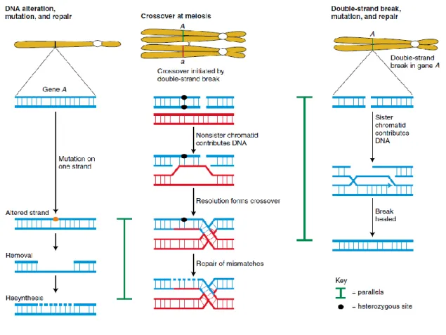
molecules are not stable in the cellular environment and each base pair in a DNA double helix is mutable with a certain probability. Mutations may affect entire chromosomes or large pieces of chromosomes. Gene alterations are the simplest form of mutation. This gene alteration is swapping of one base pair for another. Another cause of mutation can be the insertion of a transposable element from outside the genome. Most of the time the DNA damages are identified and corrected by cells. Figure 2 (page 9) shows the parallels between crossing-over and two kinds of mutational repair (excision and double-strand break repair).
9
Figure 2. Parallels between recombination and certain types of mutational repair.7
1.1.1 Gene Mutation
The gene mutation can be divided into two classes:
- Mutations affecting single base pairs;
- Mutations altering the number of copies of a small repeated sequence within a gene.
1.1.2 Mutation in Cancer Cells
Tumours occur from a sequence of mutational incidences that lead to uncontrolled proliferation and cellular immortality. The transformation of cells from the benign into the carcinogenic state has genetic origins.
1. Most of the induced carcinogens (chemical substances and radiations) are also mutagenic and they cause cancer by originating mutations into cells.
2. A large number of mutagens affiliated with cancer have been identified. Experimental models (in vivo and in vitro) help to find these associations between mutagenicity and carcinogenicity.
10
1.1.3 Base Alteration
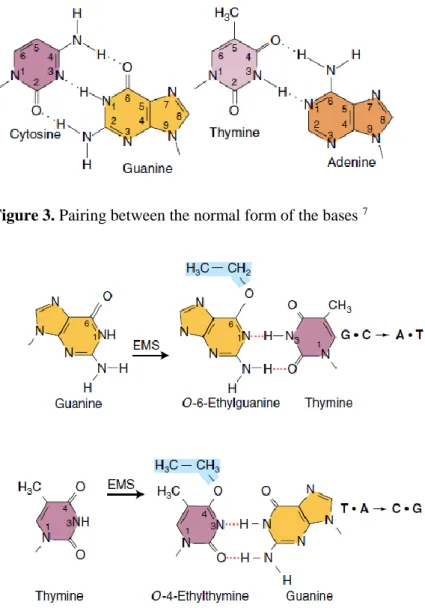
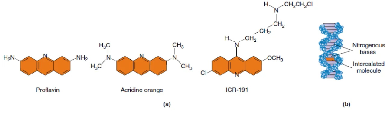
Sometimes the mutagenic agent alters a base in a DNA causing specific mispairing. Figure 3 (page 10) shows the pairing between the normal forms of the bases. Certain alkylating agents such as ethylmethanesulfonate and nitrosoguanidine, are some of the examples of these mutagens that operate by this pathway. These mutagens add alkyl groups to many positions on all four bases. Figure 4 (page 10) shows the alkylation that leads to direct mispairing and results in G.C -> A.T transitions in the next round of replication. Another important class of DNA modifiers are intercalating agents. Compounds such as proflavin, acridine orange and ICR compounds are some examples of this group (Figure 5-page 11). These agents are able to intercalate between the stacked nitrogen bases at the core of the DNA and cause single-nucleotide-pair or deletions.
Figure 3. Pairing between the normal form of the bases 7
11
Figure 5. (a) Structures of common intercalating agents and (b) their interaction with DNA 8
1.2 Genotoxic and Carcinogenic Chemicals
Absorption ways of chemical carcinogens following exposure are oral, inhalator, cutaneous and injection. Afterwards, different issues will be involved 9. All the substances absorbed orally are
distributed in the body through the liver, whereas those absorbed by the lung will enter the blood and after that will reach the liver 10. Genotoxic chemical carcinogens directly damage
DNA, non genotoxic carcinogens or procarcarcinogens require enzymatic conversion before affecting DNA 11.
Non genotoxic chemical carcinogens require bioactivation to electrophiles in order to bind covalently to DNA and often act by producing mutations. Different enzymes are involved in bioactivation reactions, such as oxidation, reduction, thiol conjugation, acetyl transfer, sulfur transfer, methyl transfer, glucuronosyl transfer, and epoxide hydrolysis. These enzymes are classified as oxidoreductases 12. Human body controls metabolic activation by phase I reactions.
Phase II reactions protect the body by transformation of activated compounds into inert products that will be eliminated from the body 13. Phase II enzymes have role in the conjugation and
inactivation of carcinogens and include transferases. Originally, these enzymes were considered to be involved only in the detoxification of biotransformation, but they can also trigger
activation of certain epigenetic carcinogens 11.
Peroxidations occur together with metabolic reactions and cause production of ROS 14,15.
Several chronic diseases are related to these radicals including chemical carcinogenesis 14.
Chemical reactions such as oxidation, nitration/nitrosation and halogenation, which are
associated with ROS trigger damage to DNA, RNA, and proteins. Consequently, mutations and alterations in the functions of important enzymes and proteins occur as a result 13. It is
12
demonstrated that excess amount of ROS created by chemical compounds stimulates initiation, promotion and progression of tomour through genotoxicity 16,17.
Although the above-mentioned metabolic methods are important for both humans and animals, differences are important to be considered. Incorrect interpretations may occur in animal models used in the assays and analysis of chemicals carcinogenicity 18,19.
1.3 The Animal Test(s)
Carcinogenicity studies cannot be limited exclusively to the epidemiological data about carcinogens. For over 40 years, long-term rodent carcinogenicity bioassay using the maximum tolerated dose in 2 species over 2 yr. has been the standard procedure for detecting potential human carcinogens20.
Data obtained from the initial 2-year carcinogenicity studies is often subjected to critiques of the screening procedures since it is inadequate for risk assessment regulatory decisions.
In laboratory experiments on animals, it is shown that most potent mutagenic chemicals are also carcinogenic21. Thus, all the chemicals that are mutagenic in animals are considered also
mutagenic or suspected mutagenic and consequently human carcinogens, until there is found some reliable evidence which shows the contrary 22.
1.3.1 Rodent Carcinogenicity Bioassay
There is increasing understanding that carcinogenesis is a multistep process 23–25. Chemicals
with positive carcinogenicity results are subjected to more accurate and detailed evaluations about their unsafe effects for humans. Verifying whether or not the “carcinogen” chemical with positive results in long-term rodent assays is also hazardous for humans needs more chemical evaluations. These evaluations are necessary to understand the dose-response relationships, the potential hazard for humans 26. It may be unrealistic to expect a basic 2 year study to provide all
the complex data needed for risk management decisions.
Even though the quality of the studies used in toxicity assessments is high, these toxicity conclusions are not sufficient for the regulatory agencies. Some results in the NCI/NTP data base and other data sources clearly show that some chemicals that cause cancer in rodent models are not carcinogenic for humans 23,27. The procedure which states that a chemical with
13
positive effect in at least one of the 4 sex/species combinations is “carcinogen”, is more adequate for selecting compounds to undergo further study than regulatory purposes. Dose extrapolation, different organ responses are among the essential factors that influence the applicability of the rodent bioassays directly to risk assessment.
1.3.2 Some Notes about Rodent Carcinogenicity Bioassay
Mechanistic considerations are essential in carcinogenicity studies especially for nongenotoxic chemicals. Use of cell culture methods will fill the gap of information about the differential metabolism between animal models and humans. According to the legislations for risk
assessment, data obtained from different sources, included experiments and mechanistic studies need to be used for the decision-making process. Many nongenotoxic chemicals are sex- or species specific, for this reason the mechanism of tumour formation has to be studied in both species and sex 28.
The reasonable solution is considering and using the whole available data rather than relying only on the most sensitive test results 29. Information about the chemical concentration used for
each animal species or sex combination is crucial and explains the sex/species specificity of the chemical effects 29. This additional information plays an important role in extrapolation of the
results to humans. Studying specific chemicals by rodent tests, produces useful mechanistic information. The methodologies for predicting carcinogenicity can be explored by conducting high quality rodent studies. These studies will lead us to developing better dose-response relationships and increasing our knowledge about interspecies extrapolation. Despite all the progressions in the animal tests, there is still lack of adequate rodent studies for identifying carcinogens 30.
The 2-year rodent studies are the most expensive tests that usually take place as the first step of the carcinogenicity assessment of a chemical. The following assessments, in case the rodent test result is positive, are more mechanistic, quicker and less expensive. The use of predictive models prior to the animal studies should be more reliable in the chemical evaluation process. This approach makes more resources available for the mechanistic studies and will accelerate the risk assessment deliberations for humans 31.
14 1.4 Ames Test
Tests for carcinogenicity are generally time consuming and are performed on small mammalian animals. Alternative tests use microbes (e.g. fungi and bacteria) and test for mutagenicity instead of carcinogenicity. Any living organism can be used for testing the mutagenicity of a chemical, this is because DNA is chemically equal in all organisms. Bacteria can be used as an alternative to mammalian models, as its life cycle is much shorter and the results can be
obtained easier and faster. The most famous mutagenicity test was developed by Bruce Ames in the 1970s, which is done using Salmonella typhimurium. Properties of the bacteria were
genetically engineered into these strains to make them suitable for mutagen detection. The genotype of the mutant strains in this assay is given as his-. In addition, they carry a mutation that eliminates the protective lipopolysaccharide coating of wild-type Salmonella to facilitate the entry of many different chemicals into the cell.
In a media lacking histidine this mutant bacteria will die. The “revertant” mutants revert the his – to his + genotype and phenotype and this will help the bacteria to grow in a media without histidine. In the Ames test the Salmonella bacteria is placed on plates with a very small amount of histidine and the chemical to be tested is added to the plate. The grown colonies on the plate indicate the number of revertants. To generate a dose-response curve, different concentrations of the chemical under study is tested.
A positive result of the mutagenicity Salmonella typhimurium test is an indication of the high probability that the tested chemical will be carcinogenic in laboratory animals and in
consequence is more likely to be a carcinogen. Not all chemicals that cause cancer in laboratory animals are mutagenic in the Ames test, but still three quarters of the chemicals with positive result in Salmonella test are carcinogenic in also animal studies. The rapidity (3-4 weeks) and low cost of the Ames test makes it an important tool for the mutagenicity screening.
1.5 Nongenotoxic Carcinogens
A great number of human carcinogens are “genotoxic” chemicals, which means their carcinogenicity effect is caused by inducing DNA damage. The rest of the carcinogens are named “nongenotoxic” chemicals, and they induce cancer in other modes of action. Nongenotoxic mechanisms are not as extensive as for genotoxic carcinogens, but evidence
15
shows that alteration in multiple pathways is responsible for their carcinogenic behaviour. Some other processes in which the nongenotoxic carcinogens act are: tumor promotion, endocrine modification, immune suppression, and tissue-specific toxicity and inflammatory responses 32,33.
Nongenotoxic carcinogens unlike genotoxic carcinogens are tissue and species specific. In the past, the unique method of identifying nongenotoxic chemicals was the 2-year carcinogenicity bioassay, but the REACH legislation recommends fewer bioassays to be used in the process of carcinogenicity assessment. The main assessment strategy of REACH for the carcinogenicity endpoint is based on Ames mutagenicity test, genotoxicity in mammalian cells (in vitro and in vivo), and germ cell mutagenicity tests. These kind of tests are unable to identify the
nongenotoxic carcinogens, the result of these tests are negative for such substances 34. Thus, it is
important to understand the mechanisms of action of these nongenotoxic carcinogens in order to help the decision makers in detecting these substances.
1.5.1 Modes of Action of Human Nongenotoxic Carcinogens
Nongenotoxic carcinogens induce cancer without altering DNA, by indirect stimulation of hyperplastic responses, or chromosome number or structure. The modes of action of these chemicals include receptor and non-receptor - mediated endocrine modulation, tumour promoting, inducers of tissue-specific toxicity and inflammatory responses,
immunosuppressants, or gap junction intercellular communication inhibitors. The identification of these substances is very challenging. Also the kinetics of human risk assessment is different from genotoxic chemicals, and a non-linear approach (threshold) is applied for nongenotoxic carcinogens. Because of the variety of the mechanisms of action of nongenotoxic carcinogens, the assessment is done on gathered data with a WOE approach. The assessment is done individually from 90-day toxicity studies, toxicokinetic and disposition studies. If any data about 2-year chronic bioassays in rodents and human epidemiological data is available they are also used in the WOE process.
1.6 Genotoxic Carcinogens
Genotoxic carcinogens involve direct damage to DNA, to which the cell responds by repair of the damages, arrest of the cell cycle or induction of apoptosis.
16
1.6.1 Modes of Action of Human Genotoxic Carcinogens
1.6.1.1 Electrophilic Chemical Reaction Mechanisms Forming Adducts with DNA
Conjunction, substitution and addition are three classical chemical reactions through which the electrophiles react with biological nucleophiles. During these mechanisms of action electron-rich component interacts with the electron-deficient one 35. Among all the known mechanisms
of covalent binding, only the following mechanisms can lead to cancer: SN1, SN2, acylation,
Schiff base formation, Michael addition, and SNAr. These mechanisms are used for the
17
Table 1. Structural alerts belonging to certain mechanistic domains 36–38
Mechanistic domains Structural alerts
SN2 Akyl esters of either phosphonic or sulphonic acids
Monohaloalkenes S- or N-mustards
Propiolactones and propiolsultones Epoxides and aziridines
Aliphatic halogens Alkyl nitrite
SN1 Aromatic nitro groups
Alkyl hydrazines
Alkyl and aryl N-nitroso groups Aliphatic N-nitro group
Aromatic nitroso group
Aromatic amines and hydroxylamine
Halogenated polycyclic aromatic hydrocarbon (PAH) Halogenated dibenzodioxins
Acylation Aromatic diazo groups Acyl halides
Schiff Base Formation Simple aldehydes N-methylol derivates Michael addition Quinones
SNAr Aromatic N-oxides,
Aromatic mono- and dialkylamino groups Halogenated benzene
1.6.1.2 Epigenetic Mechanisms of Carcinogenic Molecules
Epigenetic chemicals cause cancer without changes in the nucleotide sequences. Epigenetic factors can be found in cells under stress. The nongenotoxic (or epigenetic) carcinogens do not
18
make changes in DNA and do not form DNA adducts, but they changes the expression of certain genes 39. Epigenetic factors mainly cause cancer in two ways: by methylation or
post-translational modifications of histones (acetylation). DNA methylation happens in the promoter region 40 and results in the conversion of cytosin to 5-methylcytosine, with a high mutagenic
potential. Acetylation of histones is controlled by histone acetyl transferases, which are important in chromatin transformation and the regulation of gene transcription 41.
1.6.1.3 Other Factors Determining the Carcinogenic Potential of Chemical Compounds Carcinogenicity and mutagenicity are not caused only by SAs. The presence of a SA does not imply the mutagenic or carcinogenic property in a molecule. In fact, some SAs are not metabolically active in some chemicals. Molecular weight and the size of chemicals are important factors which may make the molecule lose its toxic property. Molecules with higher weights have less chance to be absorbed by cells. State of matter may make it difficult for the compound to reach the critical point. Solubility is another factor that affects the carcinogenic or mutagenic properties of the chemicals. High hydrophilicity leads to less absorption by the cells. Geometry of chemical and chemical reactivity are other important factors 35. There are also
other factors that cause the increase of decrease of carcinogenicity and mutagenicity of the chemical compounds, such as stability and transport through the membrane and half-life 2,42.
1.7 Carcinogenic Categories of the Substances
The substances classified in the Category 1A are known or presumed human carcinogens for which their mutagenicity has been proved in epidemiological and/or animal studies.
First category is known or presumed human carcinogens. A substance is classified in category 1 for carcinogenicity on the basis of epidemiological and/or animal data.
Category 1A
Substances known to have carcinogenic potential for humans. The classification in this category is largely based on human evidence, human studies that establish a causal relationship between human exposure to a substance and the development of cancer.
19
Substances presumed to have carcinogenic potential for humans. The classification in this category is largely based on animal evidence, animal experiments for which there is sufficient evidence to demonstrate animal carcinogenicity.
Second category: category 2
Suspected human carcinogens. The placing of a substance in category 2 is done on the basis of evidence obtained from human and/or animal studies, but is not sufficiently convincing to place the substance in category 1A or 1B.34
1.8 Structural Alert Lists for Carcinogenicity and Mutagenicity
John Ashley introduced SAs for the first time in 1985. The SAs are molecular substructures which are associated with carcinogenicity or mutagenicity properties of the molecules. These moieties represent potential mutagenicity or carcinogenicity and are the results of a long series of studies on the mechanisms of action of the mutagenic and carcinogenic chemical compounds
43. The SAs are useful in the prediction of toxicity and the classification of potential
carcinogens, as well as, in understanding the mechanism of genotoxicity 39,43–46. The
electrophilic theory of carcinogenic chemicals introduced by James and Elizabeth Miller 11,47
was the first step in rationalization of the mode of action of animal carcinogens known by the 1970s. The Miller’s hypothesis also helped to justify mutagenicity of chemicals towards
Salmonella 48. The electrophilic hypothesis has become a general theory of the carcinogens. The
epigenetic carcinogens do not bind covalently to DNA and cause carcinogenicity through a large variety of mechanisms, while the genotoxic carcinogens are either electrophiles or can be activated to electrophilic reactive intermediates. During the last decade, several chemical functional groups or SAs have been identified for genotoxic carcinogens, based on Miller’s theory. The identification of nongenotoxic carcinogens is much more challenging because there is no unifying theory for the explanation of their mechanisms of action.
John Ashby in 1985 introduced a list of SAs for carcinogenicity. This list contained eighteen SAs. The revised list of these SA can be found in Ashby and Tennant 48. Each SA in the Ashby
list has its specific mechanism of action. It is noticeable that there are some physico-chemical factors that may override the effect of these SAs in a molecule. The biological activity of a
20
molecule depends on different factors such as molecular weight, physical state, solubility and chemical reactivity. The Ashby and Tennant preliminary lists of carcinogenic SA was one of the most useful schemes to assess carcinogenic potential of substances with unknown carcinogenic properties. In 1996 Munro et al. created a functional groups list for genotoxicity based on the SAs of Ashby 49. Cheeseman et al. 50 identified SAs useful to support higher threshold levels by
using (Q)SAR, genotoxicity and short-term toxicity data. The identified SA were similar to the Ashby and Tennant list and were correlated with the TD50. The new list contained eight new more complex SAs. The list of SAs proposed by Ashby and Tennant and Cheeseman was revised by Kroes et al. 51.
Kazius et al. 46 expanded and refined Ashby’s SAs by applying modern data mining techniques
on chemical data of mutagenicity in Salmonella. Kazius et al. introduced a final set of 29 toxicophores which was able to classify the chemicals in the evaluation data set with 18% of classification error.
One of the most recent rule sets defined by human expert for mutagenic carcinogenicity has been developed by Benigni and Bossa 39,43. The updated version of this rule set 39 is
implemented in Toxtree version 2.6.13 52.
1.9 Current Hazard Identification Procedures and Related Considerations
Since the carcinogenicity is a complex process, the rodent bioassay results are insufficient for accurate human health risk assessments 53. Currently, genotoxic properties of the new chemicals
are evaluated mostly by short-term studies. The chemicals that show genotoxic effects do not undergo long-term studies. Extrapolation of the results to humans from the rodent data is possible by considering the similarities and dissimilarities of the species.
Most of the identified carcinogens in humans are genotoxic or interact directly with DNA. Genotoxic chemicals are essentially different from nongenotoxic chemicals 16,54. The
classification of a carcinogen into genotoxic or nongenotoxic category has an important effect in the choice of the further studies and the indication of the chemical for risk assessment.
Genotoxicity screening of a chemical by its metabolic information is more standardized compared to the chemical evaluation approach for defining nongenotoxic chemicals. While for evaluation of genotoxic chemicals a standard decision approach is approved 55, more effort is
21
needed for defining nongenotoxic chemicals 54. Following the scheme of genotoxicity
assessment reduces the number of animals used in carcinogenicity tests early in the evaluation process.
1.10 (Q)SAR and REACH
(Q)SARs are mathematical models that correlate the physico-chemical properties of chemicals to their biological activity such as toxicological and environmental fate properties. The (Q)SAR models are mainly statistical correlations, which describe a relationship between one or more quantitative characteristics of a chemical (descriptor) which is calculated from the chemical structure to a quantitative measure of property or activity of that chemical. These biological activities or properties for which the (Q)SAR models seek to estimate a predicted value are usually toxicological endpoints for human or environment. These prediction models can yield either continuous or categorical endpoint 56.
In other words, the information on the chemical structure of chemicals is connected to a specific property such as toxicity by means of mathematical and statistical methodologies and this relationship can be used as a predictive model for a new substance. Chemical applicability domain of each model should be defined by effective validation to make the model reliable for the new predictions. Considering the established applicability domain of each model, the reliability of the prediction of a substance by the model is decided. The chemically induced adverse effect of chemicals can be predicted by (Q)SAR models, as these models are becoming more and more robust and reliable. In addition, these models are fast and cost-effective and can replace a significant number of tests on animals and cells. The legislation on Registration, Evaluation, Authorisation and restriction of Chemicals, REACH, promotes the use of (Q)SAR models provided that, their scientific validity has been established, the substance falls within the applicability domain, the results are adequate for classification and labelling and/or risk
assessment, and adequate and reliable documentation of the method is given. The REACH guidance has not determined any fixed criteria for the acceptance of the (Q)SAR models. In case a chemical is registered by an industrial registrant using a (Q)SAR model, the (Q)SAR model must be explained by them 56. The application of (Q)SAR predictions can be useful in numerous
22
experimental tests, evaluate and improve the data obtained from experiments, classification and labelling, and persistent, bioaccumulative and toxic assessment 56.
Another important field in which (Q)SAR predictions play role is in classification, clustering and read-across. Meaningful groups of chemicals can be created by the help of (Q)SARs. (Q)SARs are mathematical models that reveal the physico-chemical properties of the chemicals associated with their biological properties or activities. Presumably, these relationships
modulate the activity giving rise to a trend development over a congeneric series of chemicals. The (Q)SAR predictions for molecular and toxicokinetic endpoints provide information for grouping and read-across process. Although, some (Q)SAR models may not provide adequate information for REACH or EU regulation CLP about the classification or the risk assessment of a compound or mixture, they can be used in a WOE approach together with other sources of data for designing a testing strategy and filling data gaps about the chemical properties 57.
According to REACH all other alternative testing options, such as (Q)SARs, should be considered before performing or requiring vertebrate testing 58. All existing information on
physico-chemical properties, toxicological and ecotoxicological data from in vivo and in vitro experiments and other non-testing methods must be gathered and put together for this end. Adding (Q)SAR and other non-testing methods, makes the information sufficient for the REACH requirements for the low tonnage substances. The REACH endpoint guidance claims that currently not all the mechanisms associated with reproductive toxicity can be identified by (Q)SAR models 59. Although REACH demands and encourages the use of non-testing and in
vitro methods to avoid vertebrate animal testing, unaccompanied (Q)SAR models do not produce reliable results that can replace whole-animal reproductive toxicity testing 59.
Supporting results from other experiments is needed to complete the negative result of the (Q)SAR and non-testing predictions for reproductive hazard assessment of a chemical.
However, results of predictions of (Q)SAR models are useful in a WOE approach for grouping and read-across models and they contribute to reduction of animal tests 60.
According to the REACH Annexes VII-X for known genotoxic carcinogens or germ cell mutagens for which sufficient risk management measures are accomplished, no testing is
23
needed for reproductive effects 58. Thus, according to the REACH guideline for chemical safety
assessment 59, (Q)SAR results may contribute to reducing testing for reproductive toxicity.
Among these points non-testing information which involves Quantitative Structure Property Relationships (QSPRs) and read-across can be used in accordance with the limitations explained for each individual endpoint. Each QSPR model has been built using a training set of substances and is more applicable to the chemicals which most closely match the samples used in the models. Therefore, the estimation of the QSPR models requires expert judgment. The predictions of such models need to be reasonable.
1.11 REACH Guidelines
Endpoint specific guidance of the REACH regulation 59 describes in what manner the WOE
approach could be used for each endpoint. This section describes how the information collected from different sources could be integrated and used so that the conclusion on this information is sufficient for regulatory purposes (i.e. risk assessment). In other words, before proposing additional animal testing, use of alternative methods and adequacy of methods for generating additional information must be considered. It is precisely emphasized that experiments on vertebrate animals should be limited to the cases that all other data sources have been exhausted
61.
There are a number of issues determined by REACH to be considered before taking decision to perform the testing. These issues help to design fit for purpose in vivo tests, also provide evidence for not performing in vivo testing under certain circumstances.
Testing requirements;
Exposure/use pattern (emissions, yes or no, consumer use, etc.); Occurrence (monitoring data);
Indications of the effect/ property based on animal or human data, in vitro data and non-testing information;
Any concern e.g. based on toxicokinetics, read-across and (Q)SAR considerations, WOE;
24 Other effects of relevance for the endpoint.62
1.12 Genotoxicity Assessments in the EU Regulations
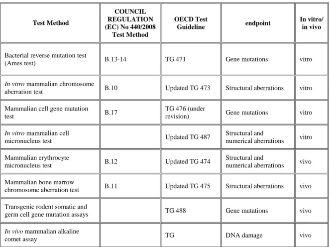
The EURL ECVAM defines 3 endpoints that need to be assessed in the process of genotoxicity for the safety assessment of chemicals and the protection of human health and environment 63.
These 3 endpoints are: gene mutation, structural chromosome aberrations, and numerical chromosome aberrations (Table 2-page 25). Classification and labelling (C&L) of chemical substances is based on the results of the genotoxicity tests of the scientific tests for toxicity assessment in the EU 34 and in the world (UN GHS).
According to the EU legislations and directives there are two different approaches for the assessment of genotoxicity to humans. According to the first approach, a chemical that is nongenotoxic in the in vitro analysis is not considered for the further in vivo assessments (e.g. REACH, CLP and Cosmetics Directive). The second approach, foresees in vitro tests of chemicals followed up by in vivo assessments (e.g. ICH for pharmaceuticals and VICH for veterinary drugs). This decision is made because the alternative methods to in vivo test for carcinogenicity cannot thoroughly replace the animal tests 64. Analyzing the regulatory
requirements, EURL ECVAM suggests an efficient approach to improve the traditional genotoxicity assessment. The new approach has the objective of reducing the animal use in genotoxicity testing, and EURL ECVAM suggests that efforts should be directed towards the improvement of the current assessments while reducing the use of animals and at the same time satisfies regulatory information approach. The identified solutions for improving the
genotoxicity assessment have the following aims:
Increasing the performance of in vitro tests in order to avoid the additional follow-up in vivo tests;
Improving the accuracy and quality of the in vivo follow-up testing to reduce unnecessary use of animals.
According to the chemical authorities the positive results of the in vitro genotoxic predictions need to be verified by in vivo tests, and this fact highlights the importance of finding solutions for reduction and refinement of genotoxicity tests. A strategy to reduce animal tests for
25
decision-making about carcinogenic or toxic compounds, can be collecting relevant data and drawing a conclusion on the basis of a data obtained from different sources. Pfuhler et al. 65
published some solutions to reduce animal tests in an ECVAM workshop report. Table 2. Test methods most commonly used for genotoxicity/mutagenicity testing 63
Test Method COUNCIL REGULATION (EC) No 440/2008 Test Method OECD Test Guideline endpoint In vitro/ in vivo
Bacterial reverse mutation test
(Ames test) B.13-14 TG 471 Gene mutations vitro
In vitro mammalian chromosome
aberration test B.10 Updated TG 473 Structural aberrations vitro
Mammalian cell gene mutation
test B.17
TG 476 (under
revision) Gene mutations vitro
In vitro mammalian cell
micronucleus test Updated TG 487
Structural and
numerical aberrations vitro
Mammalian erythrocyte
micronucleus test B.12 Updated TG 474
Structural and
numerical aberrations vivo
Mammalian bone marrow
chromosome aberration test B.11 Updated TG 475 Structural aberrations vivo
Transgenic rodent somatic and
germ cell gene mutation assays TG 488 Gene mutations vivo
In vivo mammalian alkaline
comet assay TG DNA damage vivo
1.13 Read-across
A read-across approach finds out the relevance or relationship between the properties of the chemical structures and then make assessment on the applicability of this information to another substance. It is crucial to detail the reasoning behind the inference on the substance for which the property is unknown.
A read-across process which is based on the concept of similarity can be applied in different forms: one-to-one (a similar substance can be used to make an estimation for a target substance) b) many-to-one (two or more analogues used to make a prediction for a single substance c) one-to many (one analogue used one-to make estimations for two or more substances) d) many-one-to-many (two or more similar compounds used to make estimations for two or more substances).
26
There are some important issues to be considered when using a read-across model. These characteristics are as follows:
The source substances must have the same structural features and the functional groups of the target substance;
the physico-chemical profile of the similar compounds must be comparable to those present in the target substance;
the relevant molecular descriptors must have comparable values;
the analogue substances must have approximately the same molecular weight.66
The results of a read-across should be interpreted using expert judgement and for the support of the conclusion detailed documentation is required. The read-across approach is more suitable for the physical hazard related to physico-chemical properties of the substances, as reliable test data should be available according to the CLP regulation. Therefore, if read-across is used as a unique method to generate a value to meet the endpoint data requirements, the criteria given in section 1.5 of Annex XI to REACH must be met.66
1.14 Classification and Labelling and Chemical Safety Assessment
Knowledge about physico-chemical properties of the chemicals and chemical safety assessment is important for the environment and human health. All the stages of the substances’ lifecycle must be assessed and controlled in the process of chemical safety assessment, these stages include manufacture, transfer, use and disposal of the chemical substances. Further, physico-chemical data are essential for the correct planning of (eco)toxicological studies and for the optimization of the test conditions.
The standard test and most confident assay for carcinogenicity is the 2-year rodent
carcinogenicity bioassay determined and described by OECD. The purpose of this assay is “to observe test animals for a major portion of their life span for the development of neoplastic lesions during or after exposure to various doses of a test substance by an appropriate route of administration.” Usually two species (mice and rats) and both sexes are used in this test. The chemical exposure is dosed and executed by oral, dermal or inhalation modes based on the expected human exposure. Dosing is done during two years and animal health is screened
27
throughout the test. The most important results of the test is obtained by the thorough examination of the animal tissues and organs at the termination of the assay.
The combination of carcinogenicity and chronic toxicity animal bioassays endpoints may reduce the animal use 67. A number of transgenic rodent models have been suggested as alternatives to
the standard bioassay carcinogenicity test by the ILSI HESI, but none of them was as efficient as the traditional 2-year assay for identification of carcinogens 68. Most of these models were
capable of detecting genotoxicity that can be already detected by other in vitro genotoxicity assays. Alternative models are not still suitable for the detection of nongenotoxic carcinogens 69.
1.15 Development and Optimisation of Alternative Methods
1.15.1 Importance of Mode of Action and Weight of Evidence Approach
The most appropriate key events are needed to understand the mechanisms of action of nongenotoxic carcinogens. Since there exist numerous modes of action for these substances, a WOE approach seems to be essential to deduce a reasonable conclusion out of the gathered data for a chemical.
There are nongenotoxic carcinogens in IARC group 3 (i.e. not classifiable as to its
carcinogenicity to humans) which are not carcinogenic in humans. To evaluate these group of carcinogens, the WOE approach is a useful tool. This approach helps the scientists to
understand the differences of modes of action in rodents and humans.
1.15.2 Alternative Methods for Detecting Nongenotoxic Carcinogens
In the process of the detection of the nongenotoxic carcinogens and exploring alternative methods for their assessment, it is important to consider the vast range of modes of action of these chemicals. These modes of action include: mitogenic induction, inhibition of gap-junctional intercellular communications, endocrine modifiers, oxidative stress,
immunosuppressants, regenerative proliferation and/or DNA methylation. Some of the examples of alternative methods for the nongenotoxicity detection are: (Q)SARs, measuring replicative DNA synthesis as an indication of cell proliferation, the in vitro cell transformation assays, measurements of inhibition of gap junction intercellular communication 70 and the use of
28
gene expression profiles with mechanistic networks for the identification of potential markers of nongenotoxic carcinogens.
1.16 Quantitative Structure–Activity Relationship (QSAR)
The main results of in vitro cell toxicity used by (Q)SAR models for nongenotoxic carcinogenicity are: several markers of in vitro cell toxicity including inhibition of gap-junctional intercellular communications, modulation of apoptosis and induction of cellular proliferation 71. In this study, the structural features of the nongenotoxic carcinogen associated
with toxicity or ligand binding, as in the case of estrogen, peroxisome proliferators and tubulin protein receptors, have been analyzed 71.
The (Q)SAR models for detection of carcinogenicity use information and correlate biological activity or chemical reactivity to chemical structure. These models are practically based on the assumption that similar chemicals have similar activities. The OECD has defined a number of principles for the validation of the (Q)SAR models for regulatory analysis 72,73. These principles
for (Q)SAR models are: having a defined endpoint, an unambiguous algorithm, and measures of goodness-of-fit, robustness, predictivity, and applicability domain.
A great number of nongenotoxic carcinogens are mutagenic inducers which cause cancer by increasing cellular proliferation. Hepatocyte rodent in vivo studies indicate that most of the hepatocarcinogens cause cancer by accelerating hepatocyte division 74–77.
As a result of putting into practice the REACH legislation, the number of 2-year rodent carcinogenicity bioassay is reduced and this fact can lead to the lack of detection of a large number of nongenotoxicity carcinogens. Because of the high risk of hazard associated with this group of carcinogens, there is an increasing need of alternative methods for their detection. Possible alternative methods for this purpose include: SARs and (Q)SARs, replicative DNA synthesis assay, the in vitro cell transformation assay and/or inhibition of GJICs. None of these alternative methods provide any information on the mode of action, thus further studies are needed to fill this gap of data. Using toxicogenomics to analyse multiple pathway-specific gene expression profiling is an efficient method to identify putative alerts. Additionally, statistical validation studies to examine the sensitivity, specificity and accuracy of these models play an
29
important role in improving these alternative models. It is important to discover as much as possible different modes of action of nongenotoxic carcinogens and not to depend only on the traditional nongenotoxic carcinogens identification methods (tetrachlorodibenzo-p-dioxin, carbon tetrachloride, and cyclosporine).
1.17 Software Packages for Mutagenicity and Carcinogenicity Predictions
Two major alternatives to in vivo testing are in vitro and in silico techniques. In the last decade, numerous computer software have been developed in order to replace, reduce and refine the animal tests. These software packages include also mutagenicity and carcinogenicity predicting models of the chemical compounds.
1.17.1 VEGA Platform
The VEGA platform contains a number of (Q)SAR models for predicting mutagenicity and carcinogenicity such as CAESAR. Two new carcinogenicity models have been added to the VEGA platform by the author 78. CAESAR ((Q)SAR mutagenicity models) was specifically
developed for the REACH regulation in collaboration with the United States Environmental Protection Agency (http://www.caesar-project.eu/). The mutagenicity models in the VEGA platform are based on data obtained from the Ames bacterial test. Models on carcinogenicity, developmental toxicity and etc. are freely available from the VEGA platform.
1.17.2 DEREK Nexus
The DEREK Nexus 79 is a knowledge-based expert system, developed by LHASA Limited that
predicts the genotoxicity, mutagenicity and carcinogenicity of a chemical by highlighting the SAs present in its molecular structure. Derek Nexus toxicity predictions are a result of two processes: evaluating SAs and estimating the likelihood of toxicity.
The knowledge-based SAs for in vitro mutagenicity have been implemented by experts who have assessed relevant Ames data and supporting mechanistic data (e.g. DNA adduct formation experiments). If a query compound matches a SA, the alert will fire with an associated
reasoning level (e.g. plausible, probable or certain). The reasoning levels associated with the in vitro bacterial mutagenicity alerts in Derek Nexus gives an indication of the likelihood for compounds in that class to be active in the Ames test 80.
30
Each bacterial, in vitro mutagenicity alert in the knowledge base was examined by a scientist with expertise in mutagenicity alert development. The patterns encoding the SAR for each alert were modified using the Derek Knowledge Editor if they contained features that were
implemented to prevent the pattern being activated by nonmutagenic compounds (so-called exclusion patterns). Such features were removed and the resultant ‘predictive space’ was stored within a modified knowledge base. Thus, each bacterial, in vitro mutagenicity alert in Derek had a corresponding region of predictive space 81.
1.17.3 TOPKAT
TOPKAT 82 is a (Q)SAR-based system, developed by Accelrys Inc. (http://accelrys.co m/).
Some of the TOPKAT toxicological endpoints are mutagenicity, developmental toxicity, rodent carcinogenicity, rat chronic LOAEL, rat MTD and rat oral LD50. TOPKAT models are
developed using two-dimensional molecular, electronic and spatial descriptors. The toxicity prediction is obtained from a chemical’s molecular structure. TOPKAT defines an applicability domain value which estimates the confidence in the prediction by applying the patented Optimal Predictive Space validation method. Any prediction generated for a query structure outside of the OPS space is considered unreliable.
1.17.4 MultiCASE
MultiCASE 83 (MultiCASE Inc., Cleveland, OH, USA) is a prediction model for genotoxicity
and carcinogenicity endpoints based on US FDA and US EPA. MultiCASE identifies SAs with a potential to initiate high biological activity, in addition, some statistical parameters are analysed to complete the predictions. The mutagenicity and genotoxicity models are based on the data obtained from Ames test, direct mutagenicity, base-pair mutagenicity, frameshift mutagenicity, chromosomal aberrations, and sister chromatid exchange data. The
carcinogenicity model includes different rodent assays (rate, mouse, male, female, and TD50 rats) and human epigenetic studies. All models use the statistical approach with the exception of the rule-based model for the Ames mutagenicity.
1.17.5 QSAR Toolbox
QSAR Toolbox 84 in cooperation with the ECHA is a read-across tool for grouping the
31
(http://www.qsartoolbox.org/). QSAR Toolbox systematically groups chemicals into classes according to their molecular structure, physico-chemical and biological properties. This software extracts structural characteristics and modes of action based on experimental information for the target molecule. The common mechanisms of action and common toxicological behaviour or consistent trends among results related to regulatory endpoints are results for an evaluation in this prediction software.
1.17.6 Toxtree
Toxtree 52 is a free tool for the assessment of mutagenicity and carcinogenicity of the chemicals
using decision trees. Toxtree mutagenicity and carcinogenicity model is based on the SAs of the Benigni-Bossa rule set, SAs for identification of Michael acceptors, and SAs confirmed by positive in vivo micronucleus tests. The program identifies any SA present in the target
molecule structure and concludes about the mutagenic or carcinogenic property of the chemical compound under investigation. The result of the prediction can be class I (inactive), class II (weak activity), or class III (active).
1.17.7 LAZAR
LAZAR 85 is an open source tool for the prediction of carcinogenicity and Salmonella
mutagenicity. LAZAR creates local endpoint (Q)SAR models based on a training set (only nearest neighbours) for each chemical separately. It first calculated the descriptors and determines the molecular similarity and then it builds a local (Q)SAR model based on a database of experimental toxicity data. This program meets all five OECD principles.
1.17.8 ACD/Tox Suite
The ACD/Tox Suite 86 package contains predicting models for genotoxicity and carcinogenicity.
The assessments are made based on validated (Q)SAR models in combination with expert knowledge. The software highlights and identifies the SAs which are responsible for toxic properties and extracts some similar molecules from the training set. The training set is composed of compounds that are genotoxic in Ames test.