Comparative study for determination of Root Canal working length accuracy by
different methods – an in vivo/ in vitro study
Shafait Ullah Khateeb1, Vibhuti Kaul2,*, Rudra Kaul3, Sumaya Yousuf Jeri4, Rimsha Ahmad5
1Assistant Professor, Dept. of Restorative Dentistry, King Khalid University College of Dentistry, Saudi Arabia, 2Registrar, Dept. of Oral Medicine & Radiology, 3Lecturer, Dept. of Conservative Dentistry & Endodontics, Indira Gandhi Govt. Dental College & Hospital, Jammu & Kashmir, 4General Dentist, Primary Health Care Center, Hai Aldhobat, 5Associate Dentist, Oracle 32 Dental
Clinic, Jammu & Kashmir *Corresponding Author: Email: [email protected]
Abstract
Aim: To determine the best method amongst Elements Diagnostic Unit, conventional radiography and direct digital radiography by comparing them with the actual working length calculated through anatomic measurements.
Materials and Method: Fifty adult human single rooted teeth intended for extraction with mature apices and without resorptive defects were selected. Endodontic access was prepared, the root canal orifices were enlarged by Gates Glidden drill and the pulp was then extirpated. A 4th generation electronic apex locator Elements diagnostic Unit (Sybron Endo) was used as one of the devices to measure the working length along with radiographs which were taken on conventional E – speed film and the DDR’s intraoral sensor using paralleling technique & working length was determined by Ingles method. After extraction of the teeth, the external part of the apical 3rd of the root was grounded to expose the apical constriction. File was introduced through the access cavity and the stopper was set at initial occlusal reference point as the file reached the apical constriction under X4 magnification. This served as the actual working length. The mean difference between actual working length measurement and individual group measurements were calculated. Statistical analysis of resulting mean value was done using One Sample T Test. Intergroup analysis was done using one way Analysis Of Variance (ANOVA) and Tukey HSD test.
Results: The results revealed no statistical significant difference between actual working length and working length as determined by Elements diagnostic Unit. Statistically significant difference was found between actual working length and working length as determined by conventional radiography and DDR (direct digital radiography).
Conclusion: Significant difference between Elements diagnostic Unit and both the radiographic techniques (conventional radiography and DDR). No statistical significant difference was found between conventional radiography and DDR (direct digital radiography).
Keywords: Working length determination, RVG, Apex locator
Introduction
Working length determination in endodontic therapy is essential and must be accurately obtained in order to fulfill the basic tenets of root canal therapy procedures.(1)
It establishes the apical limit of the canal preparation and permits the creation of an apical stop. It enables thorough debridement of the canal to be performed without over instrumentation, trauma to the periapical tissues or destruction of the anatomy of root apex(2) which in turn affect healing following therapy. Therefore this step ensures longevity of the tooth under treatment.
Root morphology and radiographic distortion may cause the location of the radiographic apex to vary from the anatomic apex and therefore the two have been found not to always coincide with each other.(3)
The apical foramen is the main apical opening of the root canal. It is frequently eccentrically located away from the anatomic or radiographic apex.(3,4)
The apical constriction (minor apical diameter) “is the apical portion of the root canal having the narrowest diameter” is a morphological landmark. It is usually 0.5 to 1.0 mm short of the center of the apical foramen. The
minor diameter widens apically to the foramen (major diameter) and assumes a funnel shape.(3) When obturation is till minor constriction, it can help to improve the apical seal.(5)
To achieve the highest degree of accuracy in working length determination, a combination of several methods should be used especially in canals for which working length determination is difficult. The most commonly used methods are the radiographic methods and the electronic method.(3)
Most clinicians currently utilize radiographic methods to determine working length in day to day practice. With the emergence of 4th generation electronic apex locators, a viable endometric alternative exists. However there is a paucity of research in literature on the comparison of radiographic methods and the electronic apex locators (Elements Diagnostic Unit) with the actual working length determined anatomically. Hence this study was conducted to compare and evaluate the accuracy of elements diagnostic unit, conventional radiography, and RVG (direct digital radiography) in determining the working length through an in vivo / vitro investigation.
Materials and Method
A total of 50 adult human anterior teeth and premolars indicated for extraction as a part of orthodontic treatment were taken for the study.
Criteria for Case Selection
1. Single rooted human anterior teeth and premolars. 2. Teeth which were intended for extraction.
3. Teeth with calcified canals and dilacerations were excluded.
4. Teeth with open foramina and periapical pathology were rejected.
5. Teeth with metallic restorations were excluded. 6. Patients who have non-contributing medical
histories were included.
7. Patients with artificial pacemakers were not included.
Standard intra oral periapical radiographs were taken in order to determine whether or not the sampled tooth conformed to the selection criteria adopted for the study. An informed written consent was obtained from each patient before the study was initiated.
Endodontic access was prepared after local anesthesia administration and rubber dam application. The root canal orifices were enlarged by Gates Glidden drill. The pulp was then extirpated with a barbed broach and the canal thoroughly irrigated with 0.9% saline followed by which the pulp space was dried with paper points.
For determination of working length using 4th generation electronic apex locator – Elements diagnostic unit (Sybron endo) the clip was applied to the patients lip, and the electrode was connected to the file. File No. #15 was used. The file connected to the electrode of the device was apically advanced in the canal, until the LCD displayed 0.0. The file was then withdrawn slowly counterclockwise until the reading of EAL showed a consistent 0.5 mm. which is accepted as the apical constriction. At the meter’s 0.5 reading, the length of the file is measured and the value was recorded. (Fig. 1)
Radiographic measurement was made according to Ingle’s method (which is described below) using conventional radiography and DDR.
1. Measure the tooth on the pre-operative radiograph 2. Subtract at least 1.00 mm “safety allowance” for
possible image distortion or magnification.
3. Set the endodontic ruler at this tentative working length and adjust the stop on the instrument at that level.
4. Place the instrument in the canal until the stop is at the plane of reference unless pain is felt, in which case, the instrument is left at that level and the rubber stop readjusted to this new point of reference.
5. Expose, develop and clear the radiograph.
6. On the radiograph, measure of difference between the end of the instrument and the end of the root and add this amount to the original measured
length the instrument extended onto the tooth. If, through some oversight, the exploring instrument has gone beyond the apex, subtract the difference. 7. From the adjusted length of tooth, subtract a 1.0
mm ‘safety factor’ to conform to the apical termination of the root canal at the apical constriction.
For the conventional radiographic method Kodak E-speed film was exposed at 70 kV and 8 mA for 0.6 sec. Paralleling radiographic technique was followed. The films were uniformly exposed and processed by hand. The films were developed for 30 seconds, followed by a 5 minute water wash and fixed for 60 seconds. The measure of difference between the end of the instrument and the radiographic apex is added to the original length. From the adjusted length of the tooth, 1.0 mm safety factor is subtracted to conform to the apical termination of the root canal at the apical constriction. All measurements were made by using the same dial caliper.
For direct digital radiography the intraoral sensor was exposed for radiation for 0.2 seconds. The equipment then digitizes processes and stores the image. The working length is then measured using the calibration tool available in the software using Ingle’s method.
Color, contrast and magnification of the digital radiographic images were adjusted to achieve the best possible image for viewing. Just as would be available clinically. As it was not possible to adjust conventional radiographic images, X4 magnification was used.
For determination of actual working length after careful extraction of the teeth, it was placed in 5.25% NaOCl solution to remove any remnants of periodontal tissue from the root surface. The actual length of the tooth was determined using the same reference point and the same file used previously. The file was placed into the canal until the tip was visualized from a tangential angle at the apical exit using 4X magnification. The external part of the apical 3rd of the root was grounded with diamond burs to expose the apical constriction. Endodontic file served to limit the over grinding so as not to lose the apical constriction landmark. (Fig. 2)
New file of the same size was introduced through the access cavity and the stopper was set at the occlusal reference point as the file reached the apical constriction under X4 magnification. This served as the actual length & was determined for each tooth using same dial caliper.
The working length readings recorded were then tabulated and an overall comparison of working length obtained with the radiographic methods and electronic method was done with the actual working length and the values were subjected to statistical analysis.
Results
Working length in 50 human teeth indicated for extraction were measured using three methods:
1. Group 1 - Elements diagnostic unit. 2. Group 2 - Conventional Radiography. 3. Group 3 - DDR (Schick CDR)
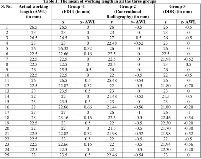
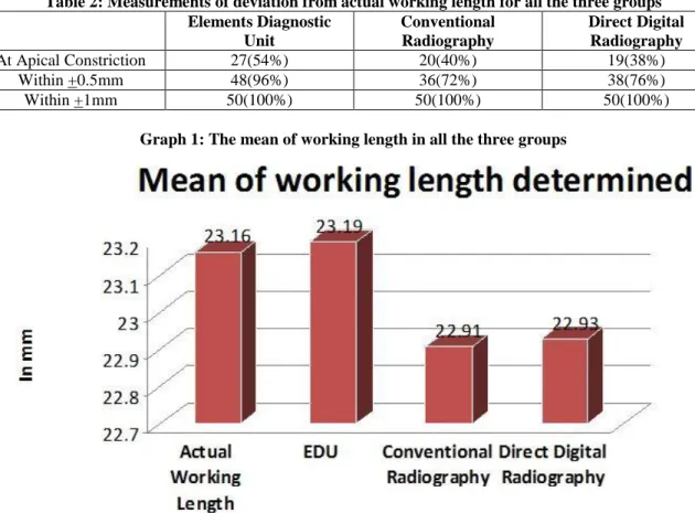
These groups were compared with actual working length. The mean of working length in all the three groups was calculated (Table 1, Graph 1) & the mean differences between actual working length measurement and individual group measurements were calculated (Table 1). The mean deviation of working length determined by Elements Diagnostic Unit from the actual working length (+0.038) was less when compared to that determined by radiographic methods (-0.268 for conventional radiography and -0.211 for DDR). A positive value for mean difference indicated that the tip of measuring instrument is beyond the actual working length and a negative value indicated
that the instrument is short of the actual working length.
The frequency histogram in (Table 2, Graph 2) demonstrates the occurrence of measurements of deviation from actual working length for all the three groups. The most frequent response for the apex locator deviation was 0.0 mm (actual working length). 27 readings (54%) coincided with the apical constriction, 48 readings (96%) were within ±0.5mm from the actual working length. Conventional radiography & DDR methods showed more deviation from the actual working length. Only 20 readings (40%) & 19 readings (38%) coincided with the apical constriction, respectively for the conventional radiography & DDR. 36(72%) and 38(76%) readings were within ± 0.5mm from the actual working length for conventional & DDR respectively. The most frequent response for the radiographic methods deviations was 0.0 to –0.5 mm (0.0 to 0.5 mm short of actual working length).
Table 1: The mean of working length in all the three groups S. No. Actual working
length (AWL) (in mm) Group -1 (EDU) (in mm) Group-2 (Conventional Radiography) (in mm) Group-3 (DDR) (in mm)
x x- AWL y y- AWL z z- AWL
1 26.5 26.5 0 26 -0.5 26 -0.5 2 23 23 0 23 0 23 0 3 26.5 26.5 0 27 0.5 26 -0.5 4 23 23 0 22.48 -0.52 23 0 5 26 26.32 0.32 26 0 26 0 6 22.5 22.66 0.16 22.5 0 22.5 0 7 22.5 22.5 0 22.5 0 21.98 -0.52 8 22.5 22.5 0 22.5 0 23 0.5 9 26 25.5 -0.5 26 0 26 0 10 22.5 22.5 0 22 -0.5 22 -0.5 11 26 26.5 0.5 25.48 -0.54 26 0 12 22.5 22.82 0.32 22 -0.5 21.80 -0.70 13 23 23.5 0.5 23 0 23 0 14 22 22 0 21.48 -0.52 21.5 -0.5 15 23 23.5 0.5 23 0 23 0 16 22 22.66 0.66 21.44 -0.56 21.80 -0.20 17 27 27 0 26 -1 26 -1 18 23 23.16 0.16 22.5 -0.5 22.46 -0.54 19 22.5 23 0.5 22 -0.5 22.30 -0.20 20 22 22 0 21.5 -0.5 21.70 -0.30 21 22.5 22.82 0.32 21.98 -0.52 21.98 -0.52 22 22.5 23 0.5 22 -0.5 22 -0.5 23 22.5 22.66 0.16 22 -0.5 21.94 -0.56 24 22.5 22.5 0 22 -0.5 22.30 -0.20 25 23 23.5 0.5 22.46 -0.54 23 0
Table 2: Measurements of deviation from actual working length for all the three groups Elements Diagnostic Unit Conventional Radiography Direct Digital Radiography At Apical Constriction 27(54%) 20(40%) 19(38%) Within +0.5mm 48(96%) 36(72%) 38(76%) Within +1mm 50(100%) 50(100%) 50(100%)
Graph 1: The mean of working length in all the three groups
Graph 2: The frequency histogram demonstrating the occurrence of measurements of deviation from actual working length for all the three groups
Statistical analysis of resulting mean values for all the three groups was done using One Sample T Test with test value kept as 0.0. The p value was set at 0.05. The p value for group-1 as determined by One Sample T Test was found to be 0.345, which is statistically not significant, compared to actual working length. The p value for group-2 as determined by One Sample T Test was found to be < 0.001, which is statistically significant, compared to actual working length.
The p value for group-3 as determined by One Sample T Test was found to be <0.001, which is statistically significant, compared to actual working length. Inter-group statistical analysis was done using one way Analysis of Variance (ANOVA) and Tukey HSD test. The p value was set at 0.05.
The p value between group 1 and 2 was found to be < 0.001, which is statistically significant.
Between group 1 and 3 p value was found to be 0.001, which is statistically significant.
Between group 2 and 3 p value was 0.688 which is statistically not significant.
Inter group statistical analysis showed significant difference between Elements diagnostic Unit and both the radiographic techniques (conventional radiography and DDR). No statistical significant difference was found between conventional radiography and DDR (direct digital radiography).
Discussion
One of the important steps in endodontic therapy is the calculation of working length. Working length establishes the apical extent of canal preparation and apical stop. Failure to accurately determine the working length may lead to apical perforation and over filling with increased incidence of postoperative pain. It may also lead to incomplete instrumentation and under filling with attendant problems. Among them should be noted persistent pain and discomfort from inflamed shreds of retained pulpal tissues. In addition, ledge formation may develop, short of the apex, making adequate treatment or retreatment extremely difficult or impossible. Finally, apical percolation may develop into the unfilled “dead space” at the apex. This could result in a prolonged healing period or continued periradicular lesion and increased incidence of failure. Therefore, an accurately determined working length is essential for endodontic success.(6,7,8)
Locating the appropriate apical stop during working length determination always has been a challenge in clinical endodontics. The cemento-dentinal junction (CDJ), where the pulp tissue changes into the apical tissue, is the most ideal physiologic apical limit of the working length. It is also referred as the minor diameter or apical constriction by some authors.(9) However, the CDJ and apical constriction do not always coincide particularly in the senile teeth as a result of cementum deposition, which alters the position of the minor diameter. Therefore, setting the apical constriction, which has the narrowest diameter as the apical limit(10) of the working length, where it is easy to clean and shape or obturate the canal is recommended.(11)
Present study was undertaken to determine the working length of tooth with the electronic apex locator (Elements Diagnostic unit) conventional radiography & DDR & to evaluate the accuracy of working length measurement of radiographic, DDR & electronic apex locator methods by comparing with the actual working length obtained after extraction & grinding of the teeth.
The apical constriction was taken as the terminating point to measure the actual canal length. A potential error of ± 0.5 from this point was accepted as a tolerable range for the clinical application. Most of the studies conducted to evaluate the accuracy of
electronic apex locators are in-vitro. The present study being in-vivo simulates the clinical situation better.
Elements Diagnostic Unit and Apex Locator (Sybron Endo, CA, USA) is a fourth generation apex locator. The device does not process the impedance information as a mathematical algorithm, but instead takes the resistance and capacitance measurements and compares them with a database to determine the distance to the apex of the root canal.(12) It uses multiple frequencies to eliminate the influence of the canal conditions, which is similar to Root ZX thus permitting less sampling error per measurement and more constant readings,(12) in addition it uses a lookup matrix rather than making any internal calculations.
The results of the study show that there is no statistical significant difference between the actual working length and working length as determined by Elements diagnostic Unit apex locator(p value:0.345). Elements diagnostic Unit measured within a narrow band (SD = 0.312 mm) near the actual working length (the mean value was 0.038 mm apical to ideal working length). 96% of time it located the canal length within ± 0.5 mm of actual working length. The results of the present study are in agreement with the studies of (Shabahang et al 1996),(13) (Plotino. G 2006).(14) Moreover results showed that mean of the absolute value of the deviation from the apical constriction for the apex locator was significantly less than that for the radiographic method, which is in agreement with the study of Pratten and McDonald.(15) Later it was seen that there is no statistical significant difference between conventional radiographic method and DDR group. These results are in agreement with the studies of Shearer et al,(16) Ong et al(17) & Leddy et al.(18)
The radiographs provide an archival image of the tooth which gives valuable information on root canal anatomy and proximity of vital structures and provides the only means by which the size of the root canal, its curvature and the number of roots may be gained.(19)
Conclusion
There is no statistical significant difference between actual working length and working length as determined by Elements diagnostic Unit. Statistically significant difference was found between actual working length and working length as determined by conventional radiography and DDR (direct digital radiography). Inter group statistical analysis showed significant difference between Elements diagnostic Unit and both the radiographic techniques (conventional radiography and DDR). No statistical significant difference was found between conventional radiography and DDR (direct digital radiography). Suggesting apex locator as a vital irreplaceable tool in dental operatory, especially for cases where conventional radiography is contraindicated.
References
1. Frank AL, Simon JHS, Abou-Rass M, Glick DH. Clinical and surgical endodontics concepts in practice. Philadelphia: JB Lippincott and 1983.
2. B.M Griffiths et al. Comparison of three imaging techniques for assessing endodontic working length. Int Endo J. 1992, 25:279-287.
3. Ingle JI and Bakland LK. Endodontics 5th ed. Elsevier, 2002, Ch: Endodontic cavity preparation and 511-515., pp.
4. 1955, Yury Kuttler. Microscopic investigation of root apexes. JADA and 50:544.
5. Cohen S, Burns RC. In Pathway of the pulp 8th ed Louis. Mosby 2002 and 690-728.
6. Samuel Seltzer, Walter Saltanoff, and Jerry Smith. Biologic aspects of endodontics. Periapical tissue reactions to root canal instrumentation beyond the apex and root canal fillings short of and beyond the apex. Oral Surg. 1973 and 36(5):725-37.
7. Swartz D B, Skidmore A E, Griffin J A. Twenty years of endodontic success and failures. J. Endod 1983 and 198-202., 9.
8. Sjogren U, Haggund B, Sundquest G, Wing K. Factors affecting the long term results of endodontic treatment. J. Endod 1990: 16 and 498-504.
9. D Ricucci, Apical limit of root canal instrumentation and obturation, part 1. Literature review. Int Endo J 1998 and 31, 384-393.
10. Guiterrez G, Aguayo P. Apical foraminal opening in human teeth. Number and location. Oral Surg Oral Med Oral Pathol 1995 and 769-77, 79.
11. Jerome W. George, Andrew E. Michanowicz & John P. Michanowicz. A method of canal preparation to control apical extrusion of low temperature thermoplastisized gutta-percha.JOE.1987 and 13(1):18-23.
12. J.2004, M.P.J. Gordon & N.P. Chandler. Review Electronic Apex Locator. Int. Endod 37:425-37.
13. Shahrokh Shabahang, William W.Y. Goon and Alan. H Gluskin. An in-vivo evaluation of Root ZX electronic apex locator. JOE1996 and 616-8., 22(11).
14. Plotino G, Grande NM, Brigante L, et al. Ex vivo accuracy of three electronic apex locators: Root ZX, Elements Diagnostic Unit and Apex Locator Pro Pex. Int Endod J 2006 and 39:408-414.
15. Pratten. D.H, and McDonald. N.J. Comparison of Radiographic and Electronic Working Lengths’. Endod.1996 APR and 22(4):173-6.
16. Shearer. AC, Horner. K, Wilson NH. Radiovisiography for imaging root canals: an invitro comparison with conventional radiography. Quint Int 1990 and 21(10):789-94.
17. Ong EY, Pittford TR. Comparison of Radiovisiography with Radiographic film in root length determination. Int Endod J 1995 and 28:25-29.
18. Leddy BJ, Miles DA. Interpretation of endodontic file length using RVG .JOE 1994,20:542-545.
19. Ashraf. F Fouad, Eric. M. Rivera and Keith. V Krell. Accuracy of Endex with variations in canal irrigants and foramen size. JOE1993,19(2):63-7.