ADRENOMEDULLIN, CHEMOKINE RECEPTORS, AND RECEPTOR ACTIVITY MODIFYING PROTEINS IN LYMPHANGIOGENESIS
Klara Rachel Klein
A dissertation submitted to the faculty of the University of North Carolina at Chapel Hill in partial fulfillment of the requirements for the degree of Doctor of Philosophy in the
Department of Cell and Molecular Physiology
Chapel Hill 2015
ABSTRACT
Klara Rachel Klein: Adrenomedullin, Chemokine Receptors, and Receptor Activity Modifying Proteins in Lymphangiogenesis
(Under the direction of Kathleen Caron)
Over the past decade, we have begun to appreciate that the lymphatic vascular system does more than simply return plasma back into the circulatory system and, in fact, contributes to a wide variety of normal and disease states. For this reason, much research has been devoted to understanding how lymphatic vessels form and function, with a particular interest in which molecules contribute to lymphatic vessel growth and
maintenance. Here, we have focused on a potent lymphangiogenic factor, adrenomedullin, and its known roles in lymphangiogenesis, lymphatic function, and lymphatic disease.
In the course of our studies, we have discovered that the decoy receptor CXCR7 is required as a molecular rheostat for controlling the concentration of AM ligand during cardiac and lymphatic vascular development. Loss of mammalian CXCR7 results in
In Part II, we investigated whether CXCR7 and related chemokine receptors (CKRs), CXCR4 and CCR5, form protein-protein interactions with one component of the AM
To my friends and family, who are endlessly supportive, and especially to my mom, who is my inspiration, my dad, who keeps my feet on the ground, and my husband, who makes every day better than the last.
ACKNOWLEDGEMENTS
I would like to thank my labmates and friends who have offered countless insights to this work over the years, and especially my mentor, Kathleen, who offers guidance and inspiration in all aspects of science.
For the work in Chapter 2, we would like to thank Dr. Nikolaus Heveker, Univ. de Montreal, for his helpful discussions and insights. We also thank former members of the Caron laboratory, including Kimberly Fritz-Six, Drs. Manyu Li, Mahita Kadmiel, and Patricia Lenhart, and Kirk McNaughton and Ashley Ezzell of the Histopathology Core for their assistance with experiments and technical guidance. This work was supported by funds from the American Heart Association EIA (0555424U) and NIH grants (HD060860 and DK099156) to KMC, F30 HL118932 to KRK and training grants HL069768 and GM008719.
For the studies presented in Chapter 5, we would like to thank Dr. Joshua Snyder and Dr. Marc Caron for their technical advice and helpful discussions. We also thank Dr. Duncan Mackie for his contribution to this work.
TABLE OF CONTENTS
LIST OF TABLES ... x
LIST OF FIGURES ... xi
LIST OF ABBREVIATIONS ... xiii
PART I Chapter 1: Adrenomedullin in Lymphangiogenesis - From Development to Disease, ... 1
Overview ... 1
Introduction ... 2
The Adrenomedullin Peptide Family and Its Receptors ... 3
Adrenomedullin During Lymphatic Vascular Development ... 5
Adrenomedullin Signaling During Adulthood ...11
Adrenomedullin and Lymphedema ...13
Adrenomedullin in Cancer ...15
Adrenomedullin-induced Lymphangiogenesis in the Reproductive System ...17
Concluding remarks ...18
Figures ...20
References ...23
Chapter 2: Decoy Receptor CXCR7 Modulates Adrenomedullin-Mediated Cardiac and Lymphatic Vascular Development, ...32
Introduction ...33
Results ...35
Experimental Procedures ...48
Supplemental Experimental Procedures...51
Figures ...56
Tables ...71
References ...76
Chapter 3: Conclusions and Future Directions ...82
Summary of Results ...82
Current State of the Field – AM, decoy receptors, and lymphangiogenesis ...83
Future Directions ...89
Concluding Remarks ...93
Figures ...95
References ...96
Part II Chapter 4: From the dish to the mouse: A glance at receptor activity modifying protein biology, ... 100
Introduction ... 100
The discovery of RAMPs ... 101
A multitude of effects: RAMP pharmacology ... 102
RAMP function in vivo ... 107
Concluding remarks and future questions ... 120
Tables ... 122
Figures ... 123
Chapter 5: The Novel Interaction of Receptor Activity Modifying Proteins
with the Family A Seven Transmembrane Chemokine Receptors ... 131
Introduction ... 131
Experimental procedures ... 133
Results ... 135
Discussion ... 137
Figures ... 141
References ... 145
Chapter 6: Conclusions and Future Directions ... 149
Summary of Results ... 149
Future Studies ... 149
Concluding Remarks ... 151
LIST OF TABLES
Table 2-S1. List of genes significantly changed following 1-hour AM treatment
LIST OF FIGURES
Figure 1-1. Early development of the lymphatic system and the effects of AM on LECs. ...20
Figure 1-2. Proper AM dosage is required for normal lymphangiogenesis during development and in tumor biology. ...21
Figure 1-3. Loss of AM signaling in adulthood results in disruption of many lymphatic beds. ...22
Figure 2-A1. Graphical Abstract ...56
Figure 2-1. CXCR7 scavenges AM, dampens AM-mediated ERK phosphorylation in vitro, and reduces AM peptide levels in vivo. ...58
Figure 2-2. Cxcr7 is dynamically expressed in lymphatic endothelium during development. ...59
Figure 2-3. Cxcr7-/- embryos have enlarged blood filled lymphatic sacs and interstitial edema. ...60
Figure 2-4. Loss of CXCR7 enhances cardiac lymphangiogenesis by promoting AM-mediated cellular migration...61
Figure 2-5. Loss of CXCR7 causesenlarged dermal lymphatics with decreased branching complexity in vivo and enhances LEC proliferation in vitro ...62
Figure 2-6. Genetic titration of Adm influences Cxcr7-/-phenotypes. ...63
Figure 2-7. Genetic reduction of AM normalizes Cxcr7-/- cardiac proliferation and size. ...64
Figure 2-S1. Supplemental data related to Figure 2-1 ...65
Figure 2-S2. LYVE1 and pERK colocalize in lymphatic vessels from Cxcr7-/- mice (related to figure 2-1). ...66
Figure 2-S3. Cxcr7-/- mice have normal lymphovenous valves and precocious lymphatic development (related to figure 2-3). ...67
Figure 2-S4. Cxcr7-/- embryos have more cardiac lymphatics with decreased branching complexity (related to figure 2-4). ...68
Figure 2-S5. Loss of CXCR7 affects AM-signaling in LECs (related to figure 2-5). ...69
Figure 2-S6. Cxcr7 is stochastically expressed in dermal lymphatic vessels (related to figure 2-5). ...70
Figure 3-1. AM expression is localized to the developing epicardium and up-regulated in Admhi/hi mice. ...95
Figure 4-2. The effects of RAMP association on GPCR pharmacology ... 124
Figure 5-1. BRET analysis of CKR-RAMP protein-protein interaction ... 141
Figure 5-2. Localization of the GPCR-RAMP3 complex in vitro ... 142
LIST OF ABBREVIATIONS
7-TM – seven-transmembrane
ACKR – atypical chemokine receptor ACTH – adrenocorticotropic hormone Adm – adrenomedullin gene
AM – adrenomedullin peptide AMY – amylin
Ang-II – angiotensin II
Assp1 – apoptosis stimulating protein of p53 B2ADR – beta-2 adrenergic receptor
BEC – blood endothelial cells
BRET – bioluminescence resonance energy transfer cAMP – cyclic AMP
CaSR – calcium sensing receptor CFA – complete Freund’s adjuvant CGRP – calcitonin gene related peptide CKR – chemokine receptor
CLR – calcitonin receptor-like receptor
CRF – corticotrophin releasing factor receptor CT – calcitonin
CV – cardinal vein
ER – endoplasmic reticulum FAP – fluorogen activating protein GPCR – G protein-coupled receptor
IMD – intermedin JV – jugular vein
LAD – left anterior descending LEC – lymphatic endothelial cell LLC – Lewis lung carcina LPS – lipopolysaccharide MHC – myosin heavy chain
NSF – N-ethylmaleimide-sensitive factor NHERF – Na+/H+ exchanger regulatory factor-1 OVA – ovalbumin
PAM – peptidylglycine alpha-amidating monooxygenase PDZ – PSD-95/Discs-large/ZO-1
PTH1R – parathyroid 1 receptor PTH2R – parathyroid 2 receptor PTHrP – parathyroid related peptide RAMP – receptor activity modifying protein Rluc – Renilla luciferase
V2R – vasopressin 2 receptor
VE-Cadherin – vascular endothelial cadherin VEGF – vascular endothelial growth factor
Chapter 1: Adrenomedullin in Lymphangiogenesis - From Development to Disease1,2
Overview
Over the past decade, we have begun to appreciate that the lymphatic vascular system does more than simply return plasma back into the circulatory system and, in fact, contributes to a wide variety of normal and disease states. For this reason, much research has been devoted to understanding how lymphatic vessels form and function, with a particular interest in which molecules contribute to lymphatic vessel growth and maintenance. In the following review, we focus on a potent lymphangiogenic factor,
adrenomedullin, and its known roles in lymphangiogenesis, lymphatic function, and human lymphatic disease. As one of the first, pharmacologically-tractable G protein-coupled receptor pathways characterized in lymphatic endothelial cells, the continued study of adrenomedullin effects on the lymphatic system may open new avenues for the modulation of lymphatic growth and function in a variety of lymphatic-related diseases that currently have few treatments.
1 Authors: Klein, K.R. and Caron, K.M.
Introduction
The lymphatic system is a vascular network in parallel with blood vessels that penetrates every tissue in the body, with the exception of bone marrow and the central nervous system [1]. Over the past dozen years, our understanding of how the lymphatic system develops, functions, and contributes to disease has markedly improved [2]. It has become apparent that this complex system is more than just a simple conduit for returning interstitial fluid to blood. The lymphatic system not only participates in maintaining fluid homeostasis, but also mediates fat absorption and provides a highway for immune cell trafficking to distant sites. In fact, the lymphatic system has now been recognized for its contribution to a wide variety of normal and pathophysiological states [3,4].
Lymphedema, the most common and poorly treated lymphatic disease, affects 140-250 million people worldwide. Characterized by debilitating swelling of one or more limbs, lymphedema is a lifelong condition that can lead to inflammation, fibrosis, infection, subcutaneous fat accumulation, and decreased mobility and function [1,5]. Despite its prevalence and morbidity, no pharmacological agents exist for the management of lymphedema.
A multitude of studies in genetic mouse models have also implicated lymphatics in other common diseases. For example, disruption of neolymphangiogenesis in the skin induces salt-sensitive hypertension in mice [6]. This model demonstrates that dermal lymphangiogenesis is required to maintain electrolyte balance and subsequently blood pressure homeostasis [6]. Further, haploinsufficiency of the essential transcription factor Prox1 leads to adult onset obesity due to abnormal fluid leakage from disrupted lymphatic
To this end, a great deal of effort has recently been devoted to identifying factors that participate in lymphangiogenesis and control lymphatic vascular function in adults. The following review will focus on one such factor, adrenomedullin (AM = peptide, Adm = gene), and will address what is currently known about its role in the lymphatic vascular system, and its potential as a novel therapeutic for the treatment of diseases linked to lymphatic
dysfunction.
The Adrenomedullin Peptide Family and Its Receptors
AM, a 52-amino acid peptide hormone, is classified as a member of the calcitonin gene related peptide (CGRP) family due to its shared secondary structure and overlapping biological activity with other peptide family members [8,9]. The four peptides of this family besides AM include amylin, calcitonin, intermedin, and CGRP. Each of these peptides has essential roles in normal physiology [10]. Similarities in their secondary structure allow for overlap in the pharmacology of the binding sites of the CGRP family and, consequently, cross-reactivity between their receptors [8,11]. Therefore, discerning which biological activities are distinct to AM proved to be challenging until the discovery of a class of single-pass transmembrane proteins called receptor activity modifying proteins (RAMPs) [12]. These proteins bind to and confer ligand specificity for G protein-coupled receptors (GPCRs) and allow cells to distinguish between members of the CGRP family.
When bound to a GPCR, RAMPs dictate ligand specificity, downstream signaling, and receptor recycling [9,12,13]. The three identified RAMPs, RAMP1, -2, and -3 share 30% sequence identity, with their nonhomologous domains modulating specificity [8]. For
example, when calcitonin receptor-like receptor (CLR) associates with RAMP2 or RAMP3, the receptor binds AM with high affinity. When complexed with RAMP1, on the other hand, CLR performs as a high affinity CGRP receptor. Thus, specific tissue and temporal
will be discussed below, recognition of the role of RAMPs in the responsiveness of AM allowed for the design of gene-targeted mouse knockout models that helped determine the physiological role of AM. The participation of RAMP2 with CLR was found to be absolutely necessary for AM response during development, as genetic deletion of Ramp2 phenocopied Adm knockout models [14]. This unusual signaling offers a unique opportunity for specific
targeting without interrupting the activity of other, closely related ligand-receptor pathways. Prior to the discovery of RAMPs, there were two additional putative AM receptors reported in the literature [15]. In 1995, Kapas and Clark suggested that two orphan GPCRs, RDC-1 and L1, served as AM receptors [16,17]. An inability to reproduce these findings led to significant controversy regarding their ability to bind AM. While L1, now known as
GPR182, remains an orphan receptor, RDC-1 has been identified as an atypical chemokine receptor, a promiscuous receptor that binds several ligands including SDF-1/CXCL12 [18], CXCL11 [19], and intermediate opioid peptide [20]. RDC-1 has since been renamed CXCR7 or atypical chemokine receptor 3 (ACKR3). The role of CXCR7 in AM signaling continues to evolve. Mounting evidence in the literature over the past decade suggests that
RDC-1/CXCR7/ACKR3 does indeed associate with adrenomedullin [15,21-24]. Although the discovery of RAMP-guided CLR responsiveness to AM overshadowed the role of these other receptors in AM biology, as will be discussed later in this review, recent studies demonstrate that AM-mediated downstream signal is indeed modulated through CXCR7 [25,26]. The identification of a second AM receptor is especially exciting, as it offers another avenue for drug discovery for the treatment of AM-mediated pathologies.
such as wall motion and infarct size [31]. Modulation of the AM signaling system, therefore, could prove to be a viable and safe treatment option not only for cardiovascular diseases, but also for other diseases in which AM plays a role. In particular, genetic models have uncovered an additional role for AM in the development and regulation of the lymphatic vascular system.
Adrenomedullin During Lymphatic Vascular Development
Initial lymphatic vessel formation begins between e9.5-10.5 with the commitment of endothelial cells of the cardinal vein (CV) to a lymphatic fate (Figure 1-1A) [1]. After polarization of lymphatic progenitor cells in the jugular vein at e11.5-12.5, lymphatic endothelial cells (LECs) begin to migrate away and form a premature lymphatic vessel. Historically, this vessel has been referred to as the lymph sac. Recently however, ultramicroscopy of whole mount mouse embryos has revealed that the lymph sac is comprised of two separate lymphatic structures, a peripheral longitudinal lymphatic vessel and a primordial thoracic duct [32]. These structures are formed by migrating LECs that originate from both the CV and additional venous sources [32]. Failure of these ECs to establish LEC fate, migrate, or proliferate results in severe edema and embryonic lethality. For example, genetic deletion of Prox1 prevents EC differentiation to lymphatic fate. Prox1-null animals are devoid of lymphatic vasculature and die by e14.5 [33-35]. Similarly, though ECs of VEGF-C-null mice can establish LEC fate, LECs fail to migrate away from the CV, lymphangiogenesis is arrested and embryonic edema ensues, demonstrating that VEGF-C is an essential lymphangiogenic factor [36-38]. The severity and timing of the edema and subsequent embryonic lethality in these mouse models provided an essential clue for determining the role of AM in lymphangiogenesis.
Several labs observed that mice globally lacking Adm exhibited profound edema, known as hydrops fetalis [40-42]. Similarly, knockouts of Calcrl, the gene for CLR, [39], or Ramp2 [14,43,44], which make up the canonical receptor for AM, have the same lethal phenotype. Furthermore, peptidylglycine alpha-amidating monooxygenase (PAM) knockout mice [45], an enzyme required for the amidation and function of AM, are also embryonic lethal due to extreme edema [27]. These models clearly demonstrate that AM is essential for life; however, the root cause of this edema was not understood.
Consistent with what was known about AM at the time and with the nascent
lymphatic vascular field, early studies of these mouse models focused on AM in cardiac and blood vascular development rather than lymphangiogenesis. Indeed, AM was found to have a significant proliferative effect during cardiac development. Adm-/-, Calcrl-/-, and Ramp2 -/-mice share a phenotype of small, hypoplastic hearts [14,39,40]. All three genes were also found to be highly expressed in the heart and the vascular endothelium, confounding whether the observed edema could be attributed to cardiac defects or vascular dysfunction. To solve this problem, Fritz-Six et al. utilized mice expressing an endothelial cell-specific Cre recombinase via expression of a Tie2-Cre transgene [46]. Though Tie2-Cre expression has only been observed in restricted regions of adult lymphatic vessels, Tie2-Cre is expressed in the venous progenitors of lymphatic endothelial cells. Thus, prior to the development of lymphatic-specific Cre drivers, Tie2-Cre mice were widely used to excise genes from venous lymphatic progenitors during embryonic lymphangiogenesis [47-51].
Using this Tie2-Cre model, Fritz-Six et al. found that specific deletion of Calcrl in the venous lymphatic progenitors results in extreme hydrops fetalis, suggesting that
onset of edema in the AM signaling-disrupted mice between e12.5-e14.5 coincides with the separation of the lymphatic sac from the jugular vein and the beginning of lymphatic flow, suggesting that the edema could be lymphatic in origin.
Fritz-Six et al. surveyed the AM system effects on developmental lymphangiogenesis by deleting Adm, Calcrl, and Ramp2 [14]. Each gene-targeted knockout model exhibited the same edematous phenotype, while no problems with blood vascular leakage were observed [14]. Quantitative PCR of both lymphatic and blood endothelial cells further revealed that CALCRL and RAMP2 were enriched in the lymphatic endothelium compared to the blood
endothelium, a finding that has since been confirmed by other groups [14,52]. Additionally, transfection of cultured LECs with a PROX1 expression plasmid led to a three-fold increase in endogenous CALCRL expression, demonstrating that the genes required for AM signaling are inducible by this master lymphatic fate regulator [14]. Later studies have also shown that AM-treatment of LECs causes an upregulation of PROX1 [53], demonstrating that not only are the genes required for AM signaling upregulated in LECs leading to preferential AM action, but that AM activity in LECs feeds back positively on PROX1 expression. Consistent with this notion, Fritz-Six et al. found that prior to embryonic lethality, the lymphatic sacs of AM-signaling null mice were significantly smaller when compared to those of wild-type littermates [14]. Careful study of proliferation in LECs revealed that loss of AM signaling results in hypoplasia of the lymphatic sacs. These hypoplastic lymphatic sacs failed to collect extravasated fluid and resulted in aberrant accumulation of interstitial fluid in mutant embryos. This finding was consistently observed in Adm-/-, Calcrl-/-, and Ramp2-/-embryos.
permeability [54]. This reduction of permeability is caused by a reorganization of the cell-cell junctions. VEGF-A treatment of LECs disrupts the junctional proteins Zonulus Occludin-1 (ZO-1) and vascular endothelial cadherin (VE-cadherin), thereby creating characteristic gaps and a zipper-like staining pattern [54]. AM treatment stabilizes ZO-1 and VE-cadherin
(Figure 1-1B) and abrogates VEGF-A-induced disruption, decreasing lymphatic permeability
[54]. Fluorescent microlymphography of adult mice confirms this finding. AM-treated mice have significantly decreased uptake of FITC-dextran when compared to control mice, demonstrating that AM stabilizes the lymphatic endothelial barrier in vivo [54]. Therefore, it follows that mice lacking Adm, Calcrl, or Ramp2 may also have increased lymphatic permeability that exacerbates the edema. Taken together, the evidence strongly suggests that in the absence of AM signaling, edema is caused by aberrant lymph sac formation, failed lymphangiogenesis, and abnormally permeable lymphatic vessels.
system throughout the body. These two vascular systems may only physically connect on either side of the body near the jugulo-subclavian junction where lymph is returned to the blood, however these systems are developmentally, genetically, and molecularly connected in many complex ways [56]. The lymphatic vasculature has effects on the blood vasculature throughout development and beyond, and vice versa. Therefore, although aberrant
lymphangiogenesis is the primary cause of AM-associated edema, the effects of AM on the blood vasculature should not be disregarded.
Likewise, it has become clear that spatial and temporal expression of receptors and their ligands in a microenvironment can affect development of surrounding tissue [59,60]. Because of the close proximity of and complex connections between the lymphatic and blood vasculatures, it is possible that expression of Adm, Calcrl, and Ramp2 by both BECs and LECs has effects not only in their own microenvironments, but also in the adjacent vascular tissue. Recent studies from our lab have substantiated this hypothesis, definitively identifying CXCR7 as a potent modulator of AM signaling and AM-mediated
lymphangiogenesis [25].
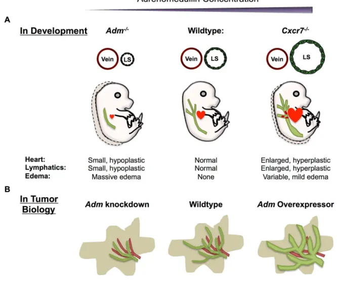
As discussed in the previous section of this review, whether CXCR7, a known non-signaling decoy receptor, acts as an AM receptor has remained unclear for over a decade. The promiscuity of the decoy receptor has also made it challenging to discern which ligand is responsible for a given phenotype. This difficulty was particularly apparent in Cxcr7-null mice, which exhibited cardiac enlargement and hyperplasia, a phenotype that was not observed in mutant mice of the canonical ligands, SDF-1 and CXCL11 [21,61,62]. However, the cardiac phenotypes closely phenocopied a genetic model of AM overexpression (known as Admhi/hi mice) which results in gross cardiac hyperplasia during embryogenesis [25,63].
signal, and a phenocopy of the Admhi/hi mice. Our lab hypothesized that if CXCR7 does
behave as an AM decoy receptor, loss of CXCR7 would also lead to lymphatic vascular defects. Because loss of AM signaling results in small, hypoplastic hearts and lymph sacs [14,39,40], and AM overexpression results in enlarged, hyperplastic hearts, we predicted that loss of Cxcr7 would result in enlarged, hyperplastic lymphatic vessels.
Indeed, as discussed extensively in Chapter 2, consistent with the notion that CXCR7 sequesters AM, Cxcr7-/- mice exhibited enlarged, blood filled lymph sacs. Careful
examination of the number of proliferating LECs in the lymph sac demonstrated that the enlarged lymph sacs were hyperplastic (Figure 1-2A). These data suggest that, in the absence of the decoy receptor, an excess of AM leads to hyperproliferation of LECs and subsequently enlarged, poorly formed lymph sacs [25]. Further examination of LECs in vitro and lymphatic beds in vivo confirmed the ability of CXCR7 to affect the lymphatic
endothelium. When treated with AM, cultured CXCR7 knockdown LECs proliferated more than wildtype LECs [25]. Moreover, dermal and cardiac lymphatic beds were disrupted in Cxcr7-null mice and displayed dysmorphic vasculature consistent with hyperplasia [25].
To definitively show that these phenotypes were caused by excessive AM signaling, we genetically titrated AM ligand onto Cxcr7-/-mice. Both genetic increase and reduction of
AM peptide on the Cxcr7 null background resulted in striking effects on the lymphatic phenotypes. Genetic reduction of Adm in Cxcr7-/- mice restored the lymphatic and cardiac
hyperplasia to wildtype levels, indicating that the hyperplasia observed in Cxcr7-/-mice
resulted from excessive AM signaling [25]. Moreover, intercross of Cxcr7mutant mice with
Admhi/hi mice resulted in an exacerbation of the lymphatic enlargement and increased
embryonic lethality to the point that it was difficult to maintain a Cxcr7+/-; Admhi/+mouse
development and that, without tight control of AM-mediated signaling, proliferation of both myocardial and lymphatic endothelial cells is disrupted (Figure 1-2A).
Interestingly, while the lymphatic endothelium dynamically expresses Cxcr7, we observed that the blood endothelium directly adjacent to the lymphatic endothelium persistently expressed Cxcr7. Cxcr7 was infrequently observed in the dermal lymphatics where Cxcr7-/-mice exhibited phenotypes consistent with dermal lymphatic hyperplasia but
was regularly observed in the dermal blood vessels [25]. Consistent with previously
published papers, therefore, we concluded that the presence of the scavenging receptor in the adjacent blood endothelium impacts development of the surrounding tissue [25]. These findings again highlight the importance that expression of the ligand and its receptors in proximity (but not necessarily directly within) the lymphatic endothelium has the ability to
impact lymphangiogenesis. Taken together, these data demonstrate that Cxcr7 modulates AM signal and identifies a new paradigm of 7-transmembrane decoy receptors as regulators of lymphatic vascular development.
Adrenomedullin Signaling During Adulthood
Mid-gestational embryonic lethality due to global knockout of the AM signaling system has made elucidation of the signaling effects of AM on the lymphatic system in adulthood difficult. The recent design of an inducible knockout model, however, has
confirmed an important role for AM in lymphatic maintenance and function following normal lymphangiogenesis. Global reduction of Calcrl in Calcrlfl/fl animals using a ubiquitously
expressed, tamoxifen-inducible Cre transgenic line (CAGGCre-ERTM) resulted in acute and
opaqueness [64]. After ruling out glaucoma, Hoopes et. al reported that Calcrl-deficient mice exhibit corneal edema, inflammation, and dilated corneoscleral lymphatic vessels.
While healthy cornea is largely avascular, lymphangiogenesis occurs in response to inflammation and can ultimately lead to blindness [65]. Though no corneal
lymphangiogenesis was reported in Calcrlfl/flCAGGCre-ERTM mice, these mice were only
evaluated after one week, which may not allow enough time for neolymphangiogenesis. Given more time, the inflammation in Calcrlfl/flCAGGCre-ERTM may lead to corneal
lymphangiogenesis. It is also possible that the absence of the AM receptor would prevent new lymphatic vessels from forming. Regardless, these findings highlight the importance of proper lymphatic function in maintenance of fluid homeostasis in and around the eye. Indeed, recent studies have demonstrated that the endothelial cells lining Schlemm’s canal express several lymphatic marker genes, are responsive to VEGFC and thereby control the aqueous humor with the anterior chamber of the eye. [66-68]. Thus, modulation of these vessels may allow for either increased hydration or drainage of the eye, allowing for a novel treatment of dry eyes, ocular edema, and glaucoma.
In addition to the ophthalmic pathology, Calcrlfl/flCAGGCre-ERTMmice also failed to
match weight gain of their wild-type littermates. Examination of the mesenteric vessels of experimental mice showed dilated lymphatic vessels filled with chyle when fed a high-fat meal. These data suggest that AM signaling is critical for absorption of fat from mesenteric lymphatic vessels. Close examination of the mesenteric lymphatic vessels following high-fat diet revealed disrupted lymphatic junctions, a finding that was consistent with previously published work demonstrating that AM stabilizes the lymphatic endothelial barrier and increases vascular permeability [54]. Indeed, further examination of the lymphatic
of the dye [64]. Importantly, no difference in blood vascular permeability was observed, reinforcing that AM loss affects primarily the lymphatic vasculature [64].
To induce edema, complete Freund’s adjuvant (CFA) was injected into the paw of the hindlimb. In contrast to tamoxifen-injected controls, Calcrlfl/flCAGGCre-ERTM mice had
prolonged edema [64], suggesting that AM is required for proper reabsorption of interstitial fluid and resolution of edema. As discussed above, previous studies demonstrate that AM treatment stabilizes the lymphatic endothelial barrier (Figure 1-1B) [54]. Therefore, similar to the mesenteric lymphatics, it follows that loss of Calcrl destabilizes the lymphatic
endothelium in the peripheral lymphatics, increases vascular permeability, allows for lymphatic fluid leakage back into the interstitium, and prevents proper edema resolution.
These findings (summarized in Figure 1-3) are particularly intriguing because they demonstrate that appropriate AM dosage following development is required for proper lymphatic maintenance and function. Loss of AM signaling results in leaky vessels, failure to resolve edema, and alterations in fat absorption. All of these data suggest that AM may be a reasonable target for the treatment of a variety of diseases for which we currently have limited therapies. Pharmaceuticals that modulate fat absorption could be a useful tool to help prevent or reduce the extent of obesity. Additionally, use of AM agonists could
decrease lymphatic vessel leakiness and increase uptake of interstitial fluid, allowing for the partial or complete resolution of lymphatic associated edema.
Adrenomedullin and Lymphedema
As mentioned above, lymphedema is the most common lymphatic disease, affecting hundreds of millions of people worldwide. Lymphedema is categorized as primary or
secondary. Primary lymphedema often results from congenital abnormalities. Mutations in nineteen genes have been associated with primary lymphedema, which have been
disrupt genes critical for downstream signaling in LECs, including mutations in the genes encoding VEGFR3, VEGFC, and CCBE1 [69]. Additionally, disruption in several
transcription factors required for LEC differentiation and specification (for example, GATA2, FOXC2, and SOX18) result in syndromic forms of primary lymphedema [1,69]. Although no
mutations in the AM signaling cascade have yet been identified in human primary
lymphedema, as whole exome sequencing studies continue, it is possible that AM will be found to associate with primary lymphedema. Secondary lymphedema, on the other hand, stems from disruption of the normal lymphatic vasculature by infection or iatrogenic
intervention [5]. Despite its prevalence and significant effect on quality of life, currently no cure exists for lymphedema. Additionally, it is not well understood why some individuals are predisposed to developing lymphedema.
Recently, AM was implicated as a molecule that might contribute to the onset of secondary lymphedema. Adm haploinsufficiency predisposed mice to developing secondary lymphedema following hind limb injury compared to wildtype mice [52]. Further, systemic injection of AM to Adm+/- mice restored proper wound healing, thereby preventing the onset
of lymphedema [52]. Here, these data demonstrate the importance of AM dosage in the resolution of edema.
Though lymphedema has yet to be associated with mutations in the AM signaling system in humans, these studies suggest that AM administration has potential as a novel therapeutic for the treatment of secondary lymphedema. The primary cause of secondary lymphedema in western countries is radical axillary lymph node dissection, with 20-30% of patients developing severe lymphedema following surgery [70-72]. The most promising treatment is the generation of new lymphatic vessels [70]. Jin and colleagues demonstrate that AM administration increases lymphatic flow, promotes angiogenesis and
formation, as both angiogenesis and lymphangiogenesis are required for wound healing [73-75]. However, the preferential effect of AM on lymphatic endothelial cells indicates that the primary cause for increased healing is neolymphangiogenesis. Therefore, AM administration following surgical resection of lymph nodes may not only promote wound healing, but also prevent the onset of lymphedema. While there are no current studies using AM infusion to treat lymphedema in humans, the potential of AM to act as a novel therapy for lymphedema is promising.
Adrenomedullin in Cancer
Originally isolated from pheochromocytoma, a rare neuroendocrine tumor, elevated AM has been associated with neoplasms [76,77]. Many cancer subtypes overexpress Adm [15], and plasma AM levels often correlate with metastasis, invasion, and poor survival [78,79]. How AM modulates cancer progression is not well understood. Many experts have speculated that AM contributes to tumor survival and progression by increasing blood flow to a hypoxic tumor environment [15,76,80]. However, the importance of lymphangiogenesis and lymphatic vessel remodeling to cancer biology [81,82] has also delineated a role for AM-mediated lymphangiogenesis in promoting distant metastasis.
Using a Lewis lung carcinoma (LLC) cell line, Karpinich et al. generated a series of tumor cells that stably over- or under-expressed Adm and demonstrated a regulatory role for AM in tumor lymphangiogenesis [83]. The LLC model was particularly attractive because LLCs do not express the canonical AM receptor, Calcrl and Ramp2. Consequently,
alterations in Adm expression did not affect tumor cell proliferation [83]. Therefore, following injection into mice, AM-mediated increases in tumor size would not confound differences in tumor metastasis.
proliferation, Adm overexpression resulted in a 3-fold elevation of LECs in both tumors and sentinel lymph nodes compared to tumors with reduced Adm expression (Figure 1-2B) [83]. Lymphatic vessels of Adm overexpressing tumors were also significantly dilated. Careful evaluation of tumor metastasis revealed that Adm overexpression increased tumor
dissemination [83]. These data recapitulate what has been observed in human tumors and suggest that Adm expression can promote metastasis via lymphangiogenesis.
Importantly, these findings extend beyond the LLC model. Studies in cervical cancer have also found associations between AM-mediated lymphangiogenesis and severity of disease. Huang et al. report that loss in the tumor stromal endothelium of miR-126, a microRNA that maintains vessel integrity during development, results in significant
upregulation of Adm and strongly associates with invasive carcinomas [84]. Upregulation of AM peptide also coupled with increases in CD31-positive endothelium (5.3% in tumors with high levels of miR-126 expression vs. 71% in invasive tumors with low miR-126 expression), suggesting that AM may promote disease progression through angiogenesis [84]. However, because lymphatic and blood vessels both stain CD31-positive, it is unclear whether this increase in vessel density is due AM-mediated effects on the blood or lymphatic
endothelium. Taken together with previous in vivo and in vitro studies that demonstrate that AM has preferential effects on the lymphatic endothelium, it is expected that much of the increase in vessel density is due to AM-mediated lymphangiogenesis. Consistently, the authors found significant co-localization of AM peptide with LYVE1-positive vessels [84].
These findings make a compelling argument for an association between increased Adm expression and tumor invasion. As such, modulation of Adm expression via AM
inhibitors could be strengthened by co-administration with AM inhibitors [87]. This hypothesis has already been tested in mouse models of prostate cancer, where anti-AM antibodies were found to disrupt tumor vasculature, decrease lymphatic vessel density, increase LEC death, and suppress tumor growth [88]. Though this anti-AM antibody has not been fully characterized or utilized in other studies, this finding supports the hypothesis that AM inhibition may improve cancer-related outcomes.
Adrenomedullin-induced Lymphangiogenesis in the Reproductive System
Perhaps the most exciting and novel aspect of AM-mediated lymphangiogenesis is its involvement in the lymphatic vasculature of the reproductive system. Our lab and others have shown that AM is critical in healthy pregnancy [9]. In a normal human pregnancy, AM increases 3-5 fold [89]. Dysregulation of this physiologic increase in both humans and mouse models has been associated with significant complications, most notably preeclampsia [90-92]. Additionally, haploinsufficiency of Adm in female mice results in reduced fertility due to implantation defects [93]. The essential role of AM in the normal reproductive system has therefore become increasingly apparent and clinically relevant over the past two decades. As such, there has been a rapidly growing interest in the role of AM-mediated angiogenesis and lymphangiogenesis in the reproductive system.
concentration of AM originates from the host and impacts the tumor environment. It will be interesting to tease out whether levels of AM originating outside of the tumor impact cancer progression via lymphangiogenesis. Adm overexpression models may prove useful in determining host AM status on tumor progression [63].
Despite the focus on AM as a tumor-promoting peptide, increased Adm expression also plays important roles during the normal reproductive cycle. Recent studies have
suggested that endometrial lymphangiogenesis is required for normal menstrual cycling and repair of damaged blood vessels during menstruation [96]. Examination of Adm during various stages of the menstrual cycle reveals that Adm expression is elevated during stages of endometrial repair [97]. Also, AM treatment results in increased lymphatic endometrial endothelial cell growth [97,98]. Taken together, these findings suggest that AM-mediated lymphangiogenesis facilitates endometrial repair and allows progression through the menstrual cycle. Dysregulation of endometrial Adm expression may lead to improper repair of damaged blood vessels and prolonged or heavy menstrual bleeding [97]. These findings again suggest that modulation of the AM signaling system could be utilized clinically for the treatment of common diseases that significantly affect quality of life.
Concluding remarks
FIGURES
Figure 1-1. Early development of the lymphatic system and the effects of AM on LECs.
Figure 1-2. Proper AM dosage is required for normal lymphangiogenesis during
development and in tumor biology. (A) Precise AM concentration is required during
Figure 1-3. Loss of AM signaling in adulthood results in disruption of many lymphatic
beds. Here, we provide a summary of the findings when AM signaling is disrupted following
REFERENCES
1. Tammela T, Alitalo K (2010) Lymphangiogenesis: Molecular mechanisms and future promise. Cell 140: 460-476.
2. Zheng W, Aspelund A, Alitalo K (2014) Lymphangiogenic factors, mechanisms, and applications. J Clin Invest 124: 878-887.
3. Kerjaschki D (2014) The lymphatic vasculature revisited. J Clin Invest 124: 874-877.
4. Mortimer PS, Rockson SG (2014) New developments in clinical aspects of lymphatic disease. J Clin Invest 124: 915-921.
5. Warren AG, Brorson H, Borud LJ, Slavin SA (2007) Lymphedema: a comprehensive review. Ann Plast Surg 59: 464-472.
6. Wiig H, Schroder A, Neuhofer W, Jantsch J, Kopp C, et al. (2013) Immune cells control skin lymphatic electrolyte homeostasis and blood pressure. J Clin Invest 123: 2803-2815.
7. Harvey NL, Srinivasan RS, Dillard ME, Johnson NC, Witte MH, et al. (2005) Lymphatic vascular defects promoted by Prox1 haploinsufficiency cause adult-onset obesity. Nat Genet 37: 1072-1081.
8. Poyner DR, Sexton PM, Marshall I, Smith DM, Quirion R, et al. (2002) International Union of Pharmacology. XXXII. The mammalian calcitonin gene-related peptides,
adrenomedullin, amylin, and calcitonin receptors. Pharmacol Rev 54: 233-246.
9. Lenhart PM, Caron KM (2012) Adrenomedullin and pregnancy: perspectives from animal models to humans. Trends Endocrinol Metab 23: 524-532.
10. Muff R, Born W, Lutz TA, Fischer JA (2004) Biological importance of the peptides of the calcitonin family as revealed by disruption and transfer of corresponding genes. Peptides 25: 2027-2038.
11. Smith DM, Coppock HA, Withers DJ, Owji AA, Hay DL, et al. (2002) Adrenomedullin: receptor and signal transduction. Biochem Soc Trans 30: 432-437.
13. Gibbons C, Dackor R, Dunworth W, Fritz-Six K, Caron KM (2007) Receptor activity-modifying proteins: RAMPing up adrenomedullin signaling. Mol Endocrinol 21: 783-796.
14. Fritz-Six KL, Dunworth WP, Li M, Caron KM (2008) Adrenomedullin signaling is necessary for murine lymphatic vascular development. J Clin Invest 118: 40-50.
15. Hay DL, Walker CS, Poyner DR (2011) Adrenomedullin and calcitonin gene-related peptide receptors in endocrine-related cancers: opportunities and challenges. Endocr Relat Cancer 18: C1-14.
16. Kapas S, Catt KJ, Clark AJ (1995) Cloning and expression of cDNA encoding a rat adrenomedullin receptor. J Biol Chem 270: 25344-25347.
17. Kapas S, Clark AJ (1995) Identification of an orphan receptor gene as a type 1 calcitonin gene-related peptide receptor. Biochem Biophys Res Commun 217: 832-838.
18. Balabanian K, Lagane B, Infantino S, Chow KY, Harriague J, et al. (2005) The
chemokine SDF-1/CXCL12 binds to and signals through the orphan receptor RDC1 in T lymphocytes. J Biol Chem 280: 35760-35766.
19. Burns JM, Summers BC, Wang Y, Melikian A, Berahovich R, et al. (2006) A novel chemokine receptor for SDF-1 and I-TAC involved in cell survival, cell adhesion, and tumor development. J Exp Med 203: 2201-2213.
20. Ikeda Y, Kumagai H, Skach A, Sato M, Yanagisawa M (2013) Modulation of circadian glucocorticoid oscillation via adrenal opioid-CXCR7 signaling alters emotional behavior. Cell 155: 1323-1336.
21. Sierro F, Biben C, Martinez-Munoz L, Mellado M, Ransohoff RM, et al. (2007) Disrupted cardiac development but normal hematopoiesis in mice deficient in the second CXCL12/SDF-1 receptor, CXCR7. Proc Natl Acad Sci U S A 104: 14759-14764.
22. Autelitano DJ, Tang F (1999) Co-expression of prepro-adrenomedullin with a putative adrenomedullin receptor gene in vascular smooth muscle. Clin Sci (Lond) 96: 493-498.
24. Ladoux A, Frelin C (2000) Coordinated Up-regulation by hypoxia of adrenomedullin and one of its putative receptors (RDC-1) in cells of the rat blood-brain barrier. J Biol Chem 275: 39914-39919.
25. Klein KR, Karpinich NO, Espenschied ST, Willcockson HH, Dunworth WP, et al. (2014) Decoy Receptor CXCR7 Modulates Adrenomedullin-Mediated Cardiac and
Lymphatic Vascular Development. Dev Cell 30: 528-540.
26. Betterman KL, Harvey NL (2014) Decoys and Cardiovascular Development: CXCR7 and Regulation of Adrenomedullin Signaling. Dev Cell 30: 490-491.
27. Karpinich NO, Hoopes SL, Kechele DO, Lenhart PM, Caron KM (2011) Adrenomedullin Function in Vascular Endothelial Cells: Insights from Genetic Mouse Models. Curr Hypertens Rev 7: 228-239.
28. Di Iorio R, Marinoni E, Letizia C, Alo P, Villaccio B, et al. (1998) Adrenomedullin, a new vasoactive peptide, is increased in preeclampsia. Hypertension 32: 758-763.
29. Nuki C, Kawasaki H, Kitamura K, Takenaga M, Kangawa K, et al. (1993) Vasodilator effect of adrenomedullin and calcitonin gene-related peptide receptors in rat mesenteric vascular beds. Biochem Biophys Res Commun 196: 245-251.
30. Hinson JP, Kapas S, Smith DM (2000) Adrenomedullin, a multifunctional regulatory peptide. Endocr Rev 21: 138-167.
31. Kataoka Y, Miyazaki S, Yasuda S, Nagaya N, Noguchi T, et al. (2010) The first clinical pilot study of intravenous adrenomedullin administration in patients with acute myocardial infarction. J Cardiovasc Pharmacol 56: 413-419.
32. Hagerling R, Pollmann C, Andreas M, Schmidt C, Nurmi H, et al. (2013) A novel multistep mechanism for initial lymphangiogenesis in mouse embryos based on ultramicroscopy. EMBO J 32: 629-644.
33. Yang Y, Oliver G (2014) Development of the mammalian lymphatic vasculature. J Clin Invest 124: 888-897.
35. Wigle JT, Oliver G (1999) Prox1 function is required for the development of the murine lymphatic system. Cell 98: 769-778.
36. Alitalo K, Tammela T, Petrova TV (2005) Lymphangiogenesis in development and human disease. Nature 438: 946-953.
37. Karkkainen MJ, Haiko P, Sainio K, Partanen J, Taipale J, et al. (2004) Vascular endothelial growth factor C is required for sprouting of the first lymphatic vessels from embryonic veins. Nat Immunol 5: 74-80.
38. Makinen T, Jussila L, Veikkola T, Karpanen T, Kettunen MI, et al. (2001) Inhibition of lymphangiogenesis with resulting lymphedema in transgenic mice expressing soluble VEGF receptor-3. Nat Med 7: 199-205.
39. Dackor RT, Fritz-Six K, Dunworth WP, Gibbons CL, Smithies O, et al. (2006) Hydrops fetalis, cardiovascular defects, and embryonic lethality in mice lacking the calcitonin receptor-like receptor gene. Mol Cell Biol 26: 2511-2518.
40. Caron KM, Smithies O (2001) Extreme hydrops fetalis and cardiovascular abnormalities in mice lacking a functional Adrenomedullin gene. Proc Natl Acad Sci U S A 98: 615-619.
41. Shindo T, Kurihara Y, Nishimatsu H, Moriyama N, Kakoki M, et al. (2001) Vascular abnormalities and elevated blood pressure in mice lacking adrenomedullin gene. Circulation 104: 1964-1971.
42. Shimosawa T, Shibagaki Y, Ishibashi K, Kitamura K, Kangawa K, et al. (2002) Adrenomedullin, an endogenous peptide, counteracts cardiovascular damage. Circulation 105: 106-111.
43. Ichikawa-Shindo Y, Sakurai T, Kamiyoshi A, Kawate H, Iinuma N, et al. (2008) The GPCR modulator protein RAMP2 is essential for angiogenesis and vascular integrity. J Clin Invest 118: 29-39.
44. Dackor R, Fritz-Six K, Smithies O, Caron K (2007) Receptor activity-modifying proteins 2 and 3 have distinct physiological functions from embryogenesis to old age. J Biol Chem 282: 18094-18099.
46. Kisanuki YY, Hammer RE, Miyazaki J, Williams SC, Richardson JA, et al. (2001) Tie2-Cre transgenic mice: a new model for endothelial cell-lineage analysis in vivo. Dev Biol 230: 230-242.
47. Eklund L, Bry M, Alitalo K (2013) Mouse models for studying angiogenesis and lymphangiogenesis in cancer. Mol Oncol 7: 259-282.
48. Tammela T, Saaristo A, Lohela M, Morisada T, Tornberg J, et al. (2005) Angiopoietin-1 promotes lymphatic sprouting and hyperplasia. Blood 105: 4642-4648.
49. Morisada T, Oike Y, Yamada Y, Urano T, Akao M, et al. (2005) Angiopoietin-1 promotes LYVE-1-positive lymphatic vessel formation. Blood 105: 4649-4656.
50. Srinivasan RS, Dillard ME, Lagutin OV, Lin FJ, Tsai S, et al. (2007) Lineage tracing demonstrates the venous origin of the mammalian lymphatic vasculature. Genes Dev 21: 2422-2432.
51. Chen L, Mupo A, Huynh T, Cioffi S, Woods M, et al. (2010) Tbx1 regulates Vegfr3 and is required for lymphatic vessel development. J Cell Biol 189: 417-424.
52. Nikitenko LL, Shimosawa T, Henderson S, Makinen T, Shimosawa H, et al. (2013) Adrenomedullin Haploinsufficiency Predisposes to Secondary Lymphedema. J Invest Dermatol.
53. Jin D, Otani K, Yamahara K, Ikeda T, Nagaya N, et al. (2011) Adrenomedullin reduces expression of adhesion molecules on lymphatic endothelial cells. Regul Pept 166: 21-27.
54. Dunworth WP, Fritz-Six KL, Caron KM (2008) Adrenomedullin stabilizes the lymphatic endothelial barrier in vitro and in vivo. Peptides 29: 2243-2249.
55. Cullen VC, Mackarel AJ, Hislip SJ, O'Connor CM, Keenan AK (2000) Investigation of vascular endothelial growth factor effects on pulmonary endothelial monolayer permeability and neutrophil transmigration. Gen Pharmacol 35: 149-157.
56. Kahn ML (2008) Blood is thicker than lymph. J Clin Invest 118: 23-26.
58. Ribatti D, Nico B, Spinazzi R, Vacca A, Nussdorfer GG (2005) The role of adrenomedullin in angiogenesis. Peptides 26: 1670-1675.
59. Moissoglu K, Majumdar R, Parent CA (2014) Cell migration: sinking in a gradient. Curr Biol 24: R23-25.
60. Venkiteswaran G, Lewellis SW, Wang J, Reynolds E, Nicholson C, et al. (2013) Generation and dynamics of an endogenous, self-generated signaling gradient across a migrating tissue. Cell 155: 674-687.
61. Yu S, Crawford D, Tsuchihashi T, Behrens TW, Srivastava D (2011) The chemokine receptor CXCR7 functions to regulate cardiac valve remodeling. Dev Dyn 240: 384-393.
62. Gerrits H, van Ingen Schenau DS, Bakker NE, van Disseldorp AJ, Strik A, et al. (2008) Early postnatal lethality and cardiovascular defects in CXCR7-deficient mice. Genesis 46: 235-245.
63. Wetzel-Strong SE, Li M, Klein KR, Nishikimi T, Caron KM (2013) Epicardial-derived adrenomedullin drives cardiac hyperplasia during embryogenesis. Dev Dyn 243: 243-256.
64. Hoopes SL, Willcockson HH, Caron KM (2012) Characteristics of multi-organ
lymphangiectasia resulting from temporal deletion of calcitonin receptor-like receptor in adult mice. PLoS One 7: e45261.
65. Regenfuss B, Bock F, Parthasarathy A, Cursiefen C (2008) Corneal
(lymph)angiogenesis--from bedside to bench and back: a tribute to Judah Folkman. Lymphat Res Biol 6: 191-201.
66. Park DY, Lee J, Park I, Choi D, Lee S, et al. (2014) Lymphatic regulator PROX1 determines Schlemm's canal integrity and identity. J Clin Invest 124: 3960-3974.
67. Aspelund A, Tammela T, Antila S, Nurmi H, Leppanen VM, et al. (2014) The Schlemm's canal is a VEGF-C/VEGFR-3-responsive lymphatic-like vessel. J Clin Invest 124: 3975-3986.
69. Brouillard P, Boon L, Vikkula M (2014) Genetics of lymphatic anomalies. J Clin Invest 124: 898-904.
70. Alitalo K (2011) The lymphatic vasculature in disease. Nat Med 17: 1371-1380.
71. Donker M, van Tienhoven G, Straver ME, Meijnen P, van de Velde CJ, et al. (2014) Radiotherapy or surgery of the axilla after a positive sentinel node in breast cancer (EORTC 10981-22023 AMAROS): a randomised, multicentre, open-label, phase 3 non-inferiority trial. Lancet Oncol 15: 1303-1310.
72. McLaughlin SA, Wright MJ, Morris KT, Giron GL, Sampson MR, et al. (2008) Prevalence of lymphedema in women with breast cancer 5 years after sentinel lymph node biopsy or axillary dissection: objective measurements. J Clin Oncol 26: 5213-5219.
73. Jin D, Harada K, Ohnishi S, Yamahara K, Kangawa K, et al. (2008) Adrenomedullin induces lymphangiogenesis and ameliorates secondary lymphoedema. Cardiovasc Res 80: 339-345.
74. Hirakawa S, Detmar M (2004) New insights into the biology and pathology of the cutaneous lymphatic system. J Dermatol Sci 35: 1-8.
75. Tonnesen MG, Feng X, Clark RA (2000) Angiogenesis in wound healing. J Investig Dermatol Symp Proc 5: 40-46.
76. Zudaire E, Martinez A, Cuttitta F (2003) Adrenomedullin and cancer. Regul Pept 112: 175-183.
77. Kitamura K, Kangawa K, Kawamoto M, Ichiki Y, Nakamura S, et al. (1993) Adrenomedullin: a novel hypotensive peptide isolated from human pheochromocytoma. Biochem Biophys Res Commun 192: 553-560.
78. Hu Z, Fan C, Livasy C, He X, Oh DS, et al. (2009) A compact VEGF signature associated with distant metastases and poor outcomes. BMC Med 7: 9.
79. Park HC, Seong J, An JH, Kim J, Kim UJ, et al. (2005) Alteration of cancer pain-related signals by radiation: proteomic analysis in an animal model with cancer bone invasion. Int J Radiat Oncol Biol Phys 61: 1523-1534.
81. Stacker SA, Williams SP, Karnezis T, Shayan R, Fox SB, et al. (2014)
Lymphangiogenesis and lymphatic vessel remodelling in cancer. Nat Rev Cancer 14: 159-172.
82. Ji RC (2014) Hypoxia and lymphangiogenesis in tumor microenvironment and metastasis. Cancer Lett 346: 6-16.
83. Karpinich NO, Kechele DO, Espenschied ST, Willcockson HH, Fedoriw Y, et al. (2013) Adrenomedullin gene dosage correlates with tumor and lymph node
lymphangiogenesis. FASEB J 27: 590-600.
84. Huang TH, Chu TY (2013) Repression of miR-126 and upregulation of adrenomedullin in the stromal endothelium by cancer-stromal cross talks confers angiogenesis of cervical cancer. Oncogene.
85. Tewari KS, Sill MW, Long HJ, 3rd, Penson RT, Huang H, et al. (2014) Improved survival with bevacizumab in advanced cervical cancer. N Engl J Med 370: 734-743.
86. Bergers G, Hanahan D (2008) Modes of resistance to anti-angiogenic therapy. Nat Rev Cancer 8: 592-603.
87. Bzowska M, Mezyk-Kopec R, Prochnicki T, Kulesza M, Klaus T, et al. (2013) Antibody-based antiangiogenic and antilymphangiogenic therapies to prevent tumor growth and progression. Acta Biochim Pol 60: 263-275.
88. Berenguer-Daize C, Boudouresque F, Bastide C, Tounsi A, Benyahia Z, et al. (2013) Adrenomedullin blockade suppresses growth of human hormone-independent prostate tumor xenograft in mice. Clin Cancer Res 19: 6138-6150.
89. Di Iorio R, Marinoni E, Scavo D, Letizia C, Cosmi EV (1997) Adrenomedullin in pregnancy. Lancet 349: 328.
90. Lenhart PM, Nguyen T, Wise A, Caron KM, Herring AH, et al. (2014) Adrenomedullin signaling pathway polymorphisms and adverse pregnancy outcomes. Am J Perinatol 31: 327-334.
92. Matson BC, Corty RW, Karpinich NO, Murtha AP, Valdar W, et al. (2014) Midregional pro-adrenomedullin plasma concentrations are blunted in severe preeclampsia. Placenta 35: 780-783.
93. Li M, Yee D, Magnuson TR, Smithies O, Caron KM (2006) Reduced maternal
expression of adrenomedullin disrupts fertility, placentation, and fetal growth in mice. J Clin Invest 116: 2653-2662.
94. Rodero MP, Prignon A, Avril MF, Boitier F, Aractingi S, et al. (2013) Increase
lymphangiogenesis in melanoma during pregnancy: correlation with the prolactin signalling pathway. J Eur Acad Dermatol Venereol 27: e144-145.
95. Khosrotehrani K, Nguyen Huu S, Prignon A, Avril MF, Boitier F, et al. (2011) Pregnancy promotes melanoma metastasis through enhanced lymphangiogenesis. Am J Pathol 178: 1870-1880.
96. Rogers PA, Donoghue JF, Girling JE (2008) Endometrial lymphangiogensis. Placenta 29 Suppl A: S48-54.
97. Maybin JA, Battersby S, Hirani N, Nikitenko LL, Critchley HO, et al. (2011) The
expression and regulation of adrenomedullin in the human endometrium: a candidate for endometrial repair. Endocrinology 152: 2845-2856.
98. Nikitenko LL, MacKenzie IZ, Rees MC, Bicknell R (2000) Adrenomedullin is an autocrine regulator of endothelial growth in human endometrium. Mol Hum Reprod 6: 811-819.
99. Yurugi-Kobayashi T, Itoh H, Schroeder T, Nakano A, Narazaki G, et al. (2006)
Adrenomedullin/cyclic AMP pathway induces Notch activation and differentiation of arterial endothelial cells from vascular progenitors. Arterioscler Thromb Vasc Biol 26: 1977-1984.
100. Guidolin D, Albertin G, Spinazzi R, Sorato E, Mascarin A, et al. (2008) Adrenomedullin stimulates angiogenic response in cultured human vascular endothelial cells:
Chapter 2: Decoy Receptor CXCR7 Modulates Adrenomedullin-Mediated Cardiac and
Lymphatic Vascular Development1,2
Overview
Atypical 7-transmembrane receptors, often called decoy receptors, act
promiscuously as molecular sinks to regulate ligand bioavailability and consequently temper the signaling of canonical G protein-coupled receptor (GPCR) pathways. Loss of
mammalian CXCR7, the most recently described decoy receptor, results in postnatal lethality due to aberrant cardiac development and myocyte hyperplasia. Here, we provide the molecular underpinning for this proliferative phenotype by demonstrating that the dosage and signaling of adrenomedullin (Adm = gene, AM = protein)—a mitogenic peptide-hormone required for normal cardiovascular development—is tightly controlled by CXCR7. To this end, Cxcr7-/- mice exhibit gain-of-function cardiac and lymphatic vascular phenotypes which can be reversed upon genetic depletion of adrenomedullin ligand. In addition to identifying a biological ligand accountable for the phenotypes of Cxcr7-/- mice, these results reveal a previously underappreciated role for decoy receptors as molecular rheostats in controlling the timing and extent of GPCR-mediated cardiac and vascular development. A graphical abstract is included in Figure 2-A1.
1Authors: Klara R. Klein, Natalie O. Karpinich Ph.D, Scott T. Espenschied, Helen H. Willcockson,
William P. Dunworth Ph.D, Samantha L. Hoopes, Ph.D, Erich J. Kushner Ph.D., Victoria L. Bautch
Ph.D, Kathleen M. Caron, Ph.D.
2 Reprinted with permission from 1. Klein KR, Karpinich NO, Espenschied ST, Willcockson HH,
Introduction
The precise spatiotemporal dosage of mitogenic and chemotactic factors is critical for the proper organization and development of organ systems. While the concentration of ligands often differs between tissues and developmental stages, the bioavailability of ligands within local microenvironments must also be controlled at a cellular level. Thus, cells can express molecular sink receptors, which in an autocrine or paracrine manner, sequester ligand away from canonical signaling receptors, thereby driving important developmental processes like neurogenesis, angiogenesis, chemotaxis and cellular proliferation [2,3]. Molecular sink receptors include atypical chemokine receptors, also known as decoy
receptors, which belong to the larger family of 7-transmembrane receptors. Decoy receptors act as molecular sinks by binding, internalizing, and degrading a wide range of ligands independent of G-protein coupling (Graham, 2012). CXCR7 is the most recently described decoy receptor and has been extensively studied for its role as a CXCL12/SDF-1 receptor [4-6], particularly in tumor cell migration and cancer progression [7,8].
However, prominent roles for CXCR7 during normal development and physiology have also been recently appreciated. In zebrafish, the expression and molecular sink functions of CXCR7 in trailing cells of the posterior lateral line primordium allows for a CXCL12 chemotactic gradient to be established and sensed by the leading primoridal germ cells which express CXCR4, the canonical SDF-1/CXCL12 receptor [9-12]. In this instance, CXCR7 exerts its decoy activities over a wide region to help coordinate and guide the migration of multicellular tissue structures.
However, the decoy activities of CXCR7 can also occur in a cell-autonomous
shown to regulate circulating levels of ligands, suggesting that CXCR7 expression in vessels may not only affect signaling events in a microenvironment, but systemically as well [15].
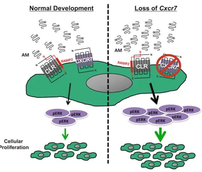
Due to these well-described roles in the CXCL12/CXCR4 signaling axis, Cxcr7 -/-mice were expected to exhibit phenotypes that might resemble gain-of-function mutations for the CXCL12/CXCR4 signaling axis. However, Cxcr7-/- mice have unexpected phenotypes including cardiomyocyte hyperplasia and postnatal lethality associated with gross cardiac enlargement and cardiac valve defects [16-18]. Because decoy receptors typically bind and sequester multiple ligands, it has been difficult to discern which ligand may be causally related to the developmental cardiac defects of Cxcr7-/- mice. In this regard, we appreciated that CXCR7 was originally identified as RDC1--a putative receptor for adrenomedullin (Adm = gene, AM = protein), a 52 amino acid mitogenic peptide hormone critical for cardiac and lymphatic vascular development [19-22]. AM binds RDC1/CXCR7 with a Kd of 1.9 x10⁻⁷ M, similar to CLR when associated with RAMPs [23]. Importantly, we have recently shown that genetic overexpression of AM ligand in Admhi/hi mice results in gross cardiac enlargement due to cardiac hyperplasia during embryogenesis [24], which closely phenocopies the dysmorphic cardiac hyperplasia of Cxcr7-/- mice.
Results
Gene expression of Cxcr7 and Adm are coupled in the heart and lymphatic endothelium
Historical ligand binding data [23] and more recent findings showing a down-regulation of Adm gene expression in Cxcr7-/-mice [17], strongly support the existence of
this ligand-receptor pair. Considering the well-established function of CXCR7 as a decoy receptor, we predicted that expression levels of Cxcr7 may homeostatically increase under conditions of increased AM peptide. To further evaluate whether this interaction exists, we measured the expression of Cxcr7 in hearts of Admhi/hi mice which have a genetically
engineered, 3-fold increase in Adm gene expression [24]. Indeed, utilizing qRT-PCR, we identified a potent 2.5-fold upregulation of Cxcr7 gene expression in Admhi/hi cardiac tissue compared to that of wildtype littermates (Figure 2-1A). Conversely, loss of Admexpression in isolated endothelial cells resulted in a nearly 5-fold reduction in Cxcr7 expression (Figure
2-1A).
We also found that Cxcr7 is expressed at high levels in isolated, adult lymphatic vessels—a tissue where AM peptide plays important roles (Figure 2-S1A). Consistently, microarray analysis of cultured, human lymphatic endothelial cells (LECs) showed that expression of the human CXCR7 gene (aka ACKR3 or CMKOR1) was one of the ten most significantly induced genes within 1 hour of 10nM AM treatment (p=2.5E-07) (Figure 2-1B
and Table 2-S1). This finding was further confirmed by qRT-PCR, revealing a 4-fold
increase in CXCR7 gene expression following 1- and 24- hours of AM treatment (Figure 2-1C). Pretreatment with AM22-52, a CLR/R2 antagonist, significantly reduced this
AM-mediated increase (Figure 2-1C), demonstrating that the upregulation of CXCR7 gene expression is modulated through the canonical AM receptor. Collectively, these data
CXCR7 expression, we next tested directly the hypothesis that CXCR7 serves as a decoy
receptor to modify AM concentration.
CXCR7 scavenges AM peptide and dampens canonical AM signaling
Using a classical scavenger assay, CXCR7-expressing HEK293T cells were treated with biotinylated-AM1-52, and aliquots of media were collected over time to determine the remaining levels of AM peptide within the media. While the levels of biotinylated-AM1-52 in the media of vector-transfected control cells remained unchanged, CXCR7-expressing cells rapidly and steadily depleted AM peptide from the media to levels that were statistically lower than control cells by the conclusion of the time course (Figure 2-1D, E). These data demonstrate the ability of CXCR7 to modulate AM ligand concentrations exogenously in a controlled in vitro system.
To determine whether this scavenging of AM peptide by CXCR7 was conserved in vivo, we compared AM staining in cardiac tissue of wildtype mice and Cxcr7-/- mice, which harbor an insertional GFP reporter within the targeted allele. Firstly, we noted that
expression of the GFP reporter was enriched within the epicardium and the endocardium surrounding the trabeculae and weakly expressed in the compact zone (Figure 2-1F). Secondly, the staining pattern of receptor expression was spatially juxtaposed and/or overlapping with the most prominent sites of AM peptide expression, including the
epicardium and trabeculae (Figure 2-1G and [24]). Remarkably, we found a significant 30% increase in the relative staining intensities of AM peptide in the epicardium and trabeculae of Cxcr7-/- mice compared to wildtype littermates (Figure 2-1H-K), but no changes within the
Activation of the canonical AM-receptor complex, CLR and receptor activity modifying protein 2 (RAMP2), elicits an increase in cAMP and subsequent downstream activation and phosphorylation of ERK [19]. Utilizing a highly-sensitive bioluminescence resonance energy transfer (BRET) reporter system [27,28], we found that HEK293 cells that overexpress CXCR7 failed to accumulate cAMP upon AM stimulation—a finding consistent with the lack of G-protein coupling by decoy receptors (Figure 2-S1B). As expected, AM treatment of CLR/RAMP2-expressing cells resulted in a potent accumulation of cAMP
(Figure 2-S1B) and pERK:tERK upregulation (Figure 2-1M, N and Figure S1C).
Importantly, while AM did not elicit a pERK:tERK upregulation in cells transfected with CXCR7 alone, the CLR-RAMP2 mediated activation of pERK:tERK was markedly abrogated
when cells were co-transfected with CXCR7 (Figure 2-1M, N). These in vitro signaling assays demonstrate that CXCR7 can act as a cell-autonomous molecular rheostat to dampen canonical AM pERK:tERK signaling.
The effects of CXCR7 on dampening pERK:tERK signaling were also confirmed in vivo, where we noted significant accumulation of pERK staining in dermal lymphatic vessels
of postnatal day 1 Cxcr7-/- tail skin compared to wildtype littermates (Figure 2-S2A-D).
Furthermore, we also observed a significant increase in the pERK staining in the lymph sac of e13.5 Cxcr7-/- embryos compared to wildtype animals (Figure 2-S2E-J). These in vivo
data from a genetic loss-of-function model aptly reciprocate the findings from the in vitro gain-of-function experiments and furthermore demonstrate that loss of Cxcr7 expression influences ERK phosphorylation on a tissue level.
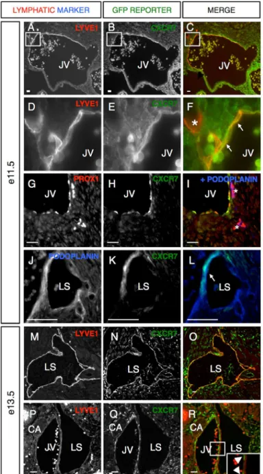
CXCR7 is dynamically expressed in lymphatic endothelium during development
Cxcr7+/- reporter allele, we found Cxcr7 expression co-localized with the lymphatic makers LYVE1 (Figure 2-2A-C), Prox1 (Figure 2-2G-I) and podoplanin (Figure 2-2I-L). At e11.5, lymphatic progenitor cells are arranged in a stereotypically-polarized fashion within the jugular vein (JV) and express Cxcr7 (Figure 2-2D-F, white arrows, Figure 2-2G, H). Interestingly, we often noted that Cxcr7 expression is temporarily reduced as the lymphatic progenitors begin to migrate away from the JV (Figure 2-2F, I asterisks)− underscoring the dynamic expression of the decoy receptor in areas of active cell migration. As lymphatic cells coalesce to form the lymph sacs (LS) between e11.5-e13.5, Cxcr7 was again
expressed in some lymphatics, which were identified by LYVE1 and podoplanin co-labeling
(Figure 2-2J-O). Cxcr7 was also persistently expressed in the JV cells directly adjacent to
the LS (Figure 2-2P-R, white arrowheads), consistent with recently published studies demonstrating a paracrine function for decoy receptors [12,30]. In summary, Cxcr7 is highly and dynamically expressed within lymphatic progenitors and early lymphatic vessels at the time of nascent lymphangiogenesis, which also spatiotemporally correlates with the
proliferative effects of AM during lymphatic development.
Cxcr7-/- mice have enlarged, blood filled lymphatic sacs
We have previously established that AM signaling is required for normal LEC proliferation at e13.5 [19]. Thus, we investigated whether loss of Cxcr7, which we hypothesize to be a molecular rheostat for AM, disrupts lymphangiogenesis at this point during embryogenesis. Indeed, approximately 10% of Cxcr7-/- mice exhibited visible
interstitial edema upon dissection at mid-gestation (Figure 2-3A, white arrows, Figure 2-3B, C, black arrows). Histological evaluation further revealed that approximately 10-15% of Cxcr7-/- mice displayed interstitial edema, particularly within the thoracic regions surrounding