Protease-activated Receptor-1 Enhances Poly I:C Induction of
the Anti-Viral Response in Macrophages and Mice
Silvio Antoniak1,2, Kohei Tatsumi1, Michael Bode1, Swetha Vanja1, Julie C. Williams1, and
Nigel Mackman1
1Department of Medicine, Thrombosis and Hemostasis Program, Division of Hematology and
Oncology, UNC McAllister Heart Institute, University of North Carolina, Chapel Hill, NC, USA
2Department of Pathology and Laboratory Medicine, University of North Carolina at Chapel Hill,
Chapel Hill, NC
Abstract
The coagulation cascade is activated during viral infections as part of the host defense system. Coagulation proteases activate cells by cleavage of protease-activated receptors (PAR). Recently, we reported that activation of PAR-1enhanced interferon (IFN)-β and CXCL10 expression in cardiac fibroblasts and in the hearts of mice infected with Coxsackievirus B3. In this study, we used the double-stranded RNA mimetic polyinosinic:polycytidylic acid (poly I:C) to induce an anti-viral response in macrophages and mice. Activation of PAR-1 enhanced poly I:C induction of IFNβ and CXCL10 expression in the murine macrophage cell line RAW264.7, bone-marrow derived mouse macrophages (BMM) and mouse splenocytes. Next, poly I:C was used to induce a type I IFN innate immune response in the spleen and plasma of wild-type (WT) and PAR-1−/− mice. We found that poly I:C treated PAR-1−/− mice and WT mice given the thrombin inhibitor dabigatran etexilate exhibited significantly less IFNβ and CXCL10 expression in the spleen and plasma compared to WT mice. These studies suggest that thrombin activation of PAR1 contributes to the anti-viral response in mice.
Keywords
protease-activated receptor 1; dsRNA; innate immune response; spleen; macrophage
Introduction
Interferons (IFNs) are divided into three groups: type I (IFNα/β), type II (IFNγ) and type III (IFNλ). Most cells express type I IFNα/β. In contrast, IFNγ synthesis is restricted to activated T cells and natural killer (NK) cells, and acts to promote the transition from the innate to the adaptive immune response.[1] Synthesis of IFNs is an essential part of antiviral innate immune responses.[1] Indeed, a deficiency of IFNs or IFN-dependent signaling is
Address for Correspondence: Silvio Antoniak, PhD, University of North Carolina at Chapel Hill, 111 Mason Farm Road Campus Box 7126, Chapel Hill, North Carolina, 27599, Telephone: (919) 843 7305, Fax : (919) 843 4585, [email protected].
HHS Public Access
Author manuscript
J Innate Immun
. Author manuscript; available in PMC 2018 January 01.Published in final edited form as:
J Innate Immun. 2017 ; 9(2): 181–192. doi:10.1159/000450853.
A
uthor Man
uscr
ipt
A
uthor Man
uscr
ipt
A
uthor Man
uscr
ipt
A
uthor Man
uscr
associated with increased susceptibility to certain virus infections, such as enteroviruses, influenza and herpes virus.[1]
Double-stranded (ds) RNA is a potent trigger of the type I IFN response.[1] dsRNA is part of the genome of dsRNA viruses and is an intermediate of ssRNA virus replication.[1] DsRNA is detected by the transmembrane receptor toll-like receptor (TLR) 3, as well as the cytosolic receptors dsRNA-dependent protein kinase, retinoic acid–inducible gene I, and melanoma differentiation-associated gene 5.[2,3] The dsRNA mimetic polyinosinic: polycytidylic acid (poly I:C) is used experimentally to stimulate TLR3 and induce the expression of type I IFNs in vitro and vivo.[1,3–5]
TLR3 is broadly expressed by myeloid and non-myeloid cells.[3,6] Within the spleen high levels of TLR3 expression are present in cells of the red pulp as well as marginal zone, while lower levels of expression are present in macrophages and dendritic cells (DCs) in the white pulp.[6] TLR3-dependent innate immune activation is essential to fight particular virus infections, including poliovirus, rhinovirus, herpes simplex virus, encephalomyocarditis virus, and Coxsackievirus group B (CVB).[3] In addition, TLR3 enhances the antiviral responses of macrophages against other viruses, such as the monkey and human immunodeficiency viruses (HIV).[7,8]
Virus infections activate the coagulation system by inducing tissue factor (TF) expression in various cell types.[9–11] Interestingly, one study found that poly I:C induced TF expression in endothelial cells but not monocytes.[12] Furthermore, administration of poly I:C to mice increased systemic activation of coagulation and fibrin deposition in the liver in a TLR3-dependent manner.[12] Thrombin is the central protease of the coagulation cascade but also activates cells by cleavage of protease-activated receptors (PAR), such as PAR-1.[13] This receptor is also activated by other proteases, such as matrix metalloproteinases (MMP) 1 and 13.[4,9,10,14,15] PAR-1 is expressed by a variety of human and murine immune cells, including monocytes/macrophages, DC, T cells, and NK cells.[16,17]
The coagulation and immune systems appear to have a common evolutionary origin.[18] Moreover, there is crosstalk between the two systems during infections.[9,10,17,18] It has been proposed that TLRs and PARs act as a cooperative sensor system to detect pathogens. [10,19] We demonstrated that PAR-1 activation enhanced poly I:C induced IFNβ and CXCL10 expression in murine cardiac fibroblasts, suggesting interaction between PAR-1 and TLR3.[4] In a CVB3 mouse model, we observed that a deficiency of PAR-1 was associated with reduced type I IFN responses leading to increased myocarditis.[4] In addition, inhibition of the PAR-1 activating proteases thrombin or MMP13 led to increased myocarditis.[4] Bone marrow transplantation experiments further revealed that PAR-1 on both non-hematopoietic and hematopoietic cells contributed to PAR-1 mediated protection against CVB3 myocarditis.[4] This finding is consistent with the observation that cardiac damage and survival of CVB4-infected TLR3-deficient mice can be improved by adaptive transfer of wild-type (WT) macrophages.[20]
A
uthor Man
uscr
ipt
A
uthor Man
uscr
ipt
A
uthor Man
uscr
ipt
A
uthor Man
uscr
In this study, we utilized poly I:C as surrogate for viral activation and PAR-1−/− mice to investigated the role of PAR-1 in the induction of an anti-viral innate immune response in macrophages and mice.
Material and Methods
Mice
We used WT and PAR-1−/− mice on a C57BL/6J background.[21] All mouse studies were performed with the approval of the University of North Carolina at Chapel Hill Institutional Animal Care and Use Committee (IACUC).
Isolation and in vitro stimulation of macrophages, DCs and splenocytes
WT and PAR-1−/− mice (4 mice each genotype, 8–12 weeks old) were used to isolate splenocytes or generate bone marrow-derived macrophages (BMMs) and dendritic cells (BMDCs). Each isolation was repeated once. For splenocyte isolation, spleens were harvest and passed through a 70 µm cell strainer to generate a single cell suspension. Red blood cells were lysed and the splenocytes washed and re-suspended at a density of 1 × 10⁶ cells per well of a 24 well plate in RPMI 1640 with 10% FBS and 1% penicillin/streptomycin. Cells were stimulated with 25 µg/mL poly I:C (γ-radiated, Sigma-Aldrich, St. Louis, MO) and/or PAR-1 agonist peptide (TFLLR-NH2, 200 µM, Abgent, San Diego, CA) for 2 hours.
BMMs were generated from bone marrow using the L929-conditioned media method as described.[22] BMDCs were derived from bone marrow using GM-CSF and IL-4 (30 ng/mL each) as described.[23] RAW264.7 cells, a murine macrophage cell line, were maintained as described.[24]
For experiments, BMM were plated at a density of 1 × 10⁶ cells per well of a 12 well plate the day before stimulation. The next day cell culture media was replaced with 0.5 mL fetal bovine serum (FBS)-free media and the cells were stimulated with poly I:C (25 µg/mL) and/or PAR-1 agonist peptide (200 µM) for different times. BMDC were seeded in FBS-containing culture media at a density of 1 × 10⁶ cells in one mL of media the day before stimulation. The next day, cells were stimulated without changing the media with poly I:C (25 µg/mL) and/or PAR-1 agonist peptide (200 µM) for 8 hours. RAW264.7 cells were seeded at a density of 5 × 10⁵ cells per well of a 12 well plate one day prior stimulation. Media was replaced with 0.5 mL FBS-free culture media the next day and the cells were stimulated with 5 µg/mL poly I:C and/or 150 µM PAR-1 agonist peptide for indicated times.
For measuring IFNβ promoter activity, RAW264.7 cells were stable transfected with the pNiFty2-IFB-SEAP plasmid (containing the Ifnb1 promoter) with Zeocin selection (Invivogen, San Diego, CA) using Lipofectamine 2000 (Life Technologies, Carlsbad, CA). [4] Cells were seeded and treated with poly I:C (5 µg/mL) and/or thrombin (5 nM, human α -thrombin, Enzyme Research Laboratories, South Bend, IN) mentioned above. The next day, media was collected, and SEAP (secreted embryonic alkaline phosphatase) activity was detected using QUANTI-blue (Invivogen) at 640 nm with a SpectraMax M5 plate reader (Molecular Devices, Sunnyvale, CA). [4]
A
uthor Man
uscr
ipt
A
uthor Man
uscr
ipt
A
uthor Man
uscr
ipt
A
uthor Man
uscr
In vivo stimulation
Male mice (8–12 weeks old) were injected intraperitoneally (i.p.) with 8 mg/kg poly I:C (Sigma-Aldrich) diluted in PBS.[25] Blood was collected from the inferior vena cava and plasma prepared as previously described.[11,26]
Thrombin inhibition
To inhibit thrombin, mice were fed mice for 7 days with a custom-made chow (AIN-93M, Dyets Inc., Bethlehem, PA) with peanut flavoring (2 g/kg chow) with or without dabigatran etexilate (10g/kg chow) as described.[4] The level of anticoagulation was measured as described.[4]
CVB3 infection model
A standard CVB3 infection model was used.[4,5,27] Briefly, 6–8 weeks old male mice were infected with an intraperitoneal injection of CVB3 Nancy Strain (105 plaque forming units in 200 µL saline). Tissue and blood was collected and analyzed 4 days after infection.
Real-time PCR
Total RNA from cultured cells or splenic tissue was isolated by using TRIzol method (Life Technologies). One microgram of RNA was reverse transcribed into cDNA using the iScript™ RT Supermix (Bio-Rad Laboratories, Hercules, CA).[28,29] Real-time PCR primer/probes for IFNβ, IFNγ, CXCL10, CCL5, CXCL1, IRF3, IRF7, viperin, Rpl4, GAPDH and 18S were purchased from Integrated DNA Technologies (Coralville, IA). CVB3 genome levels were quantified as recently described.[4,5] Real-time PCR reactions were performed using the SSoFast™ Advanced Universal Supermix in a BioRad cycler (Bio-Rad Laboratories). The housekeeping genes Rpl4 or 18S (for splenic tissue) and GAPDH (for cells) were used to correct for variations in input RNA and reaction efficiency. [11]
ELISA
IFNβ (PBL Assay Science, Piscataway, NJ), CXCL10, CCL5 and CXCL1 (Duo-Set, R&D Systems, Minneapolis, MN) ELISAs were used to measure levels of IFNβ and CXCL10, CCL5 and CXCL1 in plasma and/or cleared cell culture supernatant (500 × g, 10 min, 4°C). [4]
Statistics
All statistical analyses were performed using GraphPad Prism (version 5.0; GraphPad Software Inc., La Jolla, CA). Data are represented as mean ± SEM. For 2-group comparison of continuous data, 2-tailed Student’s t test was used. For multiple-group comparison, data were analyzed by 1- or 2-way ANOVA and were Bonferroni corrected for repeated measures over time. A P value less than 0.05 was considered significant.
A
uthor Man
uscr
ipt
A
uthor Man
uscr
ipt
A
uthor Man
uscr
ipt
A
uthor Man
uscr
Results
PAR-1 activation enhances poly I:C induction of IFNβ and CXCL10 expression in murine macrophages and splenocytes
Macrophages and DCs are known to reside in the spleen and respond to TLR3 stimulation by expressing IFNβ and CXCL10.[25,30,31] In addition, depletion of phagocytic splenic macrophages and DC of the marginal zone markedly reduced anti-viral responses.[32] We analyzed the role of PAR-1 in poly I:C induction of IFNβ and CXCL10 expression in the murine macrophage cell line RAW264.7 as well as BMM and BMDC, and splenocytes from WT and PAR-1−/− mice.
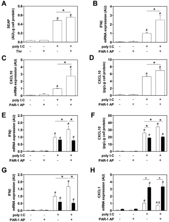
Poly I:C induction of IFNβ promotor activity as well as IFNβ and CXCL10 mRNA and protein expression in RAW264.7 cells was significantly enhanced by PAR-1 activation (Figure 1A–D). Next, we analyzed poly I:C induction of IFNβ mRNA expression and CXCL10 protein expression in BMM derived from WT and PAR-1−/− mice. Poly I:C alone induced IFNβ mRNA expression in both WT and PAR-1−/− BMM and splenocytes, whereas the PAR-1 agonist peptide alone had no effect (Figure 1E and G). Importantly,
co-stimulation of cells with poly I:C and the PAR-1 agonist peptide significantly increased IFNβ mRNA expression in WT BMM and splenocytes but not in PAR-1−/− BMM and splenocytes (Figure 1E and G). In addition, poly I:C stimulation of WT and PAR-1−/− BMM induced CXCL10 expression, which was significantly increased in WT BMM but not PAR-1−/− BMM co-stimulated with PAR-1 agonist peptide (Figure 1F). We did not detect any CXCL10 expression in splenocytes after 2 hours of poly I:C stimulation.
Poly I:C stimulated PAR-1−/− BMM and BMDC expressed lower levels of CXCL10 than WT BMM and BMDC (Figure 1F and data not shown). This result is consistent with previous studies showing that poly I:C stimulation of cardiac fibroblasts induces the expression of MMP13 that activates PAR-1.[4,10] Similar results were observed for CXCL10 protein expression (8 hours) with WT and PAR-1−/− BMDC (data not shown).
Recently, we observed increased levels of CXCL1 in lungs of influenza A infected PAR-1−/− mice compared to WT mice.[4] In addition, IL-8, the human homolog of murine CXCL1, is directly induced after TLR3 stimulation.[33]. Therefore, we analyzed CXCL1 expression after poly I:C and/or PAR-1 stimulation in vitro. Poly I:C induced CXCL1 by splenocytes and PAR-1 stimulation alone had no effect (Figure 1H). Unexpectedly, WT splenocytes exhibited 3-fold lower levels of CXCL1 mRNA after stimulation with poly I:C compared to stimulated PAR-1−/− splenocytes (Figure 1H). Furthermore, activation of PAR-1 in WT splenocytes but not PAR-1−/− splenocytes significantly reduced poly I:C induction of CXCL1 mRNA expression (Figure 1H).
Poly I:C induces an anti-viral response in wild-type mice
Poly I:C injection in mice leads to maximum type I IFN expression between 2 and 4 hours in plasma.[25,34] In addition, poly I:C-mediated synthesis of IFNβ is followed by increased CXCL10 plasma levels.[34] In our mouse studies, we focused on changes in gene expression in the spleen due to the presence of blood and lymphoid elements as well as its role in initiating anti-viral responses to blood-borne pathogens.[25] We also measured immune
A
uthor Man
uscr
ipt
A
uthor Man
uscr
ipt
A
uthor Man
uscr
ipt
A
uthor Man
uscr
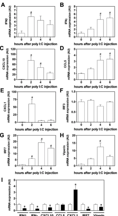
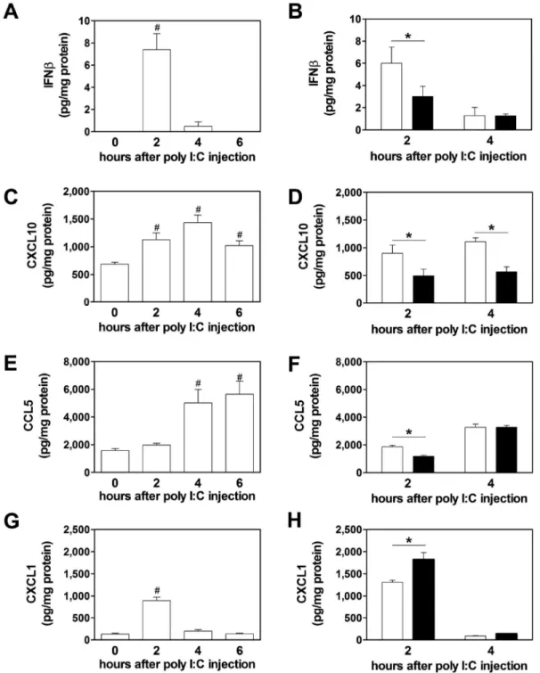
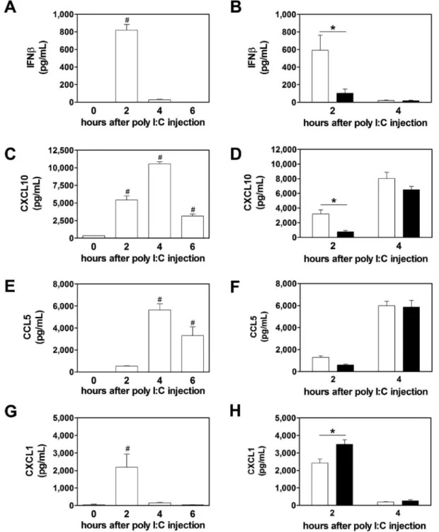
mediators in the plasma. To model virus-like infection of mice, WT mice were injected with poly I:C (8 mg/kg) i.p.. Poly I:C increased IFNβ, IFNγ, CXCL10, CCL5, CXCL1, IRF7 and viperin mRNA expression in the spleen after injection (Figure 2A–F). Maximal levels of these different immune mediators were observed at different times. The different gene expression pattern can be divided into an early (IFNβ, CXCL10, CXCL1), intermediate (IRF7, viperin) and late (IFNγ). However, we did not observe any changes in the splenic IRF3 mRNA expression after poly I:C injection (Figure 2F). Next, we analyzed the time-dependent changes in protein expression in the spleen. Maximal levels of IFNβ were observed 2 hours after administration of poly I:C injection (Figure 3A). Similar kinetics of IFNβ expression was observed in the plasma (Figure 4A). IFNβ induces several anti-viral response genes, such as CXCL10 and CCL5.[5,33,34] In addition, poly I:C induces IL8 expression.[33] Poly I:C increased levels of CXCL10, CCL5 and CXCL1 in the spleen (Figure 3C,E,G). Maximal levels of IFNβ and CXCL1 were observed at 2 hours whereas CXCL10 and CCL5 peaked at later times. Similar results were observed for the expression of these anti-viral proteins in the plasma (Figure 4C,E,G).
PAR-1 deficiency is associated with reduced poly I:C induction of ant-viral proteins in the spleen and plasma
Recently, we observed that PAR-1 expression on both non-hematopoietic and hematopoietic cells contributed to anti-viral responses against the ssRNA virus CVB3 in mice.[4] To determine if PAR-1 contributes to innate immune responses to poly I:C in vivo, WT and PAR-1−/− mice were injected with poly I:C and the immune response in the spleen and plasma was analyzed. A deficiency of PAR-1 was associated with significantly lower levels of poly I:C-dependent induction of IFNβ, IFNγ, CXCL10, IRF7 and viperin but not CCL5 mRNA expression (Figure 2I). In line with our in vitro data, we observed significant higher levels of CXCL1 mRNA in PAR-1−/− spleens compared to WT spleens (Figure 2I). In addition, PAR-1−/− mice injected with poly I:C had significant less splenic IFNβ protein levels compared to WT mice at 2 but not 4 hours (Figure 3B). The level of splenic CXCL10 protein in PAR-1−/− mice was also significantly lower at 2 and 4 hours after poly I:C injection compared to WT mice (Figure 3D). In addition, CCL5 protein levels were lower in the spleens of PAR-1−/− mice injected with poly I:C compared to WT mice (Figure 3F). Consistent with the changes in splenic CXCL1 mRNA levels, CXCL1 protein levels in the spleens of PAR-1−/− mice injected with poly I:C were significant higher compared to WT mice at 2 hours (Figure 3H).
Next, we analyzed the effect of PAR-1 deficiency on poly I:C induction of IFNβ, CXCL10, CCL5 and CXCL1 in plasma. PAR-1−/− mice had significant lower levels of IFNβ and CXCL10 in the plasma 2 hours after poly I:C treatment compared to WT mice (Figure 4B and D). CCL5 plasma levels were lower in PAR-1−/− mice compared to WT mice but this difference was not statistically significant (Figure 4F). As expected from the splenic data, the CXCL1 plasma levels were significantly higher in PAR-1−/− mice compared to WT mice 2 hours after poly I:C injection (Figure 4H).
A
uthor Man
uscr
ipt
A
uthor Man
uscr
ipt
A
uthor Man
uscr
ipt
A
uthor Man
uscr
Inhibition of thrombin is associated with reduced poly I:C induction of anti-viral proteins in the spleen and plasma
Thrombin is the primary activator of PAR-1 in vivo.[13] Furthermore, poly I:C injection into mice was associated with increased activation of coagulation.[12] We observed maximal activation of coagulation, as measured by levels of circulating thrombin-antithrombin complexes, at 4 hours post poly I:C injection (data not shown) To determine if thrombin contributes to anti-viral responses after poly I:C injection, WT mice were fed with the thrombin inhibitor dabigatran etexilate (10g/kg chow) prior poly I:C injection. Dabigatran etexilate treatment significant reduced IFNγ, CXCL10, CCL5 and viperin but not IFNβ and IRF7 mRNA expression in the spleen 4 hours after poly I:C injection in WT mice compared to placebo-treated WT mice (Figure 5A). As observed with PAR-1−/− mice, splenic CXCL1 mRNA expression was significantly increased with dabigatran etexilate compared to placebo fed WT mice (Figure 5A). Furthermore, CXCL10 protein levels in the spleen and plasma were significant lower with dabigatran etexilate compared to placebo fed WT mice 4 hours after poly I:C injection (Figure 5B,C).
PAR-1 deficiency is associated with reduced anti-viral responses in the spleen after CVB3 infection
TLR3-dependent signaling in macrophages were shown to mediate protection after CVB4 infection.[20] We found that PAR-1 deficiency was associated with reduced innate immune responses in the heart in CVB3 myocarditis.[4] In addition, CVB3 can infect and persist in the spleen and macrophages but its replication is highly suppressed.[35–37] WT and PAR-1−/− mice were infected with CVB3 and the response in the spleen and plasma analyzed. Consistent with our poly I:C data, PAR-1−/− mice expressed significantly lower levels of in IFNβ, IRF7 and CCL5 mRNA expression in the spleen compared to WT 4 days after CVB3 infection (Figure 6A). CXCL10 and viperin mRNA expression was also reduced in infected PAR-1−/− compared to WT mice spleens but did not reach significance. There was no differences in IFNγ between the genotypes. Furthermore, splenic CXCL1 mRNA was significant increased in PAR-1−/− compared to infected WT mice spleens (Figure 6A). CXCL10 protein levels were significant reduced in the spleen but not in the plasma (Figure 6B,C). As expected from the fact that CVB3 cannot replicate in macrophages, WT and PAR-1−/− mice had similar levels of viral genomes (Figure 6D) indicating that the differences in the expression of anti-viral genes was not due to different levels of virus.
Inhibition of thrombin is associated with a reduced anti-viral responses in the spleen after CVB3 infection
In our recent paper we showed that thrombin inhibition increased CVB3 myocarditis in mice due to a decrease in the innate immune response.[4] In this study, we found that thrombin inhibition reduced poly I:C induction of anti-viral gene expression in the spleen. To
determine if thrombin signaling contributes to the anti-viral responses in the spleen in CVB3 infected mice, we treated WT mice with dabigatran etexilate or placebo and then infected them with CVB3 and measured the anti-viral response at 4 days. As expected, thrombin inhibition resulted in a significant reduction of the splenic IFNγ, CXCL10, CCL5 and IRF7 but not viperin mRNA expression (Figure 7A). IFNβ mRNA was reduced but this reduction
A
uthor Man
uscr
ipt
A
uthor Man
uscr
ipt
A
uthor Man
uscr
ipt
A
uthor Man
uscr
did not reach significance (Figure 7A). However, we did not observe any differences in the CXCL10 levels in the spleen or plasma (Figure 7B,C). CXCL1 expression was increased in dabigatran compared to placebo treated mice 4 days after CVB3 infection (Figure 7A). The differences in the anti-viral response in the spleen was due to the effect of thrombin on gene expression not on virus load since the spleens of infected dabigatran etexilate and placebo treated mice showed comparable CVB3 genome levels (Figure 7D).
Discussion
The immune and blood coagulation systems combine to combat infections.[17,18] DsRNA or poly I:C activation of TLR3 induces type I IFN innate immune responses, as well as induce TF expression and activation of coagulation.[1,3,9,10,12,17] Here, we showed that PAR-1 deficiency is associated with a reduction in poly I:C induction of IFNβ and CXCL10 expression in the spleen and in the plasma. This observation is likely due to reduced TLR3-mediated responses in immune cells, including splenic macrophages and DCs. Consistent with this notion, we found that PAR-1 deficient BMM, BMDCs and splenocytes expressed lower levels of IFNβ and CXCL10 expression in vitro when stimulated with poly I:C and PAR-1 agonist compared to WT cells. Interestingly, we found that PAR-1 deficiency is associated with higher CXCL1 expression after poly I:C challenge. In addition, thrombin inhibition of WT mice produced similar results to PAR-1−/− mice suggesting that the thrombin-dependent PAR-1 activation is important for effective innate immune responses in the spleen. PAR-1−/− mice and WT mice treated with dabigatran etexilate infected with CVB3 also showed a reduction in the anti-viral response and increase in CXCL1 compared with WT mice. Together, our results indicate the PAR-1 expression and the thrombin-PAR-1 pathway contributes to innate immune responses to poly I:C and viral infection.
TLR3 is activated by viral dsRNA reaching the endosomal compartment. This can occur when cells, such as macrophages and DCs, phagocytose dsRNA released into the
extracellular space by dying virus-infected cells or when cells internalize viruses by receptor mediated endocytosis.[3] Although normally restricted to the endosome, under certain conditions TLR3 can be located on the cell surface.[3,4] TLR3 is expressed throughout the spleen.[6] In this study, we showed that PAR-1 is required for the maximal poly I:C induction of the IFN innate immune pathway in the spleen. The differences in gene expression observed in the spleen of PAR-1−/− mice may, in part, explain the decreased levels of IFNβ and CXCL10 levels in the plasma of poly I:C treated PAR-1−/− mice. This observation supports our recent findings and the proposed model of cooperative signaling between TLRs and PARs during infections.[4,5,9,10,19] Cells of the marginal zone of the spleen are the first to encounter the content of the blood, and may be the site where poly I:C is detected after i.p. injection.[6] Indeed, IFNβ was the major form of IFN produced in the red pulp and marginal zone of the spleen after poly I:C stimulation.[25,38] The differences in the splenic innate immune responses after poly I:C injection appears to be due to cooperative signaling of PAR-1 and TLR3 on immune cells within the spleen since isolated splenocytes exhibited a similar response when stimulated with poly I:C and PAR-1 agonist ex vivo. Furthermore, it was shown for some virus species that non-fibroblast like cells, such as splenic marginal zone macrophages and DCs, are the primary cells that respond with type I IFN and IFN-response gene expression after infection.[39] In addition, depletion of
A
uthor Man
uscr
ipt
A
uthor Man
uscr
ipt
A
uthor Man
uscr
ipt
A
uthor Man
uscr
phagocytic splenic macrophages and DCs of the marginal zone markedly reduced anti-viral responses including IFN-response genes CXCL10 and CCL5.[32] However, we cannot exclude the possibility of signaling from other dsRNA receptors, such as melanoma differentiation-associated gene 5, which is also dependent on PAR-1 activation in vivo. Although, this scenario seems unlikely since we used non-liposome complexed, naked poly I:C, and TLR3−/− mice showed a dramatically reduced response to poly I:C (data not shown).
Surprisingly, we observed that the CXCL1 expression was negatively regulated by PAR-1 in splenocytes and in mice. CXCL1 is the murine homolog of human IL-8. PAR-1 activation reduced CXCL1 expression in splenocytes when cells were stimulated with poly I:C, and cells from PAR-1−/− mice expression more CXCL1 after poly I:C stimulation. Furthermore, PAR-1 deficiency and thrombin inhibition increased CXCL1 expression in the spleen and plasma after poly I:C stimulation or CVB3 infection. Interestingly, it was shown that increased IRF3 activity was associated with higher IFNβ, CXCL10 and IRF7 but lower CXCL1/IL-8 and other proinflammatory cytokine expression levels in microglia and astrocytes.[40,41] This was due to protein kinase B (PKB)-dependent inhibition of NFκB. In addition, PAR-1 activation was shown to activate PKB.[42] Moreover, IFNβ was shown to suppress IL-8 gene expression by modulating NFκB p65 and therefore reduce the binding to the κB element within the IL-8 promotor.[43] These findings are in line with our recently proposed hypothesis that PAR-1 activation inhibits the NFκB pathway after TLR3 stimulation.[9]
The TLR3-IFN pathway in immune cells, such as macrophages, was shown to limit viral replication of different viruses, such as CVB, influenza, poliovirus, rhinovirus, herpes simplex and HIV.[3,7,8,20,31] Furthermore, we and others showed that viral infections lead to increased TF expression in the infected tissue and activation of coagulation.[4,9–11,27] In addition, poly I:C injection leads to activation of coagulation in mice.[12] More recently, it was shown that known PAR-1 activating proteases, such as MMP1 and MMP13, are highly expressed in respiratory syncytial virus infected mouse lungs and infected human lung epithelial cells in vitro.[44] Here, we showed that thrombin is one of the possible activators of PAR-1 after poly I:C injections. Inhibition of thrombin during in vivo poly I:C stimulation resulted in reduced IFNβ and IFNβ-response gene expression in the spleen and plasma. However, the dabigatran etexilate effect was weaker than the effect observed with a global PAR-1 deficiency which suggests that other PAR-1 activators such as MMPs may also be involved in the in vivo response to poly I:C. Furthermore, PAR-1 deficiency and thrombin inhibition led to reduced IFN responses in the spleen of mice 4 days after CVB3. The observed reduction was not due to different virus-dependent thrombin-PAR-1 signaling since both infected WT and PAR-1−/− mice as well as infected placebo and dabigatran etexilate-treated mice showed comparable splenic CVB3 genome levels.
Interestingly, PAR-1 and PAR-2 have the opposite effects on TLR3-dependent induction of IFNβ expression.[9,10,45] In contrast to PAR-1 deficient cells, PAR-2 deficient cells exhibit increased levels of IFNβ expression in response to poly I:C, and PAR-2−/− mice are
protected from CVB3 and influenza A/H1N1 infection.[4,5,33] We speculate that the innate anti-viral response is stimulated by host proteases, such as thrombin and MMP13, whereas
A
uthor Man
uscr
ipt
A
uthor Man
uscr
ipt
A
uthor Man
uscr
ipt
A
uthor Man
uscr
viral proteases may activate PAR-2 in an attempt to dampen the anti-viral response to the virus.
With regard to the potential clinical significance of our findings, it is possible that interference with the PAR-1 pathway with direct thrombin inhibitors, such as dabigatran etexilate, MMP13 inhibitors or direct PAR-1 inhibitors may increase the risk and severity of viral infection as we showed in a mouse CVB3 myocarditis model.[4] However, the importance of the PAR-1 signaling might be only applicable for an initial viral infection or in immunocompromised individuals.
A different scenario may occur with virus infections that persist in the body and lead to chronic activation of the innate immune system and clotting system as observed in HIV. [46,47] For instance, it was shown that even under highly efficient antiretroviral therapy an on-going innate immune response and coagulation activation was associated with up to a 30-fold increase in mortality in HIV patients compared to un-infected controls.[46,48] Despite undetectable virus levels, patients under antiretroviral therapy exhibit chronically elevated levels of type I IFNs, CXCL10, and coagulation markers, such as plasma TF and D-dimer. [46–48] In the case of a retroviral infection, anticoagulation and/or PAR-1 inhibition may reduce the overall mortality in HIV infected patients by direct affecting the activation of coagulation system, as well as dampen the coagulation-dependent immune system
activation.[47] In line with our proposed mechanism, there are two ongoing clinical studies to use targeted anticoagulation therapy with the FXa inhibitor Edoxaban (TACTICAL-HIV, NCT02339415) or the PAR-1 inhibitor Vorapaxar (ADVICE, NCT02394730) in patients with HIV infection who are successfully treated with combination antiretroviral therapy. Both studies will compare the safety and efficacy of either Edoxaban or Vorapaxar in reducing markers of cellular immune activation in HIV disease.
In summary, we show that PAR-1 activation contributes to poly I:C induction of TLR3/IFN responses in isolated macrophages and splenocytes and mice. Our results highlight the important crosstalk between the activation of coagulation and early innate antiviral responses in immune cells and the spleen in general.
Acknowledgments
We want to thank Ying Zhang and Wyeth Alexander for excellent technical assistance. The study was supported by grants of the Myocarditis Foundation (S.A. and M.B.), the American Heart Association (14BGIA20380134, to S.A.), the Uehara Memorial Foundation (to K.T.), and the National Institutes of Health (HL119523 to N. M.).
References
1. Nan Y, Nan G, Zhang YJ. Interferon induction by RNA viruses and antagonism by viral pathogens. Viruses. 2014; 6:4999–5027. [PubMed: 25514371]
2. Kato H, Takeuchi O, Sato S, Yoneyama M, Yamamoto M, Matsui K, Uematsu S, Jung A, Kawai T, Ishii KJ, Yamaguchi O, Otsu K, Tsujimura T, Koh CS, Reis e Sousa C, Matsuura Y, Fujita T, Akira S. Differential roles of MDA5 and RIG-I helicases in the recognition of RNA viruses. Nature. 2006; 441:101–105. [PubMed: 16625202]
3. Zhang SY, Herman M, Ciancanelli MJ, Perez de Diego R, Sancho-Shimizu V, Abel L, Casanova JL. TLR3 immunity to infection in mice and humans. Curr Opin Immunol. 2013; 25:19–33. [PubMed: 23290562]
A
uthor Man
uscr
ipt
A
uthor Man
uscr
ipt
A
uthor Man
uscr
ipt
A
uthor Man
uscr
4. Antoniak S, Owens AP 3rd, Baunacke M, Williams JC, Lee RD, Weithauser A, Sheridan PA, Malz R, Luyendyk JP, Esserman DA, Trejo J, Kirchhofer D, Blaxall BC, Pawlinski R, Beck MA, Rauch U, Mackman N. PAR-1 contributes to the innate immune response during viral infection. J Clin Invest. 2013; 123:1310–1322. [PubMed: 23391721]
5. Weithauser A, Bobbert P, Antoniak S, Bohm A, Rauch BH, Klingel K, Savvatis K, Kroemer HK, Tschope C, Stroux A, Zeichhardt H, Poller W, Mackman N, Schultheiss HP, Rauch U. Protease-activated receptor 2 regulates the Innate Immune Response to Viral Infection in a CVB3-induced Myocarditis. J Am Coll Cardiol. 2013; 62:1737–1745. [PubMed: 23871888]
6. McCartney S, Vermi W, Gilfillan S, Cella M, Murphy TL, Schreiber RD, Murphy KM, Colonna M. Distinct and complementary functions of MDA5 and TLR3 in poly(I:C)-mediated activation of mouse NK cells. J Exp Med. 2009; 206:2967–2976. [PubMed: 19995959]
7. Sang M, Liu JB, Dai M, Wu JG, Ho WZ. Toll-like receptor 3 signaling inhibits simian
immunodeficiency virus replication in macrophages from rhesus macaques. Antiviral Res. 2014; 112:103–112. [PubMed: 25453343]
8. Zhou Y, Wang X, Liu M, Hu Q, Song L, Ye L, Zhou D, Ho W. A critical function of toll-like receptor-3 in the induction of anti-human immunodeficiency virus activities in macrophages. Immunology. 2010; 131:40–49. [PubMed: 20636339]
9. Antoniak S, Mackman N. Multiple roles of the coagulation protease cascade during virus infection. Blood. 2014; 123:2605–2613. [PubMed: 24632711]
10. Antoniak S, Mackman N. Coagulation, protease-activated receptors, and viral myocarditis. J Cardiovasc Transl Res. 2014; 7:203–211. [PubMed: 24203054]
11. Antoniak S, Tatsumi K, Hisada Y, Milner JJ, Neidich SD, Shaver CM, Pawlinski R, Beck MA, Bastarache JA, Mackman N. Tissue factor Deficiency Increases Alveolar Hemorrhage and Death in Influenza A Infected Mice. J Thromb Haemost. 2016
12. Shibamiya A, Hersemeyer K, Schmidt Woll T, Sedding D, Daniel JM, Bauer S, Koyama T, Preissner KT, Kanse SM. A key role for Toll-like receptor-3 in disrupting the hemostasis balance on endothelial cells. Blood. 2009; 113:714–722. [PubMed: 18971420]
13. Coughlin SR. Thrombin signalling and protease-activated receptors. Nature. 2000; 407:258–264. [PubMed: 11001069]
14. Jaffre F, Friedman AE, Hu Z, Mackman N, Blaxall BC. Beta-adrenergic receptor stimulation transactivates protease-activated receptor 1 via matrix metalloproteinase 13 in cardiac cells. Circulation. 2012; 125:2993–3003. [PubMed: 22610965]
15. Austin KM, Covic L, Kuliopulos A. Matrix metalloproteases and PAR1 activation. Blood. 2013; 121:431–439. [PubMed: 23086754]
16. Lopez ML, Soriano-Sarabia N, Bruges G, Marquez ME, Preissner KT, Schmitz ML, Hackstein H. Expression pattern of protease activated receptors in lymphoid cells. Cell Immunol. 2014; 288:47– 52. [PubMed: 24637088]
17. Shrivastava S, McVey JH, Dorling A. The interface between coagulation and immunity. Am J Transplant. 2007; 7:499–506. [PubMed: 17229071]
18. Loof TG, Schmidt O, Herwald H, Theopold U. Coagulation systems of invertebrates and vertebrates and their roles in innate immunity: the same side of two coins? J Innate Immun. 2011; 3:34–40. [PubMed: 21051879]
19. Moretti S, Bellocchio S, Bonifazi P, Bozza S, Zelante T, Bistoni F, Romani L. The contribution of PARs to inflammation and immunity to fungi. Mucosal Immunol. 2008; 1:156–168. [PubMed: 19079173]
20. Richer MJ, Lavallee DJ, Shanina I, Horwitz MS. Toll-like receptor 3 signaling on macrophages is required for survival following coxsackievirus B4 infection. PLoS One. 2009; 4:e4127. [PubMed: 19122812]
21. Darrow AL, Fung-Leung WP, Ye RD, Santulli RJ, Cheung WM, Derian CK, Burns CL, Damiano BP, Zhou L, Keenan CM, Peterson PA, Andrade-Gordon P. Biological consequences of thrombin receptor deficiency in mice. Thromb Haemost. 1996; 76:860–866. [PubMed: 8972001]
22. Lee R, Williams JC, Mackman N. P2X7 regulation of macrophage tissue factor activity and microparticle generation. J Thromb Haemost. 2012; 10:1965–1967. [PubMed: 22759201]
23. Gohlke PR, Williams JC, Vilen BJ, Dillon SR, Tisch R, Matsushima GK. The receptor tyrosine kinase MerTK regulates dendritic cell production of BAFF. Autoimmunity. 2009; 42:183–197. [PubMed: 19301199]
24. Sparkenbaugh EM, Chantrathammachart P, Wang S, Jonas W, Kirchhofer D, Gailani D, Gruber A, Kasthuri R, Key NS, Mackman N, Pawlinski R. Excess of heme induces tissue factor-dependent activation of coagulation in mice. Haematologica. 2015; 100:308–314. [PubMed: 25596265] 25. Ishikawa R, Biron CA. IFN induction and associated changes in splenic leukocyte distribution. J
Immunol. 1993; 150:3713–3727. [PubMed: 7682583]
26. Owens AP 3rd, Edwards TL, Antoniak S, Geddings JE, Jahangir E, Wei WQ, Denny JC, Boulaftali Y, Bergmeier W, Daugherty A, Sampson UK, Mackman N. Platelet Inhibitors Reduce Rupture in a Mouse Model of Established Abdominal Aortic Aneurysm. Arterioscler Thromb Vasc Biol. 2015; 35:2032–2041. [PubMed: 26139462]
27. Antoniak S, Boltzen U, Riad A, Kallwellis-Opara A, Rohde M, Dorner A, Tschope C, Noutsias M, Pauschinger M, Schultheiss HP, Rauch U. Viral myocarditis and coagulopathy: increased tissue factor expression and plasma thrombogenicity. J Mol Cell Cardiol. 2008; 45:118–126. [PubMed: 18495150]
28. Antoniak S, Rojas M, Spring D, Bullard TA, Verrier ED, Blaxall BC, Mackman N, Pawlinski R. Protease-activated receptor 2 deficiency reduces cardiac ischemia/reperfusion injury. Arterioscler Thromb Vasc Biol. 2010; 30:2136–2142. [PubMed: 20724699]
29. Antoniak S, Sparkenbaugh EM, Tencati M, Rojas M, Mackman N, Pawlinski R. Protease activated receptor-2 contributes to heart failure. PLoS One. 2013; 8:e81733. [PubMed: 24312345]
30. Davies LC, Jenkins SJ, Allen JE, Taylor PR. Tissue-resident macrophages. Nat Immunol. 2013; 14:986–995. [PubMed: 24048120]
31. Oshiumi H, Okamoto M, Fujii K, Kawanishi T, Matsumoto M, Koike S, Seya T. The TLR3/ TICAM-1 pathway is mandatory for innate immune responses to poliovirus infection. J Immunol. 2011; 187:5320–5327. [PubMed: 21998457]
32. Ciavarra RP, Taylor L, Greene AR, Yousefieh N, Horeth D, van Rooijen N, Steel C, Gregory B, Birkenbach M, Sekellick M. Impact of macrophage and dendritic cell subset elimination on antiviral immunity, viral clearance and production of type 1 interferon. Virology. 2005; 342:177– 189. [PubMed: 16143360]
33. Nhu QM, Shirey K, Teijaro JR, Farber DL, Netzel-Arnett S, Antalis TM, Fasano A, Vogel SN. Novel signaling interactions between proteinase-activated receptor 2 and Toll-like receptors in vitro and in vivo. Mucosal Immunol. 2010; 3:29–39. [PubMed: 19865078]
34. Matsumoto T, Takahashi H, Shiva D, Kawanishi N, Kremenik MJ, Kato Y, Yano H. The reduction of voluntary physical activity after poly I:C injection is independent of the effect of poly I:C-induced interferon-beta in mice. Physiol Behav. 2008; 93:835–841. [PubMed: 18191426] 35. Klingel K, Stephan S, Sauter M, Zell R, McManus BM, Bultmann B, Kandolf R. Pathogenesis of
murine enterovirus myocarditis: virus dissemination and immune cell targets. J Virol. 1996; 70:8888–8895. [PubMed: 8971018]
36. Lindner D, Li J, Savvatis K, Klingel K, Blankenberg S, Tschope C, Westermann D. Cardiac fibroblasts aggravate viral myocarditis: cell specific coxsackievirus B3 replication. Mediators Inflamm. 2014; 2014:519528. [PubMed: 25374444]
37. Anderson DR, Wilson JE, Carthy CM, Yang D, Kandolf R, McManus BM. Direct interactions of coxsackievirus B3 with immune cells in the splenic compartment of mice susceptible or resistant to myocarditis. J Virol. 1996; 70:4632–4645. [PubMed: 8676490]
38. Scheu S, Dresing P, Locksley RM. Visualization of IFNbeta production by plasmacytoid versus conventional dendritic cells under specific stimulation conditions in vivo. Proc Natl Acad Sci U S A. 2008; 105:20416–20421. [PubMed: 19088190]
39. Barchet W, Cella M, Odermatt B, Asselin-Paturel C, Colonna M, Kalinke U. Virus-induced interferon alpha production by a dendritic cell subset in the absence of feedback signaling in vivo. J Exp Med. 2002; 195:507–516. [PubMed: 11854363]
41. Park C, Lee S, Cho IH, Lee HK, Kim D, Choi SY, Oh SB, Park K, Kim JS, Lee SJ. TLR3-mediated signal induces proinflammatory cytokine and chemokine gene expression in astrocytes: differential signaling mechanisms of TLR3-induced IP-10 and IL-8 gene expression. Glia. 2006; 53:248–256. [PubMed: 16265667]
42. Resendiz JC, Kroll MH, Lassila R. Protease-activated receptor-induced Akt activation--regulation and possible function. J Thromb Haemost. 2007; 5:2484–2493. [PubMed: 17883592]
43. Nozell S, Laver T, Patel K, Benveniste EN. Mechanism of IFN-beta-mediated inhibition of IL-8 gene expression in astroglioma cells. J Immunol. 2006; 177:822–830. [PubMed: 16818736] 44. Foronjy RF, Taggart CC, Dabo AJ, Weldon S, Cummins N, Geraghty P. Type-I interferons induce
lung protease responses following respiratory syncytial virus infection via RIG-I-like receptors. Mucosal Immunol. 2015; 8:161–175. [PubMed: 25005357]
45. Mackman N, Antoniak S. Roles of PAR1 and PAR2 in viral myocarditis. Thromb Res. 2014; 133(Suppl 1):S18–S20. [PubMed: 24759133]
46. Tenorio AR, Zheng Y, Bosch RJ, Krishnan S, Rodriguez B, Hunt PW, Plants J, Seth A, Wilson CC, Deeks SG, Lederman MM, Landay AL. Soluble markers of inflammation and coagulation but not T-cell activation predict non-AIDS-defining morbid events during suppressive antiretroviral treatment. J Infect Dis. 2014; 210:1248–1259. [PubMed: 24795473]
47. Younas M, Psomas C, Reynes J, Corbeau P. Immune activation in the course of HIV-1 infection: Causes, phenotypes and persistence under therapy. HIV Med. 2016; 17:89–105. [PubMed: 26452565]
48. Funderburg NT, Lederman MM. Coagulation and morbidity in treated HIV infection. Thromb Res. 2014; 133(Suppl 1):S21–S24. [PubMed: 24759134]
A
uthor Man
uscr
ipt
A
uthor Man
uscr
ipt
A
uthor Man
uscr
ipt
A
uthor Man
uscr
Figure 1. Effect of PAR-1 activation and deficiency on poly I:C induction of gene expression in macrophages and splenocytes
IFNβ promotor activity (A) measured by SEAP release in the media from pNiFty2-IFB-SEAP plasmid expressing RAW264.7 macrophages stimulated with poly I:C (5 µg/mL) with or without PAR-1 agonist peptide (150 µM) for 24 hours. IFNβ (B) and CXCL10 (C) mRNA expression in stimulated RAW264.7 macrophages for 2 hours. Levels of mRNA are shown relative to poly I:C-stimulated cells which were set to 1. (D) Levels of CXCL10 in the cell culture media of RAW264.7 macrophages with or without poly I:C (5 µg/mL) with or without PAR-1 agonist peptide (150 µM) for 7 hours. (E) IFNβ mRNA expression in WT
A
uthor Man
uscr
ipt
A
uthor Man
uscr
ipt
A
uthor Man
uscr
ipt
A
uthor Man
uscr
(white bars) and PAR-1−/− (black bars) BMM before and after stimulation with poly I:C (25 µg/mL) with or without PAR-1 agonist peptide (200 µM) for 2 hours. Results are shown relative to poly I:C-stimulated WT splenocytes or BMM which were set to 1. (F) Levels of CXCL10 in the cell culture media of WT (white bars) and PAR-1−/− (black bars) BMM before and after stimulation with or without poly I:C (25 µg/mL) with or without PAR-1 agonist peptide (200 µM) for 8 hours. (G) IFNβ and CXCL1 (H) mRNA expression in WT (white bars) and PAR-1−/− (black bars) splenocytes before and after stimulation with poly I:C (25 µg/mL) with or without PAR-1 agonist peptide (200 µM) for 2 hours. Results are shown relative to poly I:C-stimulated WT splenocytes or BMM which were set to 1. Data were normalized to GAPDH mRNA levels. Data (mean ± SEM; n > 4) presented as AU (arbitrary units) if not stated otherwise were analyzed by 1-way (A–D) and 2-way ANOVA (E–H). Statistical significance is shown as *P<0.05, # P<0.05 vs. unstimulated control within the same genotype or unstimulated control and $ P<0.05 vs. poly I:C alone.
A
uthor Man
uscr
ipt
A
uthor Man
uscr
ipt
A
uthor Man
uscr
ipt
A
uthor Man
uscr
Figure 2. PAR-1 deficiency leads to altered splenic mRNA expression of innate immune response genes after poly I:C injection
Wild-type (WT) mice were injected with poly I:C (8 mg/kg) and levels of IFNβ (A), IFNγ (B), CXCL10 (C), CCL5 (D), CXCL1 (E), IRF3 (F), IRF7 (G) and viperin (H) mRNA expression were quantified by real-time PCR before, 2, 4 and 6 hours after injection. Expression levels of controls at 0 hours were set to 1. (I) mRNA expression in spleens of WT (white bars) and PAR-1−/− (black bars) mice 2 hours after poly IC injection. Expression levels of WT were set to 1. Data were normalized to Rpl4 mRNA levels. Data (mean ± SEM; n > 4) presented as AU (arbitrary units) if not stated otherwise were analyzed by
A
uthor Man
uscr
ipt
A
uthor Man
uscr
ipt
A
uthor Man
uscr
ipt
A
uthor Man
uscr
analyzed by 1-way ANOVA (A–H) or Student’s t test (I). Statistical significance is shown as # P<0.05 vs. unstimulated control and *P<0.05 vs WT.
A
uthor Man
uscr
ipt
A
uthor Man
uscr
ipt
A
uthor Man
uscr
ipt
A
uthor Man
uscr
Figure 3. PAR-1 deficiency leads to altered splenic protein expression of innate immune response genes after poly I:C injection
Changes in splenic protein levels of IFNβ (A), CXCL10 (C), CCL5 (E) and CXCL1 (G) in wild-type (WT, white bars) mice before, 2, 4 and 6 hours after poly I:C injection (8mg/kg). IFNβ (B), CXCL10 (D), CCL5 (F) and CXCL1 (H) protein levels in spleens of WT (white bars) and PAR-1−/− (black bars) mice 2 and 4 hours after poly I:C injection. Data (mean ± SEM; n > 4) were analyzed by 1-way (A,C,E,G) and 2-way ANOVA (B,D,F,H). Statistical significances is shown as * P<0.05 and # P<0.05 vs. 0 hours.
A
uthor Man
uscr
ipt
A
uthor Man
uscr
ipt
A
uthor Man
uscr
ipt
A
uthor Man
uscr
Figure 4. PAR-1 deficiency leads to altered plasma protein expression after poly I:C injection
Changes in plasma protein levels of IFNβ (A), CXCL10 (C), CCL5 (E) and CXCL1 (G) in wild-type (WT, white bars) mice before, 2, 4 and 6 hours after poly I:C injection (8mg/kg). IFNβ (B), CXCL10 (D), CCL5 (F) and CXCL1 (H) protein levels in plasma of WT (white bars) and PAR-1−/− (black bars) mice 2 and 4 hours after poly I:C injection. Data (mean ± SEM; n >5) were analyzed by 1-way (A,C,E,G) and 2-way ANOVA (B,D,F,H). Statistical significances is shown as * P<0.05 and # P<0.05 vs. 0 hours.
A
uthor Man
uscr
ipt
A
uthor Man
uscr
ipt
A
uthor Man
uscr
ipt
A
uthor Man
uscr
Figure 5. Thrombin inhibition alters the immune response in the spleen and plasma to poly I:C
WT mice were treated with dabigatran etexilate (10g/kg chow, black hatched bars) or placebo (white bars) for 7 days prior poly I:C challenge (8mg/kg). (A) mRNA expression in the spleen 4 hours after poly IC injection. Data were normalized to Rpl4 mRNA levels. CXCL10 levels in the spleen (B) and plasma (C) 4 hours after poly I:C injection. Data (mean ± SEM; n >5) presented as AU (arbitrary units) if not stated otherwise were analyzed by Student’s t test. Statistical significances is shown as * P<0.05
A
uthor Man
uscr
ipt
A
uthor Man
uscr
ipt
A
uthor Man
uscr
ipt
A
uthor Man
uscr
Figure 6. PAR-1 deficiency is associated with altered immune responses in the spleen of CVB3 infected mice
WT (white bars) and PAR-1−/− (black bars) were infected with CVB3. (A) mRNA
expression in the spleen 4 days after CVB3 infection. CXCL10 levels in the spleen (B) and plasma (C) 4 days after CVB3 infection. (D) CVB3 genome levels in the spleen. Data were normalized to Rpl4 mRNA (A) or 18S (D) levels. Data (mean ± SEM; n >5) presented as AU (arbitrary units) if not stated otherwise were analyzed by Student’s t test. Statistical significances is shown as * P<0.05
A
uthor Man
uscr
ipt
A
uthor Man
uscr
ipt
A
uthor Man
uscr
ipt
A
uthor Man
uscr
Figure 7. Thrombin inhibition is associated with altered immune responses in the spleen of CVB3 infected mice
WT mice were treated with dabigatran etexilate (10g/kg chow, gray bars) or placebo (white bars) for 7 days prior CVB3 infection. (A) Levels of mRNA expression in the spleen 4 days after CVB3 infection. CXCL10 levels in the spleen (B) and plasma (C) 4 days after CVB3 infection. (D) CVB3 genome levels in the spleen. Data were normalized to Rpl4 mRNA (A) or 18S (D) levels. Data (mean ± SEM; n >5) presented as AU (arbitrary units) if not stated otherwise were analyzed by Student’s t test. Statistical significances is shown as * P<0.05