Applicability of CBCT as a Substitute for the Gold-Standard
Tooth Clearing Technique for Identification of Internal
Anatomical Variations of Mandibular Incisors
Mandana Naseri1, Zohreh Ahangari2,Nastaran Bagheri3, Leila Eftekhar41 Associate Professor, Iranian Center for Endodontic Research, Research Institute of Dental Sciences, School of Dentistry,
Shahid Beheshti University of Medical Sciences, Tehran, Iran
2 Professor Department of Endodontics, School of Dentistry, Shahid Beheshti University of Medical Sciences, Tehran, Iran 3 Postgraduate Student Department of Restorative Dentistry, Azad University of Medical Sciences, Tehran, Iran
4 Assistant Professor Department of Pediatric Dentistry, Dental School, Zanjan University of Medical Sciences, Zanjan, Iran
Corresponding author: Leila Eftekhar, Assistant Professor Department of Pediatric Dentistry, Dental School, Zanjan University of Medical Sciences, Zanjan, Iran
Received: 16 Aug 2019 Accepted: 27 Nov 2019
Abstract
Background and Aim: Knowledge about the root canal system variations is crucial for successful endodontic treatment. This study aimed to examine the applicability of cone-beam computed tomography (CBCT) as a substitute for the gold-standard tooth clearing technique in identification of internal anatomical variations of mandibular incisors.
Materials and Methods: This in-vitro study evaluated 66 extracted mandibular incisors. The CBCT scans with 0.1 mm voxel size were obtained using NewTom VGO scanner. The number and type of canals, presence/absence of accessory canals, anastomosis, apical delta, and morphology of the root apex were all determined. After clearing and staining of the samples, the parameters were re-evaluated under a stereomicroscope for further examination and comparison with the CBCT data. The agreement of the two methods was evaluated by intra-class correlation and Kappa coefficients for quantitative and qualitative data, respectively.
Results: The results of CBCT assessment were comparable with the gold standard. The most common type of root canal anatomy detected in the central and lateral incisors was the Vertucci’s type I followed by type III. Most apical foramina were buccally-deviated and centralized in mesiodistal aspect. CBCT assessment was not valid for assessment of the accessory canals in central incisors and for assessment of mesiodistal deviation, while an acceptable agreement was noted for the other parameters.
Conclusion: CBCT showed a high accuracy for identification of root canal configuration of mandibular incisors and was comparable with the clearing technique in root canal system assessment; therefore, it can serve as an acceptable substitute for this purpose.
Key Words: Tooth Apex, Cone-Beam Computed Tomography, Incisor, Mandible
Cite this article as:NaseriM,AhangariZ,BagheriN, Eftekhar L. Applicability of CBCT as a Substitute for the Gold-Standard Tooth Clearing Technique for Identification of Internal Anatomical Variations of Mandibular Incisors. J Islam Dent Assoc Iran. 2019; 31(4):218-227. DOI: 10.30699/jidai.31.4.218
Introduction
A thorough knowledge about the root canal system is crucial for successful endodontic treatment. The main objective of endodontic
treatment is complete mechanical and chemical cleaning and shaping, and complete obturation of root canals (1, 2). As pre- and intra-operative assessment of the complex three-dimensional
structure of the root canal system is limited by two dimensional radiography, knowledge about the root canal anatomy can help the clinicians in locating, negotiating, and cleaning the canals (3).
Despite the simple external morphology of mandibular incisors, their internal anatomy is complex. These teeth commonly have one canal but different studies have reported a various percentage of two canals; presence of the second canal has been reported in 11% to 45%
of the incisors (3-7).TheVertucci’s type I is the
most common pattern; however, there is no agreement on the frequency of other types (4,
8-10). In addition to the number of canals,
another feature of root canal morphology is the location of apical foramen. It has been reported that in more than 50% of incisors, the apical foramen matches the anatomical apex; while, reports on distribution of apical foramen deviation are controversial (8, 11-13). The prevalence of accessory canals and apical delta has been variable in the literature as well (4, 10, 11).
Different techniques have been used for root canal morphology assessment. Sectioning (14), canal staining and tooth clearing (15), modified canal staining and clearing (16), in vitro endodontic access cavity preparation and instrumentation (17), in vitro macroscopic examination (18), in vivo root canal therapy
with magnification (19), conventional
radiographic techniques (20), micro computed tomography (CT) (21) and cone-beam computed tomography (CBCT) (13) are different approaches used in the literature. The ideal technique is the one that is accurate, simple, non-invasive and capable of in-vivo application (22). Tooth staining followed by the clearing technique and micro-CT assessment of the internal anatomical pattern have been considered as the gold standard for tooth morphology analysis in the literature (16, 21). The clearing technique is a highly accurate technique to study the morphological variations of the extracted teeth (11).
Cone-beam computed tomography (CBCT) is an effective tool to explore the root canal system anatomy (23). CBCT is reported to be as
accurate as modified canal staining and clearing technique in identifying the root canal anatomy (16). The advantages of CBCT include producing
three-dimensional images, reducing
superimpositions of extra- and intraoral structures, and lower radiation dose and costs, in comparison with the conventional CT (24). Also, it enables in vivo evaluation of the root canal anatomy by a non-destructive method compared with the clearing technique (25). The aim of this study was to examine the applicability of CBCT as a substitute for canal staining and tooth clearing technique (as the gold standard) for assessment of the root canal and apical morphology.
Materials and Methods
This in vitro experimental study was conducted on mandibular incisors extracted due to periodontal disease within 6 months before the study onset in dental clinics of Tehran, Iran. This study was approved by the ethics committee of Shahid Beheshti University of Medical Sciences (ethical approval code: IR.SBMU.RIDS.RES.1394.30) and conducted in full accordance with the World Medical Association Declaration of Helsinki.
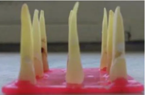
Intact mature teeth or those with minimum restoration or caries were included. Dehydrated brittle teeth due to inappropriate storage conditions, teeth with root fracture or apical resorption, teeth with root canal calcification or previous endodontic treatment were excluded. Thirty-three mandibular central and 33 mandibular lateral incisors were included. The teeth were immersed in 1% sodium hypochlorite solution (Golrang Co., Pakshou, Tehran, Iran) for 48 h and were then stored in 0.9% saline. After removing the calculus, debris and tissue residues from the tooth surfaces, they were placed upside down on a red wax sheet measuring 5 x 5 cm (Figure 1). The CBCT scans were obtained using NewTom VGI scanner (QR SRL, Verona, Italy) with 110 kVp, high resolution, 6 cm × 6 cm field of view and 0.1 mm voxel size. Image reconstruction was performed by multiplanar reformatting, cross and multiplanar features of NewTom NNT Viewer software version 3.0 (QR SRL, Verona,
Italy). The buccolingual and mesiodistal deviation of the apical foramen from the anatomical apex was assessed in the coronal and sagittal planes, respectively. The number and type of canals (according to the Vertucci’s classification) (26), presence of accessory canals, apical delta and anastomoses were determined by assessing serial axial views from the pulp chamber to the apex and also serial sagittal views obtained from the NNT Viewer. The morphology of the root apex was also assessed in the coronal and sagittal planes. The slice interval was 0.5 mm. All CBCT
examinations and interpretations were
performed by two calibrated researchers (a radiologist and an endodontist). In case of any disagreement, a consultation with another radiologist was done.
Figure 1. Samples prepared for CBCT
To carry out the tooth clearing procedures, an endodontic access cavity was prepared in all samples using ½ round bur (Ecodent, Rosdorf, Germany) with high-speed handpiece (Kavo, Werst, Germany), and complete removal of the pulp chamber roof was ensured by an endodontic explorer. The apical foramen was identified using a #8 K-file (Mani, Tochigi, Japan). The presence or absence of deviation, the distance from the anatomical apex, and the direction of deviation in the buccolingual or mesiodistal dimension were determined under a stereomicroscope (SZX-1LLB200; Olympus, Tokyo, Japan) at x16 magnification with 0.01 mm accuracy using its respective software (Olysia Zoom 3.2).
The samples were immersed in 1% sodium hypochlorite solution (Golrang Co., Pakshou, Tehran, Iran) for 24 h to facilitate the
dissolution of the pulp tissue and the remaining organic debris and then they were rinsed with tap water for 4 h and dried. For demineralization, the teeth were immersed in 5% nitric acid (Shiraz Petrochimie, Shiraz, Iran) for 48-72 h. The solution was refreshed daily and to control complete demineralization, the teeth were examined by an explorer and radiographically (Skydent, NY, USA) compared with the control teeth. Upon completion of the decalcifying process, the teeth were rinsed for another 4 h with tap water, and were dehydrated in different concentrations (70%, 80% and 96%) of ethyl alcohol (Razi, Ahvaz, Iran) for 36 h. Next, clearing of the teeth was performed by their immersion in methyl salicylate (Merck, Darmstadt, Germany) for 2 h. A 30-gauge needle (SUPA, Tehran, Iran) was used to inject ink (Pelikan, Tehran, Iran) into the root canal system through the access cavity. The injected dye was suctioned with 25 mmHg negative pressure to fill the canals to the apical foramen.
The number and type of canals were identified according to the Vertucci’s classification (26). The presence/absence of accessory canals, anastomosis, apical delta and distance of apical constriction from the apical foramen and anatomical apex were all determined using a stereomicroscope. This procedure was done by an experienced endodontist.
Statistical analysis:
Data were analyzed using SPSS version 22 (SPSS Inc., IL, and USA). Quantitative and qualitative data were analyzed after reporting the mean and frequency values. The differences in quantitative data were analyzed using one-way ANOVA and the Kruskal Wallis test, and the differences in qualitative data were analyzed using the Chi-square test or Fisher’s exact test. The intra-class correlation coefficient (ICC) was calculated for assessment of quantitative data while the kappa coefficient was used for qualitative data to ensure the validity of CBCT results.
Results
The distribution of root canal types based on the Vertucci’s classification as determined by
CBCT and clearing techniques is presented in Table 1. The most common detected root canal anatomy of central and lateral incisors was the Vertucci’s type I followed by type III in both techniques. In CBCT assessment, one of the lateral incisors could not be included in Vertucci’s classification. It was type XVІІІ according to the classification by Peiris et al (27) (Figure 2). In assessment with the clearing technique, one of the central incisors and one of the lateral incisors could not be included in the Vertucci’s classification. The central incisor was type XVІІ according to the classification by Sert et al, (11) and the lateral incisor was type XVІІІ according to the classification by Peiris et al (27) (Figure 3; Table 1).
Figure 2. Lateral incisor with type XѴІІІ according to the classification by
Peiris et al (27)
Figure 3. Lateral incisor with type XѴІІІ of the Peiris’s classification observed by the clearing
technique (27)
Table 1 also presents the number of canals, presence of lateral canals, and presence of anastomosis and apical delta as determined by the CBCT and the clearing techniques.
One canal was detected in the majority of central incisors (57.6%), while two canals were detected as the dominant finding in lateral incisors (51.1%) by the CBCT and the clearing technique. In CBCT assessment, the frequency of accessory canals, anastomosis and apical delta was 3.0%, 6% and 33.3% in the central and 12%, 18% and 18.8% in the lateral incisors, respectively while in the clearing technique, these values were 12%, 6% and 30.3% for central and 12%.18% and 24.2% for lateral incisors, respectively.
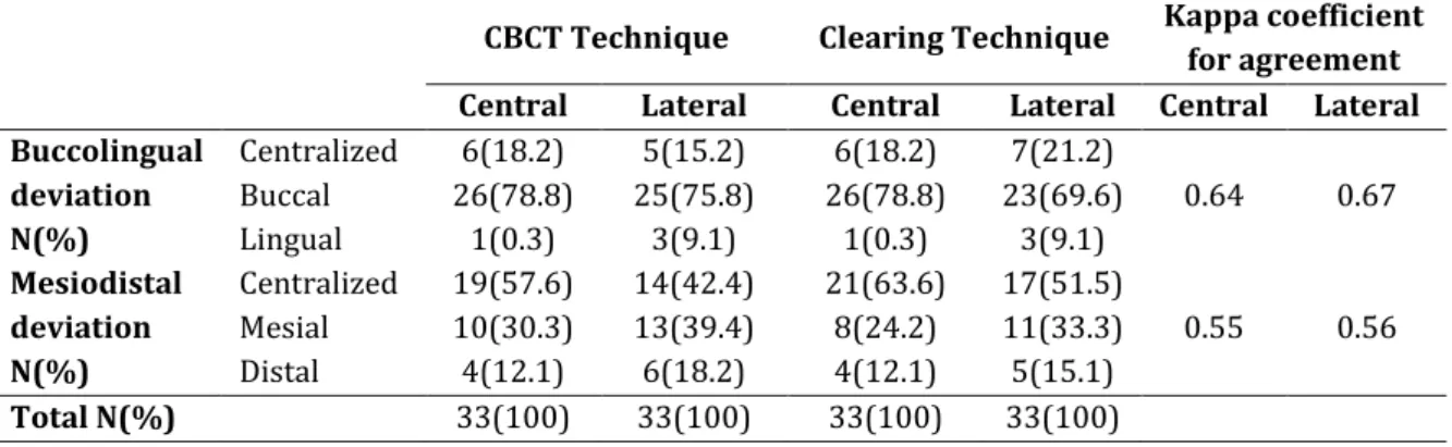
Table 2 shows the deviation of apical foramen from the anatomical apex. In buccolingual dimension, the central and lateral incisors were mostly buccally-deviated; while in the mesiodistal dimension, they were mostly centralized in both techniques.
Morphology of the root apex:
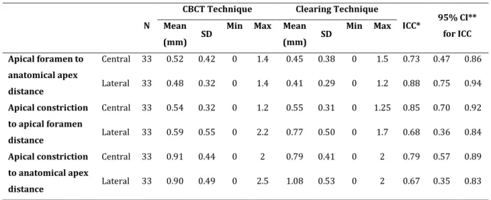
Table 3 shows the mean distance from the apical foramen to the anatomical apex, apical constriction to the apical foramen, and apical constriction to the anatomical apex determined by the CBCT and the clearing technique.
Comparison of CBCT and clearing technique:
The kappa coefficient of agreement between the two methods for canal type was 1 (>0.6) for both central and lateral incisors, which means a total agreement of 100%. The ICC was 1 regarding the number of canals, which expresses the agreement between the two methods and the reliability of CBCT in detecting the canals. The kappa coefficient in detecting the accessory canals in central incisors (0.36) showed the low validity of CBCT; while, it was acceptable in lateral incisors (0.63) (Table 1). The kappa coefficient of 1 and 0.76 in central and lateral incisors for the agreement of the two methods regarding the presence of anastomosis indicated the validity of CBCT; also, both methods were accepted for diagnosis of apical delta considering 0.79 and 0.71 kappa coefficients for central and lateral incisors, respectively (Table 1). CBCT was also acceptable for assessment of the buccolingual
Table 1. Identification of root canal configuration types, number of canals, accessory canals, anastomosis and apical delta by CBCT compared with the clearing technique
CBCT Technique Clearing Technique Agreement* Central Lateral Central Lateral Central Lateral
Canal type N(%)
I 19(57.6) 15(45.5) 19(57.5) 15(45.6)
1.0 1.0 II 0(0) 1(3.0) 0(0) 1(3.0)
III 13(39.4) 13(39.4) 13(39.3) 13(39.4) ІѴ 0(0) 1(3.0) 0(0) 1(3.0)
Ѵ 0(0) 2(6.1) 0(0) 2(6.0) ѴІІ 1(3.0) 0(0) 0(0) 0(0) XІІѴ (Sert) 0(0.0) 0(0) 1(3.0) 0(0) XѴІІІ (Pieris) 0(0) 1(3.0) 0(0) 1(3.0)
New 1(3.0) 0(0) 0(0) 0(0)
Canal number N(%)
1 19(57.6) 15(45.5) 19(57.6) 15(45.5)
1.0 1.0 2 13(39.4) 17(51.5) 13(39.4) 17(51.5)
3 1(1.8) 1(1.7) 1(3) 1(3)
Accessory canals N(%)
Absence 32(97) 29(88) 29(88) 29(88)
0.36 0.63 Presence 1(3.0) 4(12) 4(12) 4(12)
Anastomosis N(%)
Absence 31(93.9) 27(89) 31(94) 27(89)
1.0 0.76 Presence 2(6.1) 6(18) 2(6) 6(18)
Apical delta N(%)
Absence 22(66.7) 26(81.3) 23(69.7) 25(75.8)
0.79 0.71 Presence 11(33.3) 6(18.8) 10(30.3) 8(24.2)
Total N(%) 33(100) 33(100) 33(100) 33(100)
*>0.6 shows acceptable agreement between the two techniques
Table 2. Identification of apical foramen deviation by CBCT compared with the clearing technique CBCT Technique Clearing Technique Kappa coefficient
for agreement Central Lateral Central Lateral Central Lateral Buccolingual
deviation N(%)
Centralized 6(18.2) 5(15.2) 6(18.2) 7(21.2)
0.64 0.67
Buccal 26(78.8) 25(75.8) 26(78.8) 23(69.6)
Lingual 1(0.3) 3(9.1) 1(0.3) 3(9.1)
Mesiodistal deviation N(%)
Centralized 19(57.6) 14(42.4) 21(63.6) 17(51.5)
0.55 0.56
Mesial 10(30.3) 13(39.4) 8(24.2) 11(33.3)
Distal 4(12.1) 6(18.2) 4(12.1) 5(15.1)
Total N(%) 33(100) 33(100) 33(100) 33(100)
Table 3. Identification of root apex morphology, apical foramen to anatomical apex distance, apical constriction to apical foramen distance, and apical constriction to anatomical apex
distance (mm) by CBCT compared with the clearing technique
N
CBCT Technique Clearing Technique
ICC* 95% CI** for ICC Mean
(mm) SD
Min Max Mean (mm) SD
Min Max
Apical foramen to anatomical apex distance
Central 33 0.52 0.42 0 1.4 0.45 0.38 0 1.5 0.73 0.47 0.86 Lateral 33 0.48 0.32 0 1.4 0.41 0.29 0 1.2 0.88 0.75 0.94
Apical constriction to apical foramen distance
Central 33 0.54 0.32 0 1.2 0.55 0.31 0 1.25 0.85 0.70 0.92 Lateral 33 0.59 0.55 0 2.2 0.77 0.50 0 1.7 0.68 0.36 0.84
Apical constriction to anatomical apex distance
Central 33 0.91 0.44 0 2 0.79 0.41 0 2 0.79 0.57 0.89 Lateral 33 0.90 0.49 0 2.5 1.08 0.53 0 2 0.67 0.35 0.83
* Intra-class correlation coefficient (ICC) used for the assessment of quantitative data to ensure validity of CBCT results in comparison with the gold standard technique. The amounts of ICC>75 show excellent reliability and 40<ICC<75 shows good reliability.
**Confidence Interval
deviation of the apical foramen with kappa coefficients of 0.64 and 0.67 for the central and lateral incisors, respectively; while, in assessing the mesiodistal deviation, CBCT was not valid according to the kappa coefficient of 0.55 for central incisors and 0.56 for lateral incisors (Table 2).
The ICC for agreement between the two methods in assessment of the distances from the apical foramen to the anatomical apex, apical constriction to the apical foramen and apical constriction to the anatomical apex was found to be 0.73, 0.85 and 0.79 for central incisors and 0.88, 0.65 and 0.67 for lateral incisors, respectively. The reliability of CBCT was excellent (>75%) for the distance from the apical constriction to the apical foramen and apical constriction to the anatomical apex in central incisors and apical foramen to the anatomical apex in lateral incisors; while, it was good (between 40%-75%) for other parameters (ICC>0.75 shows excellent reliability and 40<ICC<75 expresses good reliability) (Table 3)
Discussion
The knowledge about root canal configurations is a prerequisite for successful endodontic treatment because it helps to achieve an appropriate treatment plan and, consequently, avoids possible technical errors in all phases of treatment (28). CBCT provides high quality, accurate, three-dimensional images and by
eliminating the surrounding tissue
superimpositions, it provides a nondestructive method for investigation of the root canal anatomy at a lower cost and shorter scanning time in comparison with micro-CT (16, 21, 28). Micro-CT and the clearing technique are generally considered as the gold standard of the root canal morphology analysis (16, 21). Due to high cost, time consuming process of micro-CT, and on the other hand, high accuracy of the clearing and staining technique, the latter was used as the classic gold standard in this study to assess the validity of CBCT results (29). However, one study declared that micro-CT was significantly more accurate in detection of root
canal morphology of mandibular mesial roots than both CBCT and the clearing technique (25). In the present study, lateral incisors showed the highest variations in root canal types based on the results of both techniques. The prevalence of the Vertucci’s type I in lateral incisors which is reportedly 36% (29) to 88% (5) in the literature was found to be 45% in both tech-niques in this study. The frequency of the Vertucci’s type III was 39%, which was higher than the reported values in the literature ranging from 0% (5) to 26% (29). Based on the results of both techniques, the most common canal type in central incisors was the Vertucci’s type I (57%), which was consistent with the prevalence rates reported in the literature
[32.5% (29) to 91% (30)]; while, this value was
lower in comparison with the studies conducted in Iran [64% (31) to 88% (32)] (31-33). The prevalence of the Vertucci’s type III, as the second most prevalent type in central incisors, was 39% in both techniques, which was higher than the reported values in studies on the Iranian population [1.5% (34) to 16% (31)] (31, 32, 34). This can be attributed to ethnic and genetic differences, age of patients, patterns of occlusion, chronic stimulations, history of trauma, different methodologies and in vitro or in vivo design of the studies. These factors also clarify the difference between the results of this study and other studies in other fields. According to the total agreement of 100% between CBCT and the clearing technique, CBCT was as accurate as the gold standard in identifying root canal configurations.
Based on the results of both methods used in this study, 57% of central incisors and 45% of lateral incisors had one canal and 39% of central incisors and 51% of lateral incisors had two canals and a few of them had three canals. The consistency of the results of CBCT with the clearing technique (ICC=1) indicates that CBCT was efficient to evaluate this variable.
Using CBCT, the prevalence of accessory canals was 3% and 12% in central and lateral incisors, respectively. This value was found to be 12% in both central and lateral incisors by the clearing technique. The results were in harmony with the results of Boruah and Bhuyan (35) and Sert
and Bayirli (29) and lower in comparison with the results of Vertucci (9), Kartal and Yanikoglu (4) and Sadr Lahijani and Sadegh (32). According to the kappa coefficient, CBCT was only reliable to evaluate the presence of accessory canals in lateral incisors.
The prevalence of anastomosis according to the results obtained from CBCT assessment was 3.4% in central incisors and 15.4% in lateral incisors, which indicated significantly higher prevalence of anastomosis in lateral incisors.
The clearing technique showed that
anastomosis was more prevalent in lateral incisors, even though it was not statistically significant. Data obtained from the present study in this respect were lower than those reported by Sert and Bayirli (29), Boruah and Bhuyan (35), and Al-Qudah and Awawdeh (10). According to the results, CBCT could be used as a substitute for the clearing technique for detection of anastomosis in mandibular incisors.
As proven by both the CBCT and the clearing technique, apical delta was more prevalent in central incisors, even though the difference was not statistically significant. The prevalence of apical delta was 33.3% and 30.3% in central and 18.8% and 24.2% in lateral incisors according to the CBCT and the clearing technique, respectively. The prevalence of apical delta in this study was similar to that
reported by Sert and Bayirli (29) (29% in
central incisors and 19% in lateral incisors) and was higher when compared with the study by
Vertucci (9)(5% in central incisors and 6% in
lateral incisors) or Boruah and Bhuyan (35)
(7.5%). The results indicated that CBCT was efficient for assessment of this variable.
The apical foramen in central and lateral incisors was most commonly buccally-deviated and had no deviation in fewer samples based on both the CBCT and the clearing technique. The prevalence of lingual deviation was the lowest. The highest frequency of buccal deviation was similar to the results of Blaskovic-Subat et al, (36) and Burch and Hulen (37); while Martos et al. (12) reported that the central position was the most common position. The result of CBCT was in harmony with the clearing technique;
thus, CBCT is a reliable technique for detection of apical foramen deviation from the anatomical apex.
The CBCT was validated for assessing the mesiodistal deviation of apical foramen from the anatomical apex in lateral incisors, while it was not reliable in central incisors. The narrow mesiodistal dimension of the root, especially in the apical region, may cause an error in detection of apical foramen orientation. Deviation of apical foramen cannot be detected on two-dimensional radiographs taken during endodontic treatment or in case of presence of deviation in the buccolingual direction. Correct determination of working length cannot be done merely based on measuring the distance from the file tip to the radiographic apex. Electronic apex locators can be helpful in such cases.
The distance from the apical foramen to the anatomical apex was 0.52 mm in central incisors, and 0.45 mm in lateral incisors, based on both techniques used in this study, which was consistent with the results of Martos et al,
(12) and Dummer et al, (8)who reported 0.32
mm and 0.36 mm values for mandibular incisors, respectively. The clearing technique and CBCT were in agreement for the assessment of the distance from the apical foramen to the anatomical apex, and CBCT could be used as a reliable measure for this variable.
The distance from the apical constriction to the apical foramen was 0.53 mm in CBCT assessment and 0.67 mm in the clearing technique (mean = 0.6 mm). Cietterio et al. (38) reported the mean distance of 0.72 mm for all teeth and Kuttler (39) reported 0.5-1.5 mm. The difference of the methods was not statistically significant in this respect; therefore, CBCT could be used as an alternative to the gold standard for this measurement.
The distance between the apical constriction and anatomical apex was 0.8-0.94 mm in CBCT assessment and 0.7-0.94 mm in the clearing technique (mean = 0.9). Dummer et al. (8) reported 0.95 mm for incisors using the sectioning technique, which was similar to this study. The ICC revealed a consistency between
CBCT and the clearing technique; thus, CBCT was accurate for measurement of this distance. The main reasons for the differences in the results of studies are morphological variations in the root canal anatomy due to ethnicity and genetic differences, age of patients, occlusion patterns, chronic stimulations, history of trauma, different methodologies and in vitro or in vivo design of the studies. Similar studies on different age groups are recommended. Also, different races and ethnic groups can be compared in future studies to better elucidate the effect of race and ethnicity in this respect.
Conclusion
In conclusion, CBCT showed high applicability for root canal configuration identification in mandibular incisors as an alternative to the complicated clearing technique for root canal system assessment except for the identification of accessory canals and mesiodistal deviation of the apical foramen in mandibular central incisors.
References
1. Vertucci F. Root canal morphology and its relationship to endodontic procedures. Endod Topics. 2005 March;10(1):3-29.
2. Wu YC, Cheng WC, Chung MP, Su CC, Weng PW, Cathy Tsai YW, et al. Complicated root canal morphology of mandibular lateral incisors is associated with the presence of distolingual root in mandibular first molars: A cone-beam computed tomographic study in a Taiwanese population. J Endod. 2018 Jan;44(1):73-79. 3. Prado MC, Gusman H, Belladonna FG, Prado M, Ormiga F. Effectiveness of three methods for evaluating root canal anatomy of mandibular incisors. J Oral Sci. 2016 Sep;58(3):347-51. 4. Kartal N, Yanikoglu FC. Root canal morphology of mandibular incisors. J Endod. 1992 Nov;18(11):562-4.
5. Madeira MC, Hetem S. Incidence of bifurcations in mandibular incisors. Oral Surg Oral Med Oral Pathol Oral Radiol. 1973 Oct;36 (4):589-91.
6. Gomes BP, Rodrigues HH, Tancredo N. The
use of a modelling technique to investigate the
root canal morphology of mandibular incisors. Int Endod J. 1996 Jan;29(1):29-36.
7. Paes da Silva Ramos Fernandes LM, Rice D,
Ordinola-Zapata R, Alvares Capelozza AL, Bramante CM, Jaramillo D, et al. Detection of various anatomic patterns of root canals in mandibular incisors using digital periapical
radiography, 3 cone-beam computed
tomographic scanners, and micro-computed tomographic imaging. J Endod. 2014 Jan; 40(1): 42-5.
8. Dummer PM, McGinn JH, Rees DG. The
position and topography of the apical canal constriction and apical foramen. Int Endod J. 1984 Oct;17(4):192-8.
9. Vertucci FJ. Root canal anatomy of the
mandibular anterior teeth. J Am Dent Assoc. 1974 Aug;89(2):369-71.
10. Al-Qudah AA, Awawdeh LA. Root canal
morphology of mandibular incisors in a Jordanian population. Int Endod J. 2006 Nov; 39 (11):873-7.
11. Sert S, Aslanalp V, Tanalp J. Investigation of
the root canal configurations of mandibular permanent teeth in the Turkish population. Int Endod J. 2004 Jul;37(7):494-9.
12. Martos J, Lubian C, Silveira LF, Suita de
Castro LA, Ferrer Luque CM. Morphologic analysis of the root apex in human teeth. J Endod. 2010 Apr;36(4):664-7.
13. Naseri M, Safi Y, Akbarzadeh Baghban A,
Khayat A, Eftekhar L. Survey of anatomy and root canal morphology of maxillary first molars regarding age and gender in an Iranian
population using cone-beam computed
tomography. Iran Endod J. 2016 Fall;11(4):298-303.
14.Weine FS, Healey HJ, Gerstein H, Evanson L.
Canal configuration in the mesiobuccal root of the maxillary first molar and its endodontic significance. Oral Surg Oral Med Oral Pathol. 1969 Sep;28(3):419-25.
15.Naseri N, Ahangari Z, Sharifi F, Sahebnasagh
Z. Assessment of root morphology and apices of first and second maxillary molars in Tehran population. J Dent Mater Tech. 2015;4(4):176-82.
16. Neelakantan P, Subbarao C, Subbarao CV.
Comparative evaluation of modified canal
staining and clearing technique, cone-beam computed tomography, peripheral quantitative computed tomography, spiral computed
tomography, and plain and contrast
medium-enhanced digital radiography in studying root canal morphology. J Endod. 2010 Sep;36(9):1547-51.
17. Kulild JC, Peters DD. Incidence and
configuration of canal systems in the mesiobuccal root of maxillary first and second molars. J Endod. 1990 Jul;16(7):311-7.
18. Pecora JD, Woelfel JB, Sousa Neto MD.
Morphologic study of the maxillary molars. 1. External anatomy. Braz Dent J. 1991;2(1):45-50.
19. Buhrley LJ, Barrows MJ, BeGole EA,
Wenckus CS. Effect of magnification on locating the MB2 canal in maxillary molars. J Endod. 2002 Apr;28(4):324-7.
20. Pineda F, Kuttler Y. Mesiodistal and
buccolingual roentgenographic investigation of 7,275 root canals. Oral Surg Oral Med Oral Pathol. 1972 Jan;33(1):101-10.
21. Szabo BT, Pataky L, Mikusi R, Fejerdy P,
Dobo-Nagy C. Comparative evaluation of cone-beam CT equipment with micro-CT in the visualization of root canal system. Ann Ist Super Sanita. 2012 Jan;48(1):49-52.
22. A Soleymani, Namaryan N, E Moudi, A
Gholinia. Root Canal Morphology of Mandibular Canine in an Iranian Population: A CBCT Assessment. Iran Endod J. 2017 Winter; 12(1): 78–82.
23. Michetti J, Maret D, Mallet JP, Diemer F.
Validation of cone beam computed tomography as a tool to explore root canal anatomy. J Endod. 2010 Jul;36(7):1187-90.
24.Zhengyan Y, Keke L, Fei W, Yueheng L, Zhi Z.
Cone-beam computed tomography study of the root and canal morphology of mandibular permanent anterior teeth in a Chongqing population. Ther Clin Risk Manag. 2016 Nov; 12: 19-25.
25. Ordinola-Zapata R, Bramante CM, Versiani
MA, Moldauer BI, Topham G, Gutmann JL, et al.
Comparative accuracy of the Clearing
Technique, CBCT and Micro-CT methods in studying the mesial root canal configuration of mandibular first molars. Int Endod J. 2017 Jan; 50(1):90-6.
26. Vertucci FJ. Root canal anatomy of the human permanent teeth. Oral Surg Oral Med Oral Pathol. 1984 Nov;58(5):589-99.
27.Peiris R, Takahashi M, Sasaki K, Kanazawa E.
Root and canal morphology of permanent mandibular molars in a Sri Lankan population. Odontology. 2007 Jul;95(1):16-23.
28. Sousa TO, Haiter-Neto F, Nascimento EHL,
Peroni LV, Freitas DQ, Hassan B. Diagnostic Accuracy of Periapical Radiography and
Cone-beam Computed Tomography in
Identifying Root Canal Configuration of Human Premolars. J Endod. 2017 Jul;43(7):1176-9.
29. Sert S, Bayirli GS. Evaluation of the root
canal configurations of the mandibular and maxillary permanent teeth by gender in the Turkish population. J Endod. 2004 Jun; 30(6): 391-8.
30. Liu J, Luo J, Dou L, Yang D. CBCT study of
root and canal morphology of permanent mandibular incisors in a Chinese population. Acta Odontol Scand. 2014 Jan;72(1):26-30.
31. Rahimi S, Shahi S, Yavari HR, Reyhani MF,
Ebrahimi ME, Rajabi E. A stereomicroscopy study of root apices of human maxillary central incisors and mandibular second premolars in an Iranian population. J Oral Sci. 2009 Sep;51(3): 411-5.
32. Sadr Lahijani M, Shariati M, Sadeghi M.
Evaluation of root canal anatomy in mandibular anterior-teeth and mandibular and maxillary premolars in vitro. J Rafsanjan Univ Med Sci.
2002 Spring;1(2):92-8.
33. Aminsobhani M, Sadegh M, Meraji N, Razmi
H, Kharazifard MJ. Evaluation of the root and canal morphology of mandibular permanent anterior teeth in an Iranian population by cone-beam computed tomography. J Dent (Tehran). 2013 May;10(4):358-66.
34. Mirzaie M, Tork Zaban P, Mohammadi v.
Cone-beam Computed Tomography Study of Root Canals in a Hamadani Population in Iran. Avicenna J Dent Res. 2012 Feb; 4(2):93-9.
35. Boruah LC, Bhuyan AC. Morphologic
characteristics of root canal of mandibular incisors in North-East Indian population: An in vitro study. J Conserv Dent. 2011 Oct;14(4):346-50.
36. Blaskovic-Subat V, Maricic B, Sutalo J.
Asymmetry of the root canal foramen. Int Endod J. 1992 May;25(3):158-64.
37. Burch JG, Hulen S. The relationship of the
apical foramen to the anatomic apex of the tooth root. Oral Surg Oral Med Oral Pathol. 1972 Aug;34(2):262-8.
38. Citterio F, Pellegatta A, Citterio CL,
Maddalone M. Analysis of the apical constriction
using micro-computed tomography and
anatomical sections. Giornale Italiano di Endodonzia. 2014 Jun;28(1):41-5.
39. Kuttler Y. Microscopic investigation of root
apexes. J Am Dent Assoc. 1955 May;50(5):544-52.