Developing a Refined Palate: Investigating the Role of the Nectin-Afadin Pathway in Mammalian Palatogenesis
Abby J Bergman
ABSTRACT
Cleft palate (CP) forms as a result of the palatal shelves failing to fuse and is one of the most common birth defects worldwide. Human genetic studies have demonstrated that CP is heritable and frequently linked to monoallelic alterations. Previous studies done in isolated human populations have identified mutations in the cell adhesion molecule NECTIN-1 in cases of both syndromic and non-syndromic cleft palate. Furthermore, its family member NECTIN-4 has been found to be mutated in patients with ectodermal dysplasia, which often co-manifests with CP in several human syndromes. Collectively, these studies imply a crucial role for the nectin family of cell adhesion molecules in proper palate formation. Formation of the hard palate is well conserved between mice and humans, making the mouse a powerful tool to study the developmental genetics of secondary palatogenesis. While germline knockout mouse models of Nectin-1, -2, and -3 exist, they do not exhibit the clefting phenotypes observed in humans, presenting a significant challenge to understanding the role of this pathway. Utilizing an RNAi-based in utero lentiviral gene expression editing technique, we have generated mice deficient in nectin-1, which present with partially-penetrant CP. We have also generated a novel model of nectin-4 loss, which similarly presents with CP. Furthermore, combined loss of both nectin-1 and nectin-4 results in higher penetrance of CP, suggesting a clear role for both nectins in palate closure. Finally, depletion of the actin-binding protein afadin (Afdn), which acts
INTRODUCTION
Palatogenesis is a complex morphogenic process occurring in early embryogenesis
whereby the palate is formed to provide the crucial barrier between the oral and nasal cavities.
There are two distinct stages of palatogenesis, primary palatogenesis which is the development
of the soft tissue of the palate, and secondary palatogenesis, which is the formation of the hard
palate at the roof of the mouth. Errors in this process result in orofacial clefting presenting as an
aberrant opening in the roof of the mouth (cleft palate, CP) and/or a split in the lip (cleft lip, CL).
While CL is often caused by defects in primary palatogenesis, CP generally arises from defects
in secondary palatogenesis. Although associated with diseases such as Pierre-Robin and
Stickler syndromes, most cases of CL/P are non-syndromic and can arise due to a number of
genetic and environmental factors 1-3. Consequently, CL/P is one of the most frequently
occurring birth defects. While orofacial clefting and its resulting complications can be remedied
through surgeries and/or speech and related therapies, CL/P represents a significant medical
and financial burden for patients and access to these therapies is unavailable in many parts of
the world. An improved understanding of the molecular mechanisms underlying palatogenesis
has the potential to support the generation of novel andpersonalized therapies, improving both
patient outcomes and long-term quality of life.
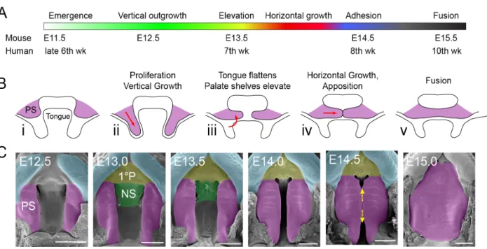
Secondary palatogenesis is a multistep developmental process, requiring intricate
coordination of proliferative, apoptotic, adhesive, and migratory cellular behaviors 4-6. At murine
embryonic day 11.5 (E11.5), the palatal shelves (PS) emerge from the maxillary prominences
and undergo a period of vertical down growth toward the floor of the mouth. This is followed by
rapid elevation of the PS and horizontal growth over the tongue. Palatogenesis concludes ~3-4
days later as the medial edge epithelia (MEE) of each shelf makes contact and adheres,
forming the medial epithelial seam (MES). The MES is then cleared by a variety of mechanisms
conserved in mice and humans, and as such, the mouse serves as useful model system in
which the mechanisms underlying mammalian secondary palate formation can be studied
(Figure 1).
Figure 1. Mammalian palatogenesis occurs during embryonic development and requires complex
morphogenic events. A) Timeline of secondary palatogenesis in mouse and human. The major events
of human palatogenesis are mirrored in mouse. B) Schematic of palatogenesis (coronal view).
C) Transverse view of the stages of murine palatogenesis. E: embryonic day, PS: palatal shelf, 1°P: primary palate, NS: nasal shelf. Figure adapted from (Lough et al., 2017).
Human studies have previously linked several mutations in the cell adhesion gene
NECTIN-1 (formerly PVRL1) and its family member NECTIN-4 (formerly PVRL4) to cases of syndromic and non-syndromic CP 7-9, underscoring a potential role for cell adhesion in proper
palatogenesis. Nectins are part of the Ig superfamily and participate in cell-cell adhesion at the
adherens junction (AJ). Each of the four nectin family members contains three Ig loops, a
transmembrane domain, and a C terminal PDZ domain-binding motif (Figure 2A). Nectins form homodimers in cis and preferentially make heterotypic interactions in trans via their extracellular loops 10, 11(Figure 2B-C). Additionally, each of the nectins binds the adapter protein afadin at its C terminus 12. This interaction with afadin promotes clustering of nectin dimers at the AJ and
components of the AJ 13(Figure 2C). It has been previously demonstrated that Nectins are
expressed in the oral epithelium 14, and nectin-1 and -4 in particular are enriched in the palatal
epithelium during secondary palatogenesis 15.
Figure 2.Nectin family members consist of Ig loops and form cis and trans dimers. A) Each nectin
consists of three Ig-loops (blue) and a C-terminal afadin binding motif (purple). Red text denotes common mutations. B) Two nectin molecules form homodimers in cis through Ig loop interactions. C) Nectin homodimers preferentially form heterotypic interactions in trans. These interactions at the AJ are facilitated by the binding of afadin (purple) at the C-terminus of nectins. The nectin-afadin interaction provides the crucial link between transmembrane nectin adhesions and the actin cytoskeleton (gray).
Several mouse models have been generated to investigate the role of nectins, including
germline knockouts of Nectin-1, -2, and -3. However, these mice show no significant orofacial defects, and do not present with CL/P 16-19. The lack of clefting phenotypes in these animals is
likely due to redundancy amongst the nectin family members, which may allow for
compensation during embryogenesis when one member is lost. Furthermore, germline knockout
of Afadin or combined loss of Nectin-1 and -3 is embryonic lethal 14, 20, preventing exploration of the role of this complex in palatogenesis via conventional mouse genetics. To date, there have
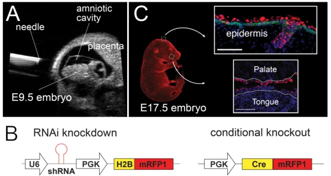
been no published Nectin-4 germline knockout mice, nor any in vivo studies of this protein. In order to bypass the obstacles associated with these models, we utilized the Lentiviral
Ultrasound Guided Gene Inactivation and Gene Editing (LUGGIGE) technique to modulate gene expression in a tissue specific and temporally restricted manner 21(Figure 3). Using this
afadin, nectin-1 and nectin-4, each of which presents with a CP phenotype with varying degrees
of penetrance.
Figure 3.LUGGIGE is an efficient method to transduce embryos during development and
knockdown proteins of interest. A) Ultrasound guides microinjection of lentivirus into the amniotic
cavity of an E9.5 embryo. B) Standard lentiviral constructs contain a hairpin sequence targeting the protein of interest followed by a fluorescent reporter. Lentiviral constructs used to target conditional alleles (such as floxed alleles) include Cre-recombinase followed by a fluorescent reporter. C) Injection with lentivirus causes efficient transduction of exposed epithelium, including epithelium in the oral cavity. Infected cells shown in red. K14 (green) demarcates the basal cell layer (upper panel). Dotted white line denotes basement membrane (lower panel). Nuclei visualized with DAPI (blue).
Taken together, our data suggests that the nectin-afadin pathway is essential for proper
secondary palatogenesis. We hypothesize the nectin-afadin complex plays an important role in
the adhesion of the MEE to form the MES and that temporal specific disruption of this function
causes improper or failed adhesion, resulting in cleft palate.
RESULTS
To address the potential for a temporal-specific role of nectins in palatogenesis, we
Delivery of lentivirus by this method efficiently transduces exposed surface epithelia and
induces knockdown of the target protein during the beginning stages of palatogenesis (E12.5).
This approach is distinct from the mechanism of traditional transgenic approaches, including
germline knockout and conditional alleles, which induce knockdown based on the normal
developmental expression of the targeted promoter. The LUGGIGE technique instead allows us
to control the time at which knockdown is induced, and also achieves epithelial specificity,
bypassing the lethality phenotypes of some traditional models. Embryos were allowed to
proceed through palatogenesis and were harvested at E16.5, after palate closure. All embryos,
including wild-type (WT) uninjected littermates, were imaged using widefield microscopy and
assessed for RFP positivity as well as orofacial clefting. Images of E16.5 palates demonstrate
that knockdown of either Nectin-1 or Nectin-4 in this temporally-controlled manner caused cleft secondary palate (Figure 4A). Data from numerous litters demonstrates that embryos with knockdown of either Nectin-1 or Nectin-4 presented with orofacial clefting with a higher penetrance than their WT littermates (Figure 4B).
Figure 4. Loss of nectin-1 or -4 causes cleft palate with low penetrance.A) E16.5 embryos with loss
of nectin-1 or nectin-4 present with CP. Red fluorescence in upper panels indicates regions of infection by lentivirus. Lower panels depict the same palates in grayscale. Dashed outlines denote hard palate.
Given the low clefting penetrance of single Nectin knockdown, we hypothesized that nectin-1 and nectin-4 may perform reciprocal compensatory function. This possibility was
interrogated by further perturbing the nectin-afadin pathway through dual loss of nectin-1 and -4.
Though afadin has not been directly implicated in CP, we also investigated a potential role for
this protein via LUGGIGE knockdown and conditional alleles. Afadin has been shown to
stabilize nectins at the cell surface and promote clustering at the AJ 13. Thus, we expected loss
of afadin to significantly decrease the adhesive function of nectins in the palatal epithelium.
Stereoscope images demonstrated clefting after dual knockdown of Nectin-1 and -4 or knockdown of Afadin(Figure 5A-B). Interestingly, Nectin-1; Nectin-4 double knockdown embryos occasionally presented with minor submucosal clefting at the posterior of the palate
(Figure 5C). This phenotype was also occasionally observed in Nectin-4 single knockdown (image not shown, quantified in Figure 4B). Partial clefting in these embryos further suggests that the nectin-afadin pathway plays an important role in palate closure while also implying roles
for other adhesion related pathways. In addition to direct shRNA targeting to Afadin via LUGGIGE, we utilized mice with a floxed Afadin allele and two Cre-recombinase delivery methods to further interrogate the role of afadin in palate closure. Upon delivery of Scramble-Cre lentivirus via LUGGIGE, we observed that Afadinfl/fl; lenti-Cre embryos presented with cleft palate at similar rates to those with Afadin-targeted shRNA knockdown. Contrastingly, Afadinfl/fl; K14-Cre mice never exhibit cleft palate (Figure 5D). The K14-Cre allele utilized in these
experiments has been shown to become active at ~E14 22, several days after lentiviral-mediated
cre-recombination occurs. This phenotype, or lack thereof, further suggests a temporal-sensitive
role for afadin during palatogenesis. Quantification of these data demonstrate that dual loss of
nectins-1 and -4 or loss of afadin (via LUGGIGE) increased the penetrance of secondary cleft
Figure 5. Loss of afadin or dual loss of nectin-1 and -4 increases clefting penetrance and leads to
diverse clefting phenotypes. A) E16.5 embryos with dual loss of nectin-1 and nectin-4 present with CP.
Red fluorescence in upper panels indicates regions of infection by lentivirus. Lower panels depict the same palates in grayscale. B) Loss of afadin causes cleft palate. C) Dual loss of nectin-1 and -4 causes diverse clefting phenotypes, including submucosal cleft. Dashed outlines denote hard palate in (A-C)
D) Clefting frequency of nectin and afadin knockdown embryos assessed at E16.5 or later. Phenotypes were scored into three categories, fully fused (white), cleft secondary palate (red), or submucosally cleft (black). Embryos were binned by lentiviral construct or genotype where applicable. n.s.: not significant, *: p < 0.05, **: p < 0.01, ***: p < 0.001, ****: p < 0.0001 as determined by c2 analysis.
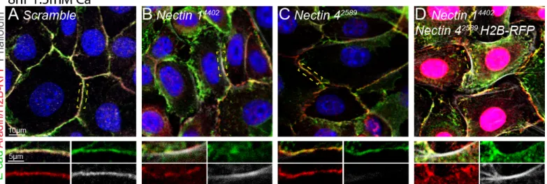
Considering the well-characterized role of the nectin-afadin pathway in cell-cell
adhesion, we hypothesized that defects in adhesion may contribute to the observed CP in these
mutants. To test this, we reduced expression of Nectin-1 and Nectin-4 in primary mouse keratinocytes via lentiviral infection and observed the formation of nascent cell adhesions upon
Ca2+ addition. Knockdown of nectin-1, nectin-4, or dual knockdown of the two led to diminished
diminished accumulation of afadin at the AJ, as well as diffuse localization of afadin and
E-cadherin around the AJ (Figure 6B). Loss of nectin-4 displayed a distinct phenotype from nectin-1 loss; while E-cadherin and afadin localize normally, actin accumulation was strikingly
reduced (Figure 6C). Interestingly, double knockdown of nectin-1 and -4 rescued the accumulation defects of afadin or actin in either single knockout but continued to exhibit
aberrant localization of E-cadherin and afadin (Figure 6D).
Figure 6. Loss of one or more nectins causes aberrant and diminished localization of adherens
junction components. A) Keratinocytes infected with non-targeting Scramble hairpin exhibit localization
of canonical adherens junction proteins E-cadherin (green), afadin (red), and actin (phalloidin, white) at the adherens junction. B) Loss of nectin-1 causes mislocalization of E-cadherin and diminished quantities of afadin. C) Loss of nectin-4 causes a failure to recruit actin to the adherens junction. D) Dual
knockdown of Nectin-1 and -4 rescues the diminished quantity phenotype of either single knockout, but does not rescue localization defects. Yellow dashed boxes in upper panel denote junction displayed in lower panel. Nuclei visualized with DAPI (blue).
DISCUSSION
Causal mutations in the cell adhesion molecule NECTIN-1 were identified in cleft palate patients in 2001, yet numerous studies have failed to generate an animal model that
recapitulates the correlation of human NECTIN mutations to clefting phenotypes 7, 8.Utilizing the
LUGGIGE technique to produce spatio-temporally restricted knockdown of components of the
nectin-afadin pathway, we have demonstrated that acute loss of these proteins can cause
The low observed penetrance, variable severity of clefting, and lack of compounding
ectodermal phenotypes in our model does not recapitulate human Cleft Lip/Palate Ectodermal
Dysplasia and Ectodermal Dysplasia Syndactyly Syndrome (CLPED/EDSS) phenotypes
observed with NECTIN mutations 7-9. The penetrance and severity observations are likely the
result of the variable infection produced using the LUGGIGE technique. Relatively little is known
about nectins’ role in digit formation, and it is possible that ectodermal phenotypes that are
observed in EDSS patients are not recapitulated in our model due to the specificity in location
and timing of knockdown. It is additionally worth noting that our technique induces knockdown of
these proteins where it is possible low concentrations of the targeted protein remain. An
additional confounding effect may be compensation by other nectins which may not accurately
mimic human NECTIN mutations. The most common human NECTIN-1 mutations are truncating mutations after the first Ig loop 7, 8, which may retain the ability to interact with
NECTIN-1, -2, and -3. If so, this may create a dominant negative affect where truncated proteins
are binding functional nectin dimers and preventing proper adhesion by way of inhibiting the
binding of two functional dimers in trans. Nonetheless, the observation that loss of nectin-1, -4, or afadin can cause cleft palate provides promising evidence that these proteins play an
important role in palatogenesis and offers a novel model that can be used to further study their
molecular role in palatogenesis.
During secondary palatogenesis, the palatal epithelium consists of two distinct cell types
organized into layers. The basal cells reside on basement membrane, nearest the
mesenchyme, while the periderm cells lie on top of the basal layer and function to prevent
premature adhesion of the palatal epithelium to other oral epithelial structures. Previous studies
have reported that the basal and periderm layers of the palatal epithelium have distinct nectin
expression profiles 15. This implies the possibility that loss of nectins may lead to improper
specification of palatal epithelium and therefore improper adhesion when the MEE meet to form
hypothesis and others in order to determine the molecular basis by which nectin or afadin loss
leads to cleft palate.
Having confirmed the role of nectins and afadin in palatogenesis via LUGGIGE, it is also
imperative to address the mechanism(s) through which this pathway contributes to proper
palatogenesis. In addition to the evidence provided by dual Nectin knockdown in vivo, single knockdown in vitro additionally suggests that nectins act in a compensatory manner in palate formation via distinct effects on AJ components. Interestingly, dual Nectin knockdown in vitro does not recapitulate the expected compounding phenotypes as observed with dual nectin loss
in vivo. Instead, dual knockdown seems to phenocopy single knockdown of Nectin-1. It is
possible that this is due to inefficient targeting of Nectin-4 via lentiviral infection. To address this, it will be important to analyze the reverse genotype (Nectin-4; Nectin-1 H2B-RFP) to corroborate our current data. Expression of nectin-2 and -3 following compound nectin loss should also be
examined in these cultures. The cell line utilized in these experiments are primary keratinocytes
from embryonic back skin, which is thought to express a larger collection of Nectins than the palatal epithelium. This presents the possibility that the phenotype observed in 1; Nectin-4 dual knockdown cells may not recapitulate dual knockdown in palatal cells. However,
upregulation of nectin-2 and/or -3 in this system would suggest similar compensatory affects to
those observed with compound nectin-1 and -4 loss.
Further experimentation should additionally aim to characterize how afadin loss affects
cellular adhesion, as well as include live imaging experiments to dynamically observe potential
sorting mechanisms based on preferential nectin binding. Previous studies have demonstrated
that heterotypic nectin adhesion supports the formation of a checkerboard pattern both in vivo and de novo in cultured cells, whereby cells expressing nectin-1 arrange themselves to adhere to nectin-3 expressing neighbors 23. If nectins are playing a similar role in the palatal epithelium,
it is plausible that disrupting one or more nectins contributes to cleft palate by way of disrupting
METHODS Animals
Mice were housed in an AAALAC certified animal facility under IACUC approved protocols.
Wild-type CD1 mice (Charles River) were used for most experiments. Alternate afadin knockout
models were generated by mating homozygous Afadinfl/fl mice 24 (gift from Louis Reichart) to
homozygous K14-Cre mice 22 or by injection of homozygous Afadinfl/fl mice with Scramble-Cre virus.Injections were performed as described below.
Lentiviral Ultrasound Guided Gene Inactivation and Gene Editing (LUGGIGE)
Nectin and Afadin knockdown mice were generated by injecting high titer lentiviral constructs containing hairpin targets to Nectin-1, Nectin-4, or Afadin and a fluorescent reporter sequence into the amniotic space of WT CD1 E9.5 embryos. Additional Afadin knockdown mice were generated by injecting a high titer lentiviral construct containing a non-targeting Scramble-Cre
hairpin sequence into the amniotic space of Afadinfl/fl E9.5 embryos 25, 26. Embryonic ages were determined using timed pregnancies, ultrasound and Theiler stages 27. Injected dames were
sacked when embryos reached E14.5-18.5. Embryos were harvested and visualized under a
stereomicroscope to determine success of injection (RFP positive or negative). Infected
embryos and wild-type littermates were fresh frozen or fixed in 4% PFA for 30 minutes and
suspended in 15% sucrose (w/v) overnight. All animals were mounted either whole or with
heads and bodies separately in OCT (Tissue Tek).
Cell Culture
Primary mouse keratinocytes were maintained in E-low media (15% FBS, 50µM CaCl2).
Infected keratinocytes were maintained in E-low media supplemented with puromycin (2µg/mL).
Viral Infection: ~1 day prior to infection, keratinocytes were plated in 6-well dishes at 150,000 cells per well. Cells were then incubated with lentivirus in the presence of polybrene
All subsequent cultures were maintained in E-low media supplemented with puromycin
(2µg/mL).
Adhesion assays: Keratinocytes were plated in 8-well chamber slides coated with collagen (Gibco, A1048301), fibronectin (Gibco, 33016-015), and poly-L-lysine (Sigma, P4832) at 40,000
cells per well. E-low media was replaced with high calcium media (1.5mM Ca2+) 16-24 hours
later. After incubation in high calcium media for the indicated amount of time, cells were fixed
with 4% paraformaldehyde for 10 minutes at room temperature and subsequently washed 3x
with PBS. Immunostaining was performed as described below.
Immunohistochemistry Staining
After fixation, slides were blocked for 1 hour in gelatin block (5% normal donkey serum,3%
BSA, 8% gelatin, 0.05% Triton X-100 in PBS). Primary antibodies were diluted in gelatin block
and incubated overnight at 4°C. After rinsing with PBS, slides were incubated 2 hours in
secondary antibodies and phalloidin (Life Technologies A22287, 1:500) at room temperature,
rinsed with PBS, and incubated with DAPI (1:2000) for 5 min. Slides were mounted using
ProLong Gold Antifade mounting medium (Invitrogen).
Primary antibodies: polyclonal goat anti-E-cadherin (R&D Systems AF748, 1:1000), polyclonal rabbit anti-l/s-Afadin (Millipore Sigma A0224, 1:500), monoclonal rat mCherry (Invitrogen
M11217, 1:1000).
Secondary Antibodies: donkey anti-goat AlexaFluor 488 (Jackson ImmunoResearch 705-545-147, 1:1000), donkey anti-rat Rhodamine Red-X (Jackson ImmunoResearch 712-295-150,
1:500).
Cloning
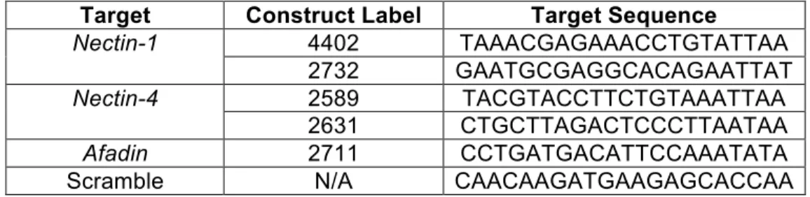
Hairpin sequences against Nectin-1, Nectin-4, Afadin, or non-targeting Scramble were identified from The Broad Institute’s Mission TRC-1 mouse library (Appendix Table 1). Oligos were annealed in New England Biosystems (NEB) buffer 2.1 and ligated into the pLKO.1 vector under
efficiency was assessed in primary keratinocytes by RT-qPCR (data not shown). Competent E. Coli cells were used for transformation. Plasmids were isolated from carbinicillin resistant clones using a QIAGEN QIAprep Minispin Kit and sequenced by GeneWiz to confirm presence of
hairpin sequences. After validating knockdown efficiency, hairpin sequences were inserted into
the pLKO H2B-RFP or pLKO H2B-YFP fluorescent reporter vector (Appendix Figure 1B) via restriction digest. Ligations, transformations, and sequencing were completed as described
above. Maxi preps of constructs were performed using a QIAGEN Endotoxin Free Maxiprep Kit
in preparation for viral production. Viral Production
HEK293TN cells were thawed and cultured in D10 media (DMEM (Lonza BioWhittaker,
12-604Q) + 10% v/v FBS (Atlanta Biosciences)+ 1% v/v Pen-strep/L-glut mix (Lonza BioWhittaker, BW17718R) + 1% v/v 100mM Sodium Pyruvate + 1% v/v 7.5% sodium bicarbonate
supplemented with 500 µg/mL G418) ~1 week prior to transfection. 24 hours prior to infection,
cells were seeded onto 500cm2 plates coated with poly-L-lysine (Sigma P4832). Cells were
transfected with pLKO H2B-RFP, pLKO H2B-YFP (Nectin-1; Nectin-4 double virus only), psPAX2, and pMD2.G (plasmids were a gift from Didier Trono; Addgene plasmid #12260,
#12259) constructs via a calcium phosphate method. 16 hours after transfection, media was
changed to Viral Production Media (UltraCulture (Lonza BioWhittaker 12-725F) + 1% v/v
Pen-strep/L-glut mix + 1% v/v 100mM Sodium Pyruvate + 1% v/v 7.5% sodium bicarbonate + 5 mM
sodium butyrate). Viral supernatant was collected 46 hours post transfection and later
Stereo Microscopy
Images were acquired using a Leica M165C Stereoscope with a PLANAPO 0.63X objective and
LAS4.1 imaging software. RFP fluorescence was visualized under a DS-Red filter.
Confocal Microscopy
Images were acquired using a Leica TCS SPE-II 4 laser confocal system on a DM5500 upright
microscope with an ACS Apochromat 40x/1.15 oil objective.
Analysis and Statistics
Statistical significance values were generated by Chi-Squared analysis using Prism 8.0.2
software. Experimental genotypes were compared to corresponding control groups (WT
REFERENCES
1. Dixon M, Marazita M, Beaty T, Murray JC. Cleft lip and Palate: synthesizing generic and enviromental influences. Nat Rev Genet. 2011;12(3):167-178. doi:10.1038/nrg2933.Cleft 2. Ma L, Shi B, Zheng Q. Targeted mutations of genes reveal important roles in palatal
development in mice. Ann Plast Surg. 2015;74(2):263-268. doi:10.1097/SAP.0b013e318295dcb8
3. Kousa Y, Mansour T, Seada H, Matoo S, Schutte B. Shared molecular networks in orofacial and neural tube development. Birth Defects Res. 2017;109(2):169-179. doi:https://doi.org/10.1002/bdra.23598
4. Bush JO, Jiang R. Palatogenesis: morphogenetic and molecular mechanisms of secondary palate development. Development. 2012;139(2):231-243.
doi:10.1242/dev.079152
5. Greene RM, Pratt RM. Developmental aspects of secondary palate formation. J Embryol Exp Morphol. 1976;36(2):225-245. http://www.ncbi.nlm.nih.gov/pubmed/1033980. 6. Lough KJ, Byrd KM, Spitzer DC, Williams SE. Closing the Gap: Mouse Models to Study
Adhesion in Secondary Palatogenesis. J Dent Res. 2017;96(11):1210-1220. doi:10.1177/0022034517726284
7. Sözen, Mehmet A., Suzuki, Koji, Tolarova, Marie M., Bustos,Tania, Fernández Iglesias, Jesús E., Spritz, Richard A. Mutation of PVRL1 is associated with sporadic,
non-syndromic cleft lip palate in northern Venezuela pp 141 - 142.pdf. 2001;29(october):141-142.
8. Suzuki K, Hu D, Bustos T, et al. Mutations of PVRL1, encoding a cell-cell adhesion molecule/herpesvirus receptor, in cleft lip/palate-ectodermal dysplasia. Nat Genet. 2000;25(4):427-430. doi:10.1038/78119
9. Brancati F, Fortugno P, Bottillo I, et al. Mutations in PVRL4, Encoding cell adhesion molecule nectin-4, cause ectodermal dysplasia-syndactyly syndrome. Am J Hum Genet. 2010;87(2):265-273. doi:10.1016/j.ajhg.2010.07.003
10. Yasumi M, Shimizu K, Honda T, Takeuchi M, Takai Y. Role of each immunoglobulin-like loop of nectin for its cell-cell adhesion activity. Biochem Biophys Res Commun.
2003;302(1):61-66. doi:10.1016/S0006-291X(03)00106-2
11. Momose Y, Honda T, Inagaki M, et al. Role of the second immunoglobulin-like loop of nectin in cell-cell adhesion. Biochem Biophys Res Commun. 2002;293(1):45-49. doi:10.1016/S0006-291X(02)00183-3
12. Takahashi K, Nakanishi H, Miyahara M, et al. Nectin/PRR: An Immunoglobulin-like Cell Adhesion Molecule Recruited to Cadherin-based Adherens Junctions through Interaction with Afadin, a PDZ Domain-containing Protein. J Cell Biol. 1999;145(3):539-549.
http://www.pubmedcentral.nih.gov/articlerender.fcgi?artid=2185068&tool=pmcentrez&ren dertype=abstract.
13. Miyahara M, Nakanishi H, Takahashi K, Satoh-Horikawa K, Tachibana K, Takai Y. Interaction of nectin with afadin is necessary for its clustering at cell-cell contact sites but not for its cis dimerization or trans interaction. J Biol Chem. 2000;275(1):613-618.
doi:10.1074/jbc.275.1.613
14. Yoshida T, Miyoshi J, Takai Y, Thesleff I. Cooperation of Nectin-1 and Nectin-3 is required for normal ameloblast function and crown shape development in mouse teeth. Dev Dyn. 2010;239(10):2558-2569. doi:10.1002/dvdy.22395
16. Barron MJ, Brookes SJ, Draper CE, et al. The cell adhesion molecule nectin-1 is critical for normal enamel formation in mice. Hum Mol Genet. 2008;17(22):3509-3520.
doi:10.1093/hmg/ddn243
17. Inagaki M, Irie K, Ishizaki H, et al. Roles of cell-adhesion molecules nectin 1 and nectin 3 in ciliary body development. Development. 2005;132(7):1525-1537.
doi:10.1242/dev.01697
18. Bouchard MJ, Dong Y, Dermott BMMC, et al. Defects in Nuclear and Cytoskeletal Morphology and Mitochondrial Localization in Spermatozoa of Mice Lacking Nectin-2, a Component of Cell-Cell Adherens Junctions. 2000;20(8):1-9.
papers3://publication/uuid/5B6624A3-038B-4321-A4F5-3F43BC0BF11E.
19. Ozaki-Kuroda K, Nakanishi H, Ohta H, et al. Nectin couples cell-cell adhesion and the actin scaffold at heterotypic testicular junctions. Curr Biol. 2002;12(13):1145-1150. doi:10.1016/S0960-9822(02)00922-3
20. Ikeda W, Nakanishi H, Miyoshi J, et al. Afadin: A key molecule essential for structural organization of cell- cell junctions of polarized epithelia during embryogenesis. J Cell Biol. 1999;146(5):1117-1131. doi:10.1083/jcb.146.5.1117
21. Beronja S, Livshits G, Williams S, Fuchs E. Rapid functional dissection of genetic networks via tissue-specific transduction and RNAi in mouse embryos. Nat Med. 2010;16(7):821-827. doi:10.1038/nm.2167
22. Dassule HR, Lewis P, Bei M, Maas R, McMahon AP. Sonic hedgehog regulates growth and morphogenesis of the tooth. Development. 2000;127(22):4775-4785.
doi:10.1186/1471-2199-12-38
23. Togashi H, Kominami K, Waseda M, et al. Nectins Establish a Checkerboard-Like Cellular Pattern in the Auditory Epithelium. Science (80- ). 2011;333(6046):1144-1148. doi:10.1126/science.1208467
24. Beaudoin GMJ, Schofield CM, Nuwal T, et al. Afadin, A Ras/Rap Effector That Controls Cadherin Function, Promotes Spine and Excitatory Synapse Density in the Hippocampus. J Neurosci. 2012;32(1):99-110. doi:10.1523/JNEUROSCI.4565-11.2012
25. Beronja S, Fuchs E. RNAi-Mediated Gene Function Analysis in Skin. Methods Mol Biol. 2013;961:351-361. doi:10.1007/978-1-62703-227-8
26. Williams SE., Beronja S, Pasolli HA, Fuchs E. Asymmetric cell divisions promote Notch-dependent epidermal differentiation. 2011;470(7334):353-358.
doi:10.1038/nature09793.Asymmetric
27. Theiler K. The House Mouse: Atlas of Embryonic Development. New York: Springer-Verlag; 1989.
ACKNOWLEDGEMENTS
All in utero injections were performed by Kendall Lough. Hairpins for viral cloning were identified by Kendall Lough and preliminary cloning was completed by Danielle Spitzer. Many thanks to all members of the Scott Williams lab for their technical support, advice, and inspiration. Special thanks to Kendall Lough for direct supervision and support of this project, Danielle Spitzer for essential preliminary experiments, and Scott Williams for invaluable expertise and
APPENDIX
Target Construct Label Target Sequence
Nectin-1 4402 TAAACGAGAAACCTGTATTAA
2732 GAATGCGAGGCACAGAATTAT
Nectin-4 2589 TACGTACCTTCTGTAAATTAA
2631 CTGCTTAGACTCCCTTAATAA
Afadin 2711 CCTGATGACATTCCAAATATA
Scramble N/A CAACAAGATGAAGAGCACCAA
Table 1. Target sequences form hairpin loop structures to induce the endogenous RNAi pathway to knockdown target genes.