Adjuncts for the evaluation of
potentially malignant disorders
in the oral cavity
Diagnostic test accuracy systematic review and
meta-analysis
—
a report of the American Dental
Association
Mark W. Lingen, DDS, PhD, FRCPath; Malavika P. Tampi, MPH; Olivia Urquhart, MPH; Elliot Abt, DDS, MS, MSc; Nishant Agrawal, MD; Anil K. Chaturvedi, PhD; Ezra Cohen, MD, FRCPSC; Gypsyamber D’Souza, PhD; JoAnn Gurenlian, RDH, PhD; John R. Kalmar, DMD, PhD; Alexander R. Kerr, DDS, MSD; Paul M. Lambert, DDS; Lauren L. Patton, DDS; Thomas P. Sollecito, DMD, FDS, RCS; Edmond Truelove, DDS, MSD; Laura Banfield, MLIS, MHS; Alonso Carrasco-Labra, DDS, MSc
I
n
2017
, an estimated
49
,
670
new cases
of cancer in the oral cavity and
pharynx will be diagnosed in the
United States, with
9
,
700
disease-associated deaths.
1Estimates for cancer in
the oral cavity alone include
32
,
670
new
cases and
6
,
650
deaths.
1Most of these
cancers will be squamous cell carcinomas.
Survival
is highly
stage
depen-dent, with
83
.
7
% of people surviving
5
years after diagnosis of localized cancer
and
64
.
2
% and
38
.
5
% of people surviving
with regional and distant metastases.
2Approximately
70
% of all new cases
are diagnosed at a late stage,
under-scoring the importance of proper patient
Copyrightª2017 American Dental Association. All rights reserved.
ABSTRACT
Background.
Oral squamous cell carcinoma is the most common
manifestation of malignancy in the oral cavity. Adjuncts are available for
clinicians to evaluate lesions that seem potentially malignant. In this
sys-tematic review, the authors summarized the available evidence on
patient-important outcomes, diagnostic test accuracy (DTA), and patients
’
values
and preferences (PVPs) when using adjuncts for the evaluation of clinically
evident lesions in the oral cavity.
Types of Studies Reviewed.
The authors searched for preexisting
systematic reviews and assessed their quality using the Assessing the
Methodological Quality of Systematic Reviews tool. The authors updated
the selected reviews and searched MEDLINE, Embase, and the Cochrane
Central Register of Controlled Trials to identify randomized controlled
trials and DTA and PVPs studies. Pairs of reviewers independently
con-ducted study selection, data extraction, and assessment of the certainty in
the evidence by using the Grading of Recommendations Assessment,
Development and Evaluation approach.
Results.
The authors identified 4 existing reviews. DTA reviews included
37 studies. The authors retrieved 7,534 records, of which 9 DTA and 10
PVPs studies were eligible. Pooled sensitivity and specificity of adjuncts
ranged from 0.39 to 0.96 for the evaluation of innocuous lesions and from
0.31 to 0.95 for the evaluation of suspicious lesions. Cytologic testing used
in suspicious lesions appears to have the highest accuracy among adjuncts
(sensitivity, 0.92; 95% confidence interval, 0.86 to 0.98; specificity, 0.94; 95%
confidence interval, 0.88 to 0.99; low-quality evidence).
Conclusions and Practical Implications.
Cytologic testing appears
to be the most accurate adjunct among those included in this review. The
main concerns are the high rate of false-positive results and serious issues of
risk of bias and indirectness of the evidence. Clinicians should remain
skeptical about the potential benefit of any adjunct in clinical practice.
Key Words.
Oral squamous cell carcinoma; potentially malignant
dis-orders; diagnostic test accuracy; patients
’
values and preferences.
JADA 2017:148(11):797-813
http://dx.doi.org/
10
.
1016
/j.adaj.
2017
.
08
.
045
evaluation for the prevention or early detection of
dis-ease.
1Clinicians detect and assess oral potentially
ma-lignant disorders (PMDs) and oral squamous cell
carcinomas (OSCCs) by using the combination of an
intra- and extraoral conventional visual and tactile
examination and the detection of dysplasia through
tis-sue biopsy. However, although as many as
10
% of
pa-tients will have some type of oral mucosal abnormality,
only a small fraction of these abnormalities or lesions will
be biologically and clinically signi
fi
cant.
3Conventional visual and tactile examination in the
oral cavity is limited in its ability to help discriminate
between similar-appearing lesions or disorders that may
require considerably different treatments. To address
analogous challenges at other anatomic sites, clinicians
have used adjunctive tests or devices, simply known as
adjuncts
, such as mammography, the Papanicolaou
smear, and colonoscopy, to assist in the detection and
evaluation of disease. A number of adjuncts have become
commercially available to aid in the evaluation and
discrimination of oral mucosal lesions.
4-8These adjuncts
can be divided into
3
broad categories: lesion detection or
discrimination, lesion assessment, and risk assessment.
-
Lesion detection or discrimination. This category is
composed mostly of light-based handheld adjuncts
pro-posed to aid clinicians in the detection and margin
discrimination of lesions by using the principles of
auto
fl
uorescence and tissue re
fl
ectance. Some also would
classify vital staining within this category.
-
Lesion assessment. This category of adjuncts is
intended to assist clinicians in assessing the biological or
clinical relevance of a mucosal abnormality through
cytomorphologic analysis of disaggregated epithelial cells
(cytologic testing). Some also would classify vital staining
within this category.
-
Risk assessment. This category is composed of
saliva-based adjuncts that involve using a number of
bio-markers, including proteins, RNAs, and DNAs.
The purpose of this systematic review was to address
the potential bene
fi
ts and limitations of commercially
available adjuncts to aid in the detection, discrimination,
and assessment of oral mucosal lesions, particularly
PMDs and OSCC in adult patients. This article is an
update and major revision of the
2010
review
6which was
performed by an expert panel of clinical and subject
matter experts convened by the American Dental
Asso-ciation (ADA) Council on Scienti
fi
c Affairs. The ADA
Center for Evidence-Based Dentistry and the Cochrane
Collaboration provided methodological support for the
development and authorship of this review.
Adjuncts can be incorporated in the diagnostic
pathway to triage before an existing test, replace an
existing test, or add on to an existing test to increase
accuracy.
9For this systematic review, we interpreted data
from the included studies in the context of using
adjuncts to triage the need for biopsy and not as
replacement for biopsy.
10Clinicians typically use triage
tools in an early stage of the diagnostic process to
identify patients with a particular
fi
nding that will be
informative for subsequent steps in the testing pathway.
These
fi
ndings informed the development of a
2017
evidence-based clinical practice guideline by the ADA
Center for Evidence-Based Dentistry,
11which contains
recommendation statements to guide the clinical
decision-making process (
eTable
1
).
METHODS
This report follows the guidance of the Preferred
Reporting Items for Systematic Reviews and
Meta-Analyses
12statement and other methodological
rec-ommendations from the Cochrane Screening and
Diagnostic Tests Methods Group.
13Selection criteria for the studies in this review.
Type of studies.
We included cross-sectional and cohort
diagnostic test accuracy (DTA) studies and randomized
controlled trials (RCTs) in which the investigators
assessed the effectiveness or accuracy of adjuncts. We
excluded study designs such as case-control studies, case
reports, case series, abstracts, and uncontrolled reports.
Type of participants and target conditions.
Studies
eligible for inclusion involved adult patients (aged
18
years or older), ideally in the context of primary care
settings, seeking care with or without clinically evident
lesions in the oral cavity, encompassing the labial
mucosae, buccal mucosae, gingival or alveolar ridge
mucosae, tongue,
fl
oor of mouth, hard and soft palate,
and retromolar trigone. If clinically evident, lesions could
manifest as seemingly innocuous or nonsuspicious,
sus-picious, or seemingly malignant. We excluded studies
involving patients seeking care for cancers of the lips,
oropharynx, and salivary glands.
Index tests and the criterion standard.
De
fi
nitive
diagnosis of PMDs and OSCC requires using a criterion
standard wherein the patient undergoes a biopsy of the
lesion followed by a histopathologic assessment. Studies
not specifying any criterion standard were ineligible for
inclusion in this systematic review. Other tests, devices,
techniques, or technologies intended to facilitate clinical
decision making are index tests. The aforementioned
adjuncts act as index tests in the context of this review
and are used as triage tools in practice. Adjuncts can
have either a positive (with suspicion of target condition)
or negative (without suspicion of target condition) test
result.
We de
fi
ned several adjuncts of interest a priori and
assessed them regarding their DTA and effectiveness
when evaluating patients with
-
no clinically evident lesions in the oral cavity;
-
clinically evident seemingly innocuous or
nonsuspi-cious lesions in the oral cavity;
-
clinically evident suspicious lesions or seemingly
malignant lesions in the oral cavity.
Adjuncts include the following:
-
cytologic testing (for example, OralCDx [OralScan
Laboratories, Inc.], OralCyte [ClearCyte Diagnostics
Inc.], ClearPrep OC [Resolution Biomedical]);
-
auto
fl
uorescence (for example, VELscope [LED
Dental], OralID [Forward Science]); tissue re
fl
ectance
(for example, ViziLite Plus [DenMat Holdings, LLC],
Microlux DL [AdDent Inc.]);
-
vital staining (for example, toluidine blue);
-
salivary adjuncts (for example, OraRisk [Oral DNA
Labs], SaliMark [PeriRx LLC], OraMark [OncAlert
Labs], MOP genetic oral cancer screening [PCG
Molec-ular], OraGenomics);
-
additional adjuncts of interest (for example, Identa
fi
[StarDental]).
We also included combinations of aforementioned
adjuncts if
1
adjunct informed the use of the second
adjunct. We reported results separately if the
in-vestigators used
2
index tests in a study independently of
each other. We excluded adjuncts not commercially
available in the United States at the date of the search.
Types of outcomes and estimates.
Patient-important
outcomes are de
fi
ned as
“
outcomes for which
—
even if it
were the only outcome improved by the intervention
—
the patient would still consider receiving the intervention
in face of some adverse events, costs, and burden.
”
14-16In
the context of adjuncts, patients will prioritize outcomes
such as morbidity and mortality and serious adverse
events over other surrogate outcomes such as DTA
estimates. We de
fi
ned the following patient-important
outcomes a priori and included all-cause mortality,
OSCC mortality, survival, quality of life, unnecessary
biopsy, costs, incidence of OSCC, and anxiety and stress.
DTA estimates de
fi
ned a priori included sensitivity,
speci
fi
city, and positive and negative likelihood ratios.
We used the proportion of true-positive, true-negative,
false-positive, and false-negative results to calculate DTA
estimates. We excluded studies when reporting made it
impossible to create a contingency table.
Positivity thresholds.
As stated in the
Cochrane
Handbook for Diagnostic Test Accuracy Reviews
,
“
binary
test outcomes are de
fi
ned on the basis of a threshold for
test positivity and change if the threshold is altered.
”
13Whenever possible, we considered all levels of oral
epithelial dysplasia (mild, moderate, and severe) assessed
during biopsy or histopathologic assessment as positive
for the target condition and absence of dysplasia assessed
during biopsy or histopathologic assessment as negative
for the target condition. For cytologic testing adjuncts,
we grouped any atypical results with dysplastic results
when possible and considered them positive for the
target condition.
Using preexisting evidence.
As a way to optimize the
development of systematic reviews to inform ADA
guidelines, we established a collaboration with the
Cochrane Oral Health Group. The purpose of this
collaboration was to increase ef
fi
ciency in the use of
secondary evidence for the development of clinical
practice guidelines by using preexisting high-quality
systematic reviews. In the event that no Cochrane
reviews were available, we searched for non-Cochrane
systematic reviews.
The eligible reviews had to meet
3
criteria. The
fi
rst
was being assessed as having moderate to high
meth-odological quality. The second was being as current as
possible. The third was meeting the selection criteria in
relation to the type of study design, patient
characteris-tics, index tests, criterion standard, and outcomes.
Identifying relevant systematic reviews.
We
identi-fi
ed eligible systematic reviews through our collaboration
with the Cochrane Oral Health Group. Members of the
group suggested Cochrane reviews that potentially met
our selection criteria. When no Cochrane reviews were
available for a speci
fi
c clinical question, we searched for
non-Cochrane reviews by using the PubMed Clinical
Queries tool and prioritized the most current ones (from
2010
to the present). To determine
fi
nal eligibility, we
used the Assessing the Methodological Quality of
Sys-tematic Reviews tool to assess their methodological
quality.
17Literature search to update existing reviews and
linked evidence on patient-important outcomes.
With
the purpose of updating potentially eligible existing
re-views, we searched MEDLINE via Ovid, Embase via
Ovid, and the Cochrane Central Register of Controlled
Trials. We included all study designs in the initial search.
We also added economic analysis and patients
’
values
and preferences (PVPs). After reviewing the results, we
deemed it necessary to rerun the related Cochrane
searches. We rebuilt the Cochrane searches for Embase,
MEDLINE, and the Cochrane Central Register of
Controlled Trials. We then restricted that language to
RCTs, systematic reviews, and meta-analyses as a means
of ensuring the update of the Cochrane review and to
inform the patient-important outcomes (linked
evi-dence) of interest. Given that literature related to salivary
adjuncts was limited within the bounds of the existing
searches, we removed study design considerations to
open up the possibilities of
fi
nding relevant language. We
restricted the updated Cochrane searches from April
2013
(latest update by Cochrane) to December
2016
. We
ran the search on economic analysis and PVPs from
inception to November
2016
. The amended search for
salivary adjuncts was run from April
2013
(latest update
by Cochrane) to February
2017
(
Appendix
1
, available
online at the end of this article). We did not apply
re-strictions on language or publication status.
Selection of primary studies for update of
system-atic reviews and data extraction.
We conducted the
study selection process in
3
phases. In the
fi
rst phase,
2
reviewers (M.P.T., O.U.) independently reassessed
eligi-bility of all included studies in the
2015
4and
2013
5Cochrane reviews. In the second phase, the same
2
reviewers independently screened titles and abstracts of
retrieved references from the updated search strategy for
both DTA studies and RCTs. In the third phase,
reviewers independently screened the full text of all
potentially eligible studies. We resolved any
disagree-ments at full-text level via discussion and consensus.
When consensus was elusive, a third reviewer (A.C.L.)
arbitrated and decided
fi
nal eligibility. For information
about the data extraction process, see
Appendix
2
(available online at the end of this article).
Summary measures of DTA and patient-important
outcomes at a study level.
DTA studies included in
this review reported results in contingency tables as a
cross-classi
fi
cation of target condition status (condition
present or absent determined by using the criterion
standard) and the adjunct
’
s outcome (condition positive
or negative determined by means of the index test).
13We
presented data as positive, false-positive,
true-negative, and false-negative results. We then calculated
summary measures of DTA such as sensitivity,
speci-fi
city, and positive and negative likelihood ratios along
with their
95
% con
fi
dence intervals (CIs). Sensitivity and
speci
fi
city are measures de
fi
ned as conditional on the
disease status, whereas likelihood ratios can be used to
update the pretest probability of disease to the posttest
probability once the test result is known.
18We planned
on obtaining the prevalence of PMDs and OSCC in the
US adult population and using sensitivity and speci
fi
city
to calculate absolute measures. For patient-important
outcomes reported dichotomously, we planned to
pre-sent their results by using relative risks and their
95
%
CIs. For continuous outcomes, we considered the use of a
mean difference, the standard deviation, and the
95
% CI
as summary measures.
Assessment of the risk of bias of included studies.
Similar to methods used in other Cochrane systematic
reviews on DTA, we used a modi
fi
ed version of the
QUADAS-
2
tool
19to assess the risk of bias and
appli-cability of primary diagnostic accuracy studies included
in our review. Two reviewers (M.P.T., O.U.) used the
tool independently and in duplicate. We assessed the
following domains in each study: patient selection, index
test, criterion standard, and
fl
ow and timing. We
assessed all domains in terms of the risk of bias by using
signaling questions to assist in the judgments. We also
assessed the
fi
rst
3
domains in terms of their
applica-bility. Other important considerations for the quality
assessment included representativeness of the study
sample, extent of veri
fi
cation bias, use of blinded
methods for interpreting test results, and presence of
missing data.
13Data synthesis and meta-analysis.
We recorded the
number of true-positive, false-positive, true-negative, and
false-negative results by using software (Review
Man-ager, Version
5
.
3
, Cochrane Collaboration). We recorded
all new events at the lesion level to mirror the data
presented in the
2015
Cochrane review.
4For each study,
we displayed estimates of DTA, sensitivity, and
speci-fi
city, along with their
95
% CIs, in coupled forest plots, as
well as plotted in summary receiver operating
charac-teristic curve space according to index test. We
per-formed meta-analysis to obtain pooled estimates for
sensitivity, speci
fi
city, and positive and negative
likeli-hood ratios for each adjunct by using the bivariate
approach
13(SAS, Version
9
.
4
, SAS Institute). When too
few studies were available for pooling by using the
bivariate approach, we obtained the pooled estimate by
combining their contingency tables for the associated
comparison. We acknowledge that this method may have
a tendency to create arti
fi
cially narrower CIs. However,
considering that this review is informing a clinical
practice guideline, we prioritized the presentation of
pooled estimates to facilitate decision making.
Assessment of the quality of the evidence.
We
assessed the quality of the evidence for all included
outcomes by using the Grading of Recommendations
Assessment, Development and Evaluation (GRADE)
approach with speci
fi
cation for the diagnostic test
context.
20The GRADE approach provides a framework
to assess the degree of con
fi
dence we can place in DTA
and patient-important outcomes. In GRADE,
cross-sectional or cohort studies in patients with diagnostic
uncertainty and a comparison with an appropriate
cri-terion standard start as high-quality evidence (high
certainty in the evidence). Our certainty is reduced,
however, when these studies have serious issues such as
risk of bias or limitations in study design, indirectness,
inconsistency, imprecision, or high probability of
publi-cation bias (
eTable
2
).
21Such issues move the quality of
the evidence from high to moderate, low, or very low
certainty. We presented data in summary-of-
fi
ndings
tables created using software (GRADEpro Guideline
Development Tool, McMaster University and Evidence
Prime). For a detailed description of the methods used to
assess heterogeneity, publication bias, and the planned
sensitivity analysis, see
Appendix
2
(available online at
the end of this article).
RESULTS
Results of the search.
We identi
fi
ed
2
Cochrane
adjuncts in patients both
with and without
clini-cally evident lesions
developed by the
Cochrane Oral Health
Group. In addition,
we identi
fi
ed
2
non-Cochrane reviews
covering the use of
salivary adjuncts.
22,23From the
2015
Cochrane review, we
identi
fi
ed
37
studies that
were eligible.
4From the
2013
Cochrane review, no
primary studies met our
selection criteria.
5The
other
2
non-Cochrane
systematic reviews were
published in
2016
and
2017
and covered salivary
adjuncts for the early
diagnosis of OSCC, and
no updating process was
required.
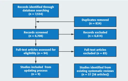
22,23During the updating process of the evidence from
these reviews, we identi
fi
ed
7
,
534
references from the
electronic databases. After eliminating duplicates, we
screened the titles and abstracts of
6
,
708
citations. We
selected
94
potentially eligible articles that we then
screened using full texts. Of the
94
full-text articles, we
selected
9
studies as part of the updating process and
excluded the remaining
85
(
eTable
3
,
4available online at
the end of this article). This resulted in a total of
46
included studies (
47
reports) (
Figure
1
).
4,12No studies on
salivary adjuncts met our selection criteria, so we
per-formed a comprehensive search to identify published
systematic reviews.
During the process of identifying studies on PVPs, we
identi
fi
ed
2
,
616
citations and included
59
of those for
full-text screening. Finally,
10
studies were eligible.
Investigators in none of the studies reported on the
relative importance of outcomes in the context of the use
of adjuncts for the evaluation of PMDs.
Characteristics of included studies.
DTA studies.
In
the
46
included studies, the investigators enrolled a total
of
4
,
543
participants ranging in age from
18
through
80
years, conducted the studies between
1980
and
2016
, and
reported data on the diagnostic accuracy of the following
adjuncts: auto
fl
uorescence,
24-31cytologic testing,
32-47vital
staining,
42,48-61tissue re
fl
ectance,
24,62-66tissue re
fl
ectance
and vital staining,
28,62,65,67,68and cytologic testing and vital
staining.
69,70Investigators had conducted most studies
in secondary
24,26-28,30-34,36,37,41,44-47,49-51,53,55-62,65,67,68,70or
tertiary
25,29,35,39,40,43,48,54,57,63,64care settings and in the
United Kingdom,
24,49,66Italy,
30,39,40,48Germany,
26,27,31,34,35,43Spain,
45,50Taiwan,
52China,
53,54Iran,
32the United
States,
44,46,55,58,62,67Australia,
25,63,64Turkey,
69India,
28,36,37,42,47,51,56,59,61,65,70Poland,
68Japan,
29Brazil,
33,57Canada,
41Sri Lanka,
38,60or Pakistan.
60The target
con-dition for all studies encompassed PMDs or OSCC
(
eTable
4
).
24-70Investigators in many of the included primary
studies did not disclose any con
fl
icts of interest and
sources of funding, though a few provided information
regarding links to industry funding and grants for
research.
33,40,44,46,52,54,60,67,69We identi
fi
ed no studies in
which the investigators assessed patient-important
outcomes such as all-cause mortality, OSCC mortality,
survival time, quality of life, costs, incidence of OSCC,
and anxiety or stress, and none met our selection
criteria.
PVPs studies.
One systematic review
71and
9
primary
studies
72-80including
1
,
950
participants provided
infor-mation about patients
’
perspective, barriers, and
facili-tators during the evaluation of PMDs. For a detailed
description of the included studies and results, see
eTable
5
71-80and
Appendix
2
(available online at the end
of this article).
Determination of prevalence of disease.
We were
unable to identify data on the prevalence of PMDs and
OSCC in the US population in the published literature. We
contacted the Centers for Disease Control and Prevention,
National Institute for Dental and Craniofacial Research,
and National Cancer Institute to determine whether
they had this information. Although these agencies were
unable to give us an accurate estimate, we built our
Records identified through
database searching
(n = 7,534)
Records screened
(n = 6,708)
Records excluded
(n = 6,614)
Duplicates removed
(n = 824)
Full-text articles
excluded (n = 85)
Studies identified from
existing systematic reviews
(n = 37 [38 articles])
Full-text articles assessed for
eligibility (n = 94)
Studies included from
updating process
(n = 9)
Figure 1.Preferred Reporting Items for Systematic Reviews and Meta-Analyses12flow chart of the screening and study selection process.
prevalence estimate by using the
2013
Surveillance,
Epidemiology, and End Results Program data from the
National Cancer Institute and
2010
census data for people
45
years or older to calculate and obtain an estimated
prevalence of OSCC in the United States of
0
.
25
%.
81,82We
recognized that this estimate did not include PMDs, so we
used an estimate of
2
.
0
% to illustrate the potential
preva-lence of PMDs and OSCC in an attempt to account for this
limitation in current available data.
Risk of bias of included reviews.
We identi
fi
ed
4
preexisting systematic reviews meeting the selection
criteria for the clinical questions included in this
review.
4,5,22,23For more information, see
eTables
6
through
9
4,5,16,22,23and
Appendix
2
(available online at
the end of this article).
Risk of bias of primary studies.
Poor reporting did
not allow us to conduct a complete risk of bias
assessment for many of the included studies. Across
the domains of patient selection, index test, and
cri-terion standard, we determined that approximately
50
% of the included studies were unclear. For the
fl
ow
and timing domains, reporting quality was much
higher, and we considered them as the domains of
least concern from a risk of bias perspective. There
were almost no applicability issues among the studies
(
eFigure
1
25-70and
eFigure
2
, available online at the
end of this article).
DTA of adjuncts.
Because no studies in which the
investigators assessed patient-important outcomes met
our selection criteria, we used DTA estimates as
surro-gate outcomes.
Evidence assessing the use of adjuncts to evaluate
patients with no clinically evident lesions.
The authors
of the
2013
Cochrane review
5found no studies informing
the accuracy and effect of adjuncts. In our update of this
preexisting review, we also failed to identify studies
meeting our selection criteria. The panel thought it was
important to include the best available evidence for this
patient scenario and thus decided to amend the selection
criteria for salivary adjuncts to include case-control
studies. Systematic reviews conducted in
2016
and
2017
met this new selection criterion and summarized the
available evidence on the potential use of salivary adjuncts
for the early diagnosis of OSCC and malignant
disor-ders.
22,23Most of the studies we identi
fi
ed were
diagnostic-test case-control studies, followed by a few
cross-sectional and prospective studies. The sampling
methods to collect saliva varied across studies
TABLE 1Auto
fl
uorescence adjuncts to evaluate clinically evident, seemingly innocuous,
or nonsuspicious lesions.
*
TEST RESULT DOWNSTREAM CONSEQUENCES EFFECT PER 100,000 PATIENTS TESTED
(95% CONFIDENCE INTERVAL [CI])
NUMBER OF LESIONS (STUDIES)
QUALITY OF THE EVIDENCE
(GRADE)§
Prevalence 0.25%† Prevalence 2%‡
True Positives (Patients With Need for Biopsy)
Patients will be correctly identified as having a potentially malignant or malignant disorder and a timely referral to a specialist or biopsy will be carried out.
125 (53 to 198) 1,000 (420 to 1,580)
156 (1) Low¶,#,**
False Negatives (Patients Incorrectly Classified as Not Having Need for Biopsy)
Appropriate diagnostic would be missed, worsening the prognosis of the disease.
125 (52 to 197) 1,000 (420 to 1,580)
True Negatives (Patients Without Need for Biopsy)
Patients will receive reassurance that they do not have a potentially malignant or malignant disorder.
38,903 (30,923 to 46,883) 38,220 (30,380 to 46,060)
156 (1) Low¶,#,**
False Positives (Patients Incorrectly Classified as Having Need for Biopsy)
Patients would be incorrectly identified as having a potentially malignant or malignant disorder and would undergo additional unnecessary testing and biopsy.
60,847 (52,867 to 68,827) 59,780 (51,940 to 67,620)
* Setting: primary care. Sensitivity, 0.50 (95% confidence interval [CI], 0.21 to 0.79). Specificity, 0.39 (95% CI, 0.31 to 0.47). Positive likelihood ratio, 0.82 (95% CI, 0.46 to 1.46). Negative likelihood ratio, 1.29; (95% CI, 0.70 to 2.35). Source: Mehrotra and colleagues.28
†We estimated the prevalence by using data from the National Cancer Institute Surveillance, Epidemiology, and End Results Program (300,682 people living with oral cavity and pharynx cancer in the United States in 2013) and the 2010 census data for adults 45 years or older collected by the US Census Bureau.
‡The panel provided illustrative prevalence as an estimation of the number of histopathologic diagnoses from dysplasia to cancer. § GRADE: Grading of Recommendations Assessment, Development and Evaluation.
¶ We judged the patient selection and index test domains as being at high risk of bias.
# The investigators conducted the study in a secondary care setting. Most patients had a higher probability of having a malignant or potentially malignant disorder.
(unstimulated saliva or oral rinse), and most of them were
assessed as being of low or moderate methodological
quality.
23Most studies had small sample sizes with fewer
than
100
participants, although a few studies were
larger.
22,23Most biomarkers showed a wide range of DTA results
(sensitivity ranging from
0
.
5
-
0
.
9
and speci
fi
city ranging
from
0
.
63
-
0
.
90
).
22Some biomarkers were clearly shown
not to be associated with the presence of early PMDs and
did not suggest the ability to inform disease
progres-sion.
22In contrast, other biomarkers were elevated
signi
fi
cantly in those with OSCC compared with those
without OSCC.
23We acknowledge that people with no clinically
evident lesions and those with clinically evident lesions
deemed seemingly innocuous or nonsuspicious
(as opposed to populations with suspicious lesions,
which primarily were included in these reviews) are
the ones who may bene
fi
t the most if these adjuncts
show improved DTA in the future.
Evidence assessing the use of adjuncts to evaluate
patients with clinically evident, seemingly innocuous
(nonsuspicious) lesions or symptoms.
We identi
fi
ed
2
studies
28,36in which the investigators addressed the DTA
of auto
fl
uorescence, cytologic testing, and tissue re
fl
ec-tance and vital staining in patients with seemingly
innocuous or nonsuspicious lesions. Pooled sensitivity
and speci
fi
city of adjuncts ranged from
0
.
39
to
0
.
96
for
the evaluation of innocuous lesions.
eTable
4
24-70sum-marizes the characteristics of the included populations,
and investigators conducted all studies in a secondary or
tertiary care setting.
Auto
fl
uorescence.
One study informed this
compari-son with the investigators evaluating data from
156
lesions.
28The positivity threshold for the criterion
standard was unclear (
eTable
10
,
24-70available online at
the end of this article). When a clinician uses
auto-fl
uorescence,
50
% of lesions with the target condition will
be identi
fi
ed correctly as positive by using the adjuncts
(sensitivity,
0
.
50
;
95
% CI,
0
.
21
to
0
.
79
). However,
39
% of
lesions without the target condition will be identi
fi
ed
correctly as negative by using the adjuncts (speci
fi
city,
0
.
39
;
95
% CI,
0
.
31
to
0
.
47
) (
eFigure
3
,
28available online at
the end of this article). See
Table
1
,
28which includes
additional absolute measures calculated using an
illustrative PMD and OSCC prevalence of
2
.
0
%.
TABLE 2
Cytologic adjuncts to evaluate clinically evident, seemingly innocuous, or
nonsuspicious lesions.
*
TEST RESULT DOWNSTREAM CONSEQUENCES§ EFFECT PER 100,000 PATIENTS TESTED
(95% CONFIDENCE INTERVAL [CI]) NUMBEROF
LESIONS (STUDIES)
QUALITY OF THE EVIDENCE
(GRADE)
Prevalence 0.25%† Prevalence 2%‡
True Positives (Patients With Need for Biopsy)
Patients will be correctly identified as having a potentially malignant or malignant disorder and a timely referral to a specialist or biopsy will be carried out.
240 (203 to 250) 1,920 (1,620 to 2,000)
79 (1) Low¶,#,**
False Negatives (Patients Incorrectly Classified as Not Having Need for Biopsy)
Appropriate diagnostic would be missed, worsening the prognosis of the disease.
10 (0 to 47) 80 (0 to 380)
True Negatives (Patients Without Need for Biopsy)
Patients will receive reassurance that they do not have a potentially malignant or malignant disorder.
89,775 (78,803 to 96,758) 88,200 (77,420 to 95,060)
79 (1) Low¶,#,**
False Positives (Patients Incorrectly Classified as Having Need for Biopsy)
Patients would be incorrectly identified as having a potentially malignant or malignant disorder and would undergo additional unnecessary testing and biopsy.
9,975 (2,992 to 20,947) 9,800 (2,940 to 20,580)
* Setting: primary care. Sensitivity, 0.96 (95% confidence interval [CI], 0.81 to 1.00). Specificity, 0.90 (95% CI, 0.79 to 0.97). Positive likelihood ratio, 10.01 (95% CI, 4.34 to 23.12). Negative likelihood ratio, 0.04 (95% CI, 0.01 to 0.28). Source: Mehrotra and colleagues.36
†We estimated the prevalence by using data from the National Cancer Institute Surveillance, Epidemiology, and End Results Program (300,682 people living with oral cavity and pharynx cancer in the United States in 2013) and the 2010 census data for adults 45 years or older collected by the US Census Bureau.
‡The panel provided illustrative prevalence as an estimation of the number of histopathologic diagnoses from dysplasia to cancer. § GRADE: Grading of Recommendations Assessment, Development and Evaluation.
¶ The sampling method, the positivity threshold for dysplasia in regard to the reference standard, and to what extent examiners were calibrated during interpretation of the index test are unclear.
# The investigators conducted the study in a secondary care setting. Most patients had a higher probability of having a malignant or potentially malignant disorder.
** The positivity threshold for the index test included atypical results.
Cytologic testing.
One study informed this
compari-son with the investigators evaluating data from
79
lesions.
36The positivity threshold for the criterion
stan-dard was unclear (
eTable
10
,
24-70available online at the
end of this article). When clinicians use cytologic testing,
96
% of lesions with the target condition will be identi
fi
ed
correctly as positive by using the adjunct (sensitivity,
0
.
96
;
95
% CI,
0
.
81
to
1
.
00
). However,
90
% of lesions
without the target condition will be identi
fi
ed correctly as
negative by using the adjunct (speci
fi
city,
0
.
90
;
95
% CI,
0
.
79
to
0
.
97
) (
eFigure
4
,
36available online at the end of
this article). See
Table
2
,
36which includes additional
absolute measures calculated using an illustrative PMD
and OSCC prevalence of
2
.
0
%.
Tissue re
fl
ectance and vital staining.
One study
informed this comparison with the investigators
evalu-ating data from
102
lesions.
28The positivity threshold for
the criterion standard was unclear (
eTable
10
,
24-70avail-able online at the end of this article). When a clinician
uses tissue re
fl
ectance and vital staining,
0
% of lesions
with the target condition will be identi
fi
ed correctly as
positive by using the adjunct (sensitivity,
0
.
00
;
95
% CI,
0
.
00
to
0
.
60
). However,
76
% of lesions without the
disorder will be identi
fi
ed correctly as negative by using
the adjunct (speci
fi
city,
0
.
76
;
95
% CI,
0
.
66
to
0
.
84
)
(
eFigure
5
,
28available online at the end of this article).
See
Table
3
,
28which includes additional absolute
mea-sures calculated using an illustrative PMD and OSCC
prevalence of
2
.
0
%.
We did not recover any studies on the DTA of vital
staining, auto
fl
uorescence and tissue re
fl
ectance,
cyto-logic testing and vital staining, and tissue re
fl
ectance
adjuncts. Therefore, we could not include any for the
evaluation of seemingly innocuous lesions in the oral
cavity.
Evidence on the use of adjuncts in patients
with clinically evident lesions suspected to be
potentially malignant or malignant.
We identi
fi
ed
44
studies
27,28,30,32-38,40-68,70-74in which the investigators
addressed the DTA of auto
fl
uorescence, cytologic testing,
vital staining, tissue re
fl
ectance, cytologic testing and vital
staining, and tissue re
fl
ectance and vital staining.
eTable
3
24-70summarizes the characteristics of the
included populations. Investigators conducted all studies
in a secondary or tertiary setting with the exception
of Rahman and colleagues
42. Pooled sensitivity and
spec-i
fi
city of adjuncts ranged from
0
.
31
to
0
.
95
for the
evalua-tion of these type of lesions.
TABLE 3
Tissue re
fl
ectance and vital staining adjuncts to evaluate clinically evident,
seemingly innocuous, or nonsuspicious lesions.
*
TEST RESULT DOWNSTREAM CONSEQUENCES EFFECT PER 100,000 PATIENTS TESTED (95%
CONFIDENCE INTERVAL [CI])
NUMBER OF LESIONS (STUDIES)
QUALITY OF THE EVIDENCE
(GRADE)§
Prevalence 0.25%† Prevalence 2%‡
True Positives (Patients With Need for Biopsy)
Patients will be correctly identified as having a potentially malignant or malignant disorder and a timely referral to a specialist or biopsy will be carried out.
0 (0 to 150) 0 (0 to 1,200)
102 (1) Low¶,#,**
False Negatives (Patients Incorrectly Classified as Not Having Need for Biopsy)
Appropriate diagnostic would be missed, worsening the prognosis of the disease.
250 (100 to 250) 2,000 (800 to 2,000)
True Negatives (Patients Without Need for Biopsy)
Patients will receive reassurance that they do not have a potentially malignant or malignant disorder.
75,810 (65,835 to 83,790) 74,480 (64,680 to 82,320)
102 (1) Low¶,#,**
False Positives (Patients Incorrectly Classified as Having Need for Biopsy)
Patients would be incorrectly identified as having a potentially malignant or malignant disorder and would undergo additional unnecessary testing and biopsy.
23,940 (15,960 to 33,915) 23,520 (15,680 to 33,320)
* Setting: primary care. Sensitivity, 0.00 (95% confidence interval [CI], 0.00 to 0.60). Specificity, 0.76 (95% CI, 0.66 to 0.84). Positive likelihood ratio, not available. Negative likelihood ratio, 1.32 (95% CI, 1.18 to 1.48). Source: Mehrotra and colleagues.28
†We estimated the prevalence by using data from the National Cancer Institute Surveillance, Epidemiology, and End Results Program (300,682 people living with oral cavity and pharynx cancer in the United States in 2013) and the 2010 census data for adults 45 years or older collected by the US Census Bureau.
‡The panel provided illustrative prevalence as an estimation of the number of histopathologic diagnoses from dysplasia to cancer. § GRADE: Grading of Recommendations Assessment, Development and Evaluation.
¶ We judged the patient selection and index test domains as being at high risk of bias.
# The investigators conducted the study in a secondary care setting. Most patients had a higher probability of having a malignant or potentially malignant disorder.
Auto
fl
uorescence.
Seven studies informed this
com-parison with the investigators evaluating data from
616
lesions.
24-27,29-31The positivity threshold for the criterion
standard included from mild dysplasia to OSCC, except
for the study by Farah and colleagues,
25in which we were
unable to elucidate how the authors classi
fi
ed a positive
test result.
When a clinician uses auto
fl
uorescence,
90
% of
lesions with the target condition will be identi
fi
ed
correctly as positive by using the adjunct (sensitivity,
0
.
90
;
95
% CI,
0
.
76
to
1
.
00
). However,
72
% of lesions
without the target condition will be identi
fi
ed
correctly as negative by using the adjunct (speci
fi
city,
0
.
72
;
95
% CI,
0
.
35
to
1
.
00
) (
eFigures
6
24-27,29-31and
7
,
available online at the end of this article). See
Table
4
,
24-27,29-31which includes additional absolute
measures calculated using an illustrative PMD and
OSCC prevalence of
2
.
0
%.
Cytologic testing.
Fifteen studies informed this
com-parison with the investigators evaluating data from
2
,
148
lesions.
32-35,37-47The positivity threshold for the criterion
standard included from mild dysplasia to OSCC in most
of the studies (
eTable
10
,
24-70available online at the end
of this article). It was unclear how dysplasia was classi
fi
ed
in the study by Navone and colleagues,
39and Rahman
and colleagues
42classi
fi
ed mild dysplasia as negative for
the target condition.
When a clinician uses cytologic testing,
92
% of lesions
with the target condition will be identi
fi
ed correctly as
positive by using the adjunct (sensitivity,
0
.
92
;
95
% CI,
0
.
86
to
0
.
98
). However,
94
% of lesions without the target
condition will be identi
fi
ed correctly as negative by using
the adjunct (speci
fi
city,
0
.
94
;
95
% CI,
0
.
88
to
0
.
99
)
(
eFigures
8
32-35,37-47and
9
, available online at the end of
this article). See
Table
5
,
32-35,37-47which includes
addi-tional absolute measures calculated using an illustrative
PMD and OSCC prevalence of
2
.
0
%.
Vital staining.
Fifteen studies informed this
comparison with the investigators evaluating data from
1
,
453
lesions.
42,48-61The positivity threshold for the
criterion standard included from mild dysplasia to OSCC
in all studies except for those of Rahman and
col-leagues,
42Singh and Shukla,
61and Cheng and Yang,
53(
eTable
10
,
24-70available online at the end of this article).
Rahman and colleagues
42classi
fi
ed mild dysplasia as
negative, and Singh and Shukla
61considered all dysplasia
TABLE 4
Auto
fl
uorscence adjuncts to evaluate clinically evident suspicious lesions.
*
TEST RESULT DOWNSTREAM CONSEQUENCES EFFECT PER 100,000 PATIENTS TESTED (95%
CONFIDENCE INTERVAL [CI]) NUMBEROF
LESIONS (STUDIES)
QUALITY OF THE EVIDENCE
(GRADE)§
Prevalence 0.25%† Prevalence 2%‡
True Positives (Patients With Need for Biopsy)
Patients will be correctly identified as having a potentially malignant or malignant disorder and a timely referral to a specialist or biopsy will be carried out.
225 (190 to 250) 1,800 (1,520 to 2,000)
616 (7) Low¶,#,**
False Negatives (Patients Incorrectly Classified as Not Having Need for Biopsy)
Appropriate diagnostic would be missed, worsening the prognosis of the disease.
25 (0 to 610) 200 (0 to 480)
True Negatives (Patients Without Need for Biopsy)
Patients will receive reassurance that they do not have a potentially malignant or malignant disorder.
71,820 (34,913 to 99,750) 70,560 (34,300 to 98,000)
616 (7) Low¶,#,**
False Positives (Patients Incorrectly Classified as Having Need for Biopsy)
Patients would be incorrectly identified as having a potentially malignant or malignant disorder and would undergo additional unnecessary testing and biopsy.
27,930 (0 to 64,837) 27,440 (0 to 63,700)
* Setting: Primary care. Pooled sensitivity, 0.90 (95% confidence interval [CI], 0.76 to 1.00). Pooled specificity, 0.72 (95% CI, 0.35 to 1.00). Positive likelihood ratio, 3.17 (95% CI, 0.85 to 11.80). Negative likelihood ratio, 0.14; (95% CI, 0.03 to 0.64). Sources: Awan and colleagues,24Farah and colleagues,25Hanken and colleagues,26Koch and colleagues,27Onizawa and colleagues,29Petruzzi and colleagues,30and Scheer and colleagues.31
†We estimated the prevalence by using data from the National Cancer Institute Surveillance, Epidemiology, and End Results Program (300,682 people living with oral cavity and pharynx cancer in the United States in 2013) and the 2010 census data for adults 45 years or older collected by the US Census Bureau.
‡The panel provided illustrative prevalence as an estimation of the number of histopathologic diagnoses from dysplasia to cancer. § GRADE: Grading of Recommendations Assessment, Development and Evaluation.
¶ Patient selection and exclusion from analysis were inappropriate. Poor-quality reporting did not provide sufficient information to judge key risk of bias domains.
# The investigators conducted most studies in secondary and tertiary care settings. Most patients had a higher probability of having a malignant or potentially malignant disorder.
** The positivity threshold for the reference test included from mild dysplasia to cancer in all studies except for that of Awan and colleagues24and Farah and colleagues.25
negative. It was unclear how Cheng and Yang
53classi
fi
ed
the varying grades of dysplasia.
When a clinician uses vital staining,
87
% of lesions
with the target condition will be identi
fi
ed correctly as
positive by using the adjunct (sensitivity,
0
.
87
;
95
% CI,
0
.
80
to
0
.
94
). However,
71
% of lesions without the target
condition will be identi
fi
ed correctly as negative by using
the adjunct (speci
fi
city,
0
.
71
;
95
% CI,
0
.
61
to
0
.
82
)
(
eFigures
10
42,48-61and
11
, available online at the end of
this article). See
Table
6
,
42,48-61which includes additional
absolute measures calculated using an illustrative PMD
and OSCC prevalence of
2
.
0
%.
Tissue re
fl
ectance.
Five studies informed this
com-parison with the investigators evaluating data from
390
lesions.
62-66The positivity threshold for the criterion
standard included from mild dysplasia to OSCC in all
studies with the exception of those of Chainani-Wu and
colleagues,
62Ujaoney and colleagues,
65and Farah and
McCullough
63(
eTable
10
,
24-70available online at the end
of this article). Ujaoney and colleagues
65classi
fi
ed mild
dysplasia as negative, and Chainani-Wu and colleagues
62classi
fi
ed mild and moderate dysplasia as negative. It
was unclear how Farah and McCullough
63classi
fi
ed
dysplasia.
When a clinician uses tissue re
fl
ectance,
72
% of
lesions with the target condition will be identi
fi
ed
correctly as positive by using the adjunct (sensitivity,
0
.
72
;
95
% CI,
0
.
62
to
0
.
81
). However,
31
% of lesions
without the target condition will be identi
fi
ed
correctly as negative by using the adjunct (speci
fi
city,
0
.
31
;
95
% CI,
0
.
25
to
0
.
36
) (
eFigures
12
62-66and
13
,
available online at the end of this article). See
Table
7
,
62-66which includes additional absolute
mea-sures calculated using an illustrative PMD and OSCC
prevalence of
2
.
0
%.
Cytologic testing and vital staining.
Two studies
informed this comparison with the investigators
eval-uating data from
139
lesions.
69,70The positivity
threshold for the criterion standard included from
mild dysplasia to OSCC in Gupta and colleagues,
70but
TABLE 5
Cytologic adjuncts to evaluate clinically evident suspicious lesions.
*
TEST RESULT DOWNSTREAM CONSEQUENCES EFFECT PER 100,000 PATIENTS TESTED (95%
CONFIDENCE INTERVAL [CI])
NUMBER OF LESIONS (STUDIES)
QUALITY OF THE EVIDENCE
(GRADE)§
Prevalence 0.25%† Prevalence 2%‡
True Positives (Patients With Need for Biopsy)
Patients will be correctly identified as having a potentially malignant or malignant, and timely referral to a specialist or biopsy will be performed.
230 (215 to 245) 1,840 (1,720 to 1,960)
2,148 (15) Low¶,#,**
False Negatives (Patients Incorrectly Classified as Not Having Need for Biopsy)
Appropriate diagnostic would be missed, worsening the prognosis of the disease.
20 (5 to 35) 160 (40 to 280)
True Negatives (Patients Without Need for Biopsy)
Patients will receive reassurance that they do not have a potentially malignant or malignant disorder.
93,765 (87,780 to 98,753) 92,120 (86,240 to 97,020)
2,148 (15) Low¶,#,**
False Positives (Patients Incorrectly Classified as Having Need for Biopsy)
Patients would be incorrectly identified as having a potentially malignant or malignant disorder and would undergo additional unnecessary testing and biopsy.
5,985 (997 to 11,970) 5,880 (980 to 11,760)
* Setting: primary care. Pooled sensitivity, 0.92 (95% confidence interval [CI], 0.86 to 0.98). Pooled specificity, 0.94 (95% CI, 0.88 to 0.99). Positive likelihood ratio, 14.18 (95% CI, 5.82 to 34.59). Negative likelihood ratio, 0.08 (95% CI, 0.04 to 0.18). Sources: Delavarian and colleagues,32Fontes and colleagues,33Kammerer and colleagues,34Koch and colleagues,35Mehrotra and colleagues,37Nanayakkara and colleagues,38Navone and col-leagues,40Navone and colleagues,39Ng and colleagues,41Rahman and colleagues,42Scheifele and colleagues,43Sciubba,44Seijas-Naya and col-leagues,45Svirsky and colleagues,46and Trakroo and colleagues.47
†We estimated the prevalence by using data from the National Cancer Institute Surveillance, Epidemiology, and End Results Program (300,682 people living with oral cavity and pharynx cancer in the United States in 2013) and the 2010 census data for adults 45 years or older collected by the US Census Bureau.
‡The panel provided illustrative prevalence as an estimation of the number of histopathologic diagnoses from dysplasia to cancer. § GRADE: Grading of Recommendations Assessment, Development and Evaluation.
¶ Patient selection and exclusion from analysis were inappropriate, index and reference tests were conducted in an unblinded fashion, and in some cases the time between index and reference test was greater than 2 weeks. It was unclear whether all participants received the reference test. Poorquality reporting did not provide sufficient information to judge key risk of bias domains.
# Investigators conducted most studies in secondary and tertiary care settings. Most patients had a higher probability of having a malignant or potentially malignant disorder.
** The positivity threshold for the reference test included from mild dysplasia to cancer in all studies except for those of Kammerer and colleagues,34
Navone and colleagues,39and Rahman and colleagues.42The positivity threshold included atypia for Rahman and colleagues,42Scheifele and
colleagues43(10/96), Sciubba (52/298), and Svirsky and colleagues.46Parentheses indicate the number of atypical results out of the total (atypical
Guneri and colleagues
69classi
fi
ed only severe dysplasia
as positive (
eTable
10
,
24-70available online at the end
of this article).
When a clinician uses cytologic testing and vital
staining,
95
% of lesions with the target condition will be
identi
fi
ed correctly as positive by using the adjunct
(sensitivity,
0
.
95
;
95
% CI,
0
.
86
to
0
.
99
). However,
68
% of
lesions without the target condition will be identi
fi
ed
correctly as negative by using the adjunct (speci
fi
city,
0
.
68
;
95
% CI,
0
.
56
to
0
.
78
) (
eFigures
14
69,70and
15
,
available online at the end of this article). See
Table
8
,
69,70which includes additional absolute measures calculated
using an illustrative PMD and OSCC prevalence of
2
.
0
%.
Tissue re
fl
ectance and vital staining.
Four studies
informed this comparison with the investigators
eval-uating data from
307
lesions.
62,65,67,68The positivity
threshold for the criterion standard included from mild
dysplasia to OSCC in all studies with the exception of
those of Ujaoney and colleagues
65and Chainani-Wu
and colleagues.
62Ujaoney and colleagues
65classi
fi
ed
mild dysplasia as negative, and Chainani-Wu and
col-leagues
62classi
fi
ed mild and moderate dysplasia as
negative (
eTable
10
,
24-70available online at the end of
this article).
When a clinician uses tissue re
fl
ectance and vital
staining,
81
% of lesions with the target condition will be
identi
fi
ed correctly as positive by using the adjunct
(sensitivity,
0
.
81
;
95
% CI,
0
.
71
to
0
.
89
). However,
69
% of
lesions without the target condition will be identi
fi
ed
correctly as negative by using the adjunct (speci
fi
city,
0
.
69
;
95
% CI,
0
.
63
to
0
.
75
) (
eFigures
16
62,65,67,68and
17
, available online at the end of this article). See
Table
9
,
62-68which includes additional absolute
mea-sures calculated using an illustrative PMD and OSCC
prevalence of
2
.
0
%.
Sensitivity analyses.
eTables
11
through
14
32-35,37-61,69and
Appendix
2
(available online at the end of this
article) provide information about the sensitivity
analyses.
DISCUSSION
Summary of main results.
We planned this review
and analysis assuming that all commercially available
adjuncts may have the potential to assist primary care
TABLE 6Vital staining adjuncts to evaluate clinically evident suspicious lesions.
*
TEST RESULT DOWNSTREAM CONSEQUENCES EFFECT PER 100,000 PATIENTS TESTED
(95% CONFIDENCE INTERVAL [CI]) NUMBEROF
LESIONS (STUDIES)
QUALITY OF THE EVIDENCE
(GRADE)§
Prevalence 0.25%† Prevalence 2%‡
True Positives (Patients With Need for Biopsy)
Patients will be correctly identified as having a potentially malignant or malignant disorder and a timely referral to a specialist or biopsy will be carried out.
217 (200 to 235) 1,740 (1,600 to 1,880)
1,453 (15) Low¶,#,**
False Negatives (Patients Incorrectly Classified as Not Having Need for Biopsy)
Appropriate diagnostic would be missed, worsening the prognosis of the disease.
33 (15 to 50) 260 (120 to 400)
True Negatives (Patients Without Need for Biopsy)
Patients will receive reassurance that they do not have a potentially malignant or malignant disorder.
70,823 (60,848 to 81,795) 69,580 (59,780 to 80,360)
1,453 (15) Low¶,#,**
False Positives (Patients Incorrectly Classified as Having Need for Biopsy)
Patients would be incorrectly identified as having a potentially malignant or malignant disorder and would undergo additional unnecessary testing and biopsy.
28,927 (17,955 to 38,902) 28,420 (17,640 to 38,220)
* Setting: primary care. Pooled sensitivity, 0.87 (95% confidence interval [CI], 0.80 to 0.94). Pooled specificity, 0.71 (95% CI, 0.61 to 0.82). Positive likelihood ratio, 3.04 (95% CI, 2.06 to 4.48). Negative likelihood ratio, 0.18 (95% CI, 0.10 to 0.32). Sources: Allegra and colleagues,48Awan and colleagues,49Cancela-Rodriguez and colleagues,50Chaudhari and colleagues,51Chen and colleagues,52Cheng and Yang,53Du and colleagues,54 Mashberg,55Nagaraju and colleagues,56Onofre and colleagues,57Rahman and colleagues,42Silverman and colleagues,58Singh and Shukla,61
Upadhyay and colleagues,59and Warnakulasuriya and Johnson.60
†We estimated the prevalence by using data from the National Cancer Institute Surveillance, Epidemiology, and End Results Program (300,682 people living with oral cavity and pharynx cancer in the United States in 2013) and the 2010 census data for adults 45 years or older collected by the US Census Bureau.
‡The panel provided illustrative prevalence as an estimation of the number of histopathologic diagnoses from dysplasia to cancer. § GRADE: Grading of Recommendations Assessment, Development and Evaluation.
¶ Patient selection and exclusion from analysis were inappropriate. It was unclear whether all participants received the reference test. Poor-quality reporting did not provide sufficient information to judge key risk of bias domains.
# Investigators conducted most studies in secondary and tertiary care settings. Most patients had a higher probability of having a malignant or potentially malignant disorder.
** The positivity threshold for the reference test included from mild dysplasia to cancer in all studies except for those of Cheng and Yang,53Rahman and colleagues,42and Singh and Shukla.61