Stolonization in Myrianida
6
0
0
Full text
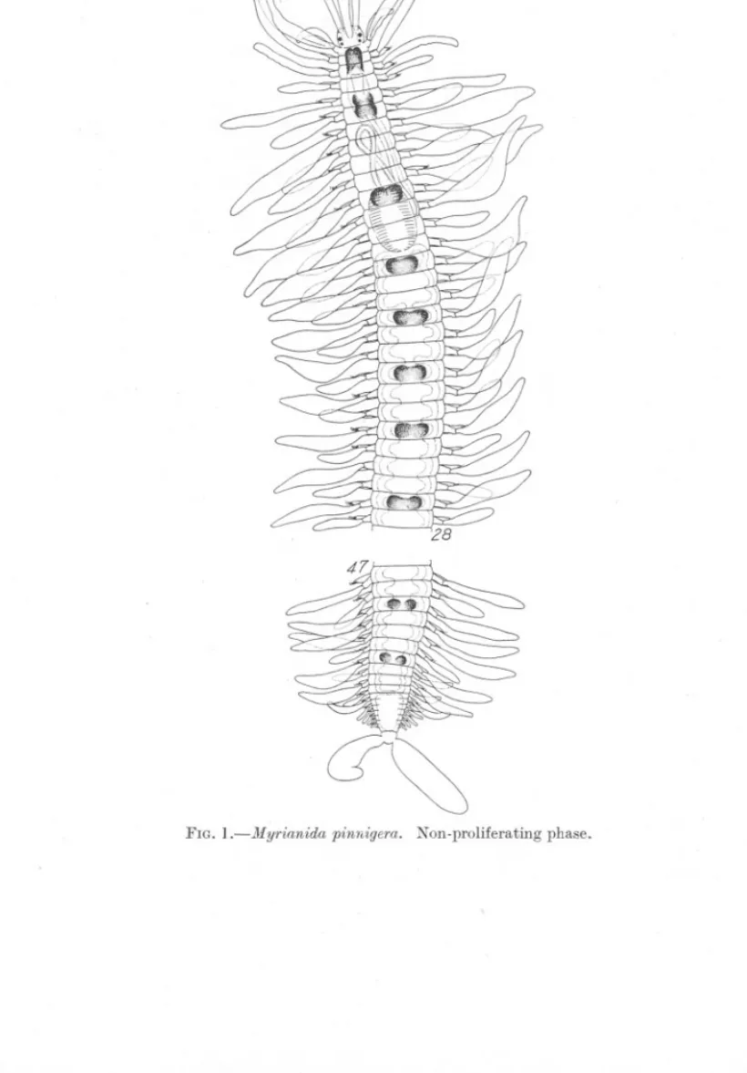
(2) 94. YO K. OKADA.. 471,(. FIG. l.-Myrianida. pinnigera.. Non-proliferating phase..
(3) STOLONIZATION. IN MYRIANIDA.. 95'. and 77. There is therefore no reason to believe that the stolonization of the present species takes place always at the posterior end of the 66th setigerous segment as A. Malaquin (1893) has stated in his monograph (p. 289). If we draw two graphs on the same horizontal plane, one for the segmental variation of the resting stage and the other for that of the parent body in the budding individuals, in such a way that the number of worms examined is plotted against the number of segments counted, the first part of the curve of stolonization evidently overlaps the posterior part of the curve that represents the segments of the resting stage. This fact at once reminds us that in the stolonization of Myrianida, as in most species of Autolytus (gemmiparous forms), there is, at least at the beginning of a chain formation of stolons, the process of simple schizogamy producing a single stolon by the separation of the posterior segments from the anterior ones; first of all there appears a new embryonic segment between two old ones at a certain distance from the posterior end at the point of separation. Actual observation shows that this distance is more than 16 but less than 22 segments. But in no case does the schizogamous stolon separate from the parent body before the appearance of the second, third and more stolons of higher order. Therefore schizogamy in the strict sense, forming a single stolon, in this case exists only temporarily. Malaquin (l.c., p. 312) describes in his case 3 of schizogamy in Autolytus Edwarsi a number of newly formed segments already intervening between the head of the stolon and the posterior end of the parent body. In case 4 he figures a stage long after the detachment of the first stolon, when gemmiparous proliferation has become well established, the embryonic segment being followed by a chain of 4 stolons in gradually increasing development. It is supposed that the process of stolonization in Myrianida is simply a combination of these two cases, case 3 and case 4 of Autolytus Edwarsi, and Malaquin (l.c., p. 314) has actually described such in case 7, where a parent body of 28 segments is followed by a chain of sexual individuals which is produced by gemmation succeeding schizogamy, passing from case 3 to case 4 above mentioned. In Myrianida such a transition from schizogamy to gemmiparity takes place at the very beginning of stolonization; that is to say before the separation of the first schizogamous stolon a number of secondary ones are quickly produced from the embryonic segment by successive stolonization. It is true that some specimens are observed carrying a chain of stolons in such anterior positions as the 34th and 37th setigerous segments, while others carry it in such posterior positions as the 73rd and 76th segments. Nevertheless, we need not necessarily suppose here a forward movement of the position of stolonization such as Malaquin has claimed for the origin.
(4) 96. yo K. OKADA.. ~2. 3. ... FIGs. 2-6.-Regeneration and stolon-formation in Myrianida. Abbreviated terms used in the figures: 11,head of stolon; p, zone of proliferation; S], S" S3' stolons of 1st, ~nd and ilrd order; aIrOW in Fig. 5 indicating the original position of cut..
(5) 97. STOLONIZATION IN MYRIANIDA.. of those specimens of Autolytus Edwarsi which carry the chain of stolons in an anterior position, as for example between segments 25 and 30 (most often on 27 or 28), since there are in Myrianida pinnigera individuals in the resting generation possessing only 50 segments. It is most interesting to notice that the positions of separation, which show so wide a range of variation, are never continuous even in two consecutive segments. On the contrary, they are entirely discontinuous. But in spite of that the position is almost always fixed after the 34th setigerous segment at one of the following segments: 34, 37, 40, 44, 48, 52, 56, 60, 64, 67, 70, 73, 76 and so on. To find the reason for this let us return to a former paper (Okada, 1929, p. 587) and see the rule of fragmentation of the Syllid which is represented by the formula H13-3-3-34-4-4-3-3-4-4-;-4 1. 1. 1. 3-3-3-3- - - - - - - - - - - - - - xp. In accordance with this formula the segmental arrangement of Myrianida pinnigera after the 34th setigerous segment can be divided into the followIng groups: segments 34-37, segments 38-40, segments 41-44, segments 45-48, segments 49-52, segments 53-56, segments 57-60, segments 61-64, segments 65-67, segments 68-70, segments 71-73, segments 74-76 and so on. A further characteristic point is that the most anterior segment of each group bears a special pigment pattern on the median dorsal surface (see Fig. 1), and in the range of stolonization which covers the median part of the body from behind about the 30th setigerous segme:q.tto about the 20th segment from the posterior extremity, the formation of a chain of stolons takes place always at the posterior boundary of each segmental group. In the part of the body just mentioned, if a worm in the process of budding is cut at a given position and the longer posterior part is removed, including the chain of stolons, the missing segments are able at once to regenerate and after a certain while they directly develop into a new chain of stolons, leaving the most anterior segment for the source of the further growth. The posterior cut end of the parent body remains as before. Therefore, the positions of stolonization are by no means absolutely restricted to certain segments and can be experimentally altered to any segment within the range of stolonization mentioned above. Figures 2-6 show 3 developmental stages of the posterior segments experimentally induced to regenerate from a segment which in the normal condition does not constitute a position of separation. Figure 2 represents a still very young stage before indication of the stolon formation. In Figure 3 the first stolon is about to be produced through direct metamorphosis of the regenera ted segments leaving one or two at the proximal side for the future proliferation of new segments. Thus in Figure 4 already 2 stolons beside the first one are produced. On the other hand if the same cut is done outside the range of stolonizaNEW. SERIES.-VOL.. XX.. NO. 1.. FEBRUARY, 1935.. G.
(6) 98. YO K. OKADA.. tion, i.e. in front of some 30th setigerous segment, for example, there is an easy regeneration of the posterior segments as in the preceding case, but there is no immediate formation of stolons (Fig. 5). In Figure 6 the beginning of natural stolonization in Myrianida pinnigera is reproduced from a microphotograph taken at the Plymouth Laboratory. Here schizog~my, with the production of a single stolon, is distinctly shown. REFERENCES. OKADA,Y& K. 1929. Regeneration and Fragmentation in the Syllidian Polychmtes. Arch. f. Ent.-Mech., 115. MALAQUIN, A. 1893. Recherches sur les Syllidiens. Mem. soc. sci. Art., Lille.. ".
(7)
Figure
Related documents
• Il est de la responsabilité de l’installateur/du propriétaire de vérifier que le site, le sous-plancher, ainsi que toutes autres conditions relatives à l’installation
The scattergram represents the distribution with age of 69 determinations of concentration of potassium in serum of 39 premature infants with respiratory distress syndrome (Table
19% serve a county. Fourteen per cent of the centers provide service for adjoining states in addition to the states in which they are located; usually these adjoining states have
On the Saudi Gulf coast the major environmental effects of coastal and marine use.. are concentrated in and around, Jubayl and
“USER INTERFACE DESIGN AND INFORMATION SYSTEMS USAGE: A CASE STUDY OF TVET INFORMATION SYSTEM AT IPRC KIGALI.” International Journal of Engineering Technologies and
The Supplement has three parts: our recommendation for enhancing the CLM5 optical properties table (S1_CLM5.pdf), and two source files (S2_OP.csv and S3_LIA.csv), which
can be recognised from all so far known members of the subgenus Sun- dodrupeus predominantly by the shape of aedeagus: the combination of narrowly lanceolate median lobe;
Several parameters affecting gene electrotransfer efficiency have been identified so far [ 7 , 30 , 31 ]: electric field distribution, which is related to the electrode
