Dr Mamidi V Sandeep Kumar et al JMSCR Volume 08 Issue 01 January 2020 Page 1032
A Devil in disguise- An interesting case of Non secretory Multiple myeloma
Authors
Dr Mamidi V Sandeep Kumar
1, Dr Vinu Boopathi
2, Dr V.R. Mohan Rao
31
Postgraduate, Department of General Medicine, Chettinad academy of Research and Education 2
Assistant Professor, Department of General Medicine, Chettinad academy of Research and Education 3
Professor, Department of General Medicine, Chettinad academy of Research and Education
Abstract
Non Secretory Multiple Myeloma is an alternate of Multiple myeloma which is a plasmacytosis involving uninhibited proliferation. Patients with NSMM presents with clinical picture of multiple myeloma, but negative for monoclonal immunoglobulins in serum, urine. We present a 57 year-old female patient who was investigated extensively for bone pain, dyspnoea, weight loss, and anaemia. She was eventually diagnosed to have Non secretory multiple myeloma based on histology of bone marrow trephine biopsy.
Case Report
A 57 year old female patient presented to the outpatient department with complaints of tiredness, generalized myalgia and bone pain since 6 months, she developed grade2 NYHA (New York Heart Association) dyspnoea since 1month. She also had history of loss of appetite & loss of weight around 10 kgs in 6 months, history of decreased vision of left eye for 1 year.
No history of fever, cough, chest pain, palpitation, abdominal pain, vomiting, loose stools. No history of bowel &bladder disturbances.
She is not a known case of Diabetes mellitus, Hypertension, Bronchial Asthma, Epilepsy, Tuberculosis, Thyroid disorder. Patient was diagnosed with macular hole and macular edema in left eye since 6 months. There is no significant family history.
On examination patient was poorly built and nourished, conscious, oriented to time, place, person, afebrile. Patient had pallor, no icterus,
cyanosis, clubbing, lymphadenopathy, pedal edema, JVP was not elevated.
Pulse rate was 86 / min, regular, radio radial delay was present, left subclavian was feeble, no bruit present, Adson’s test was positive on the left side, all other peripheral pulses were felt. Blood pressure in Right upper arm -130/70 mm Hg, Left upper arm -80/60 mmHg in supine position, Respiratory rate- 20 breaths /min thoraco abdomen type, spo2-95% in room air.
On Cardiovascular examination S1 and S2 heard in all the areas, haemic murmur heard in the pulmonary area.
Respiratory, Abdominal, Central nervous system examination were found to be normal.
Based on the above findings the provisional diagnosis was made in favour of Anaemia with Left thoracic outlet syndrome and further workup were initiated.
http://jmscr.igmpublication.org/home/ ISSN (e)-2347-176x ISSN (p) 2455-0450
Dr Mamidi V Sandeep Kumar et al JMSCR Volume 08 Issue 01 January 2020 Page 1033
Investigations
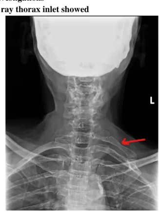
X ray thorax inlet showed
Fig 1: X Ray Thoraxic Inlet
1. Vertebral alignment appears normal
2. Vertebral bodies and posterior elements appear normal
3. Vertebral disc spaces appears normal • Facet articulations appear normal
4. Cervical rib in left side noted.
Complete blood count showed TLC was 4,700/cmm, Hb- 5.5 g/dl, platelet count – 1.22 lakhs/cmm, corrected reticulocyte count -0.99. Peripheral smear showed erythropenia, microcytic hypochromic, few macrocytes & macro ovalocytes, tear drop cells, platelets are just adequate, leucopenia with normal distribution, final impression was Pancytopenia with dimorphic anaemia.
ESR was 105 mm, RFT (BUN-28mg/dl,Sr. Creatinine-2.9mg/dl),Serum Na+- 141 mEq/L, K+- 4.4 mEq/L, Serum calcium level was 10.6 mg/dl, serum phosphorous level was 3.6 mg/dl. Urine routine showed albumin was trace.
LFT showed Total protein -6.5, Albumin – 3.9, Globulin- 2.6, A/G -1.5:1, Total bilirubin- 0.2, Direct bilirubin- 0.04 , AST-21, ALT-15,ALP-58, GGT-28, LDH-166.
USG abdomen showed – bilateral mild increased renal cortical echoes, no other abnormalities noted.
Table 1: Complete blood picture
Total WBC – 4700/cmm N-50.6,L-38.5,E-1.5,M- 8.8,B-0.6 .
Hb- 5.5g/dl MCV- 95.1,MCH- 31.1,
MCHC- 32.7 , RDW- 21.6 . PlateletCount-1.22
lakhs/cmm.
ESR- 105mm.
Reticulocyte Count- 0.1 Corrected reticulocyte count- 0.09
Summary
A 57 year old female patient with generalized body pain, tiredness, breathlessness, loss of appetite, weight loss, with no comorbidities, with macular hole in left eye and a left cervical rib, investigations showed hypoproliferative anaemia, pancytopenia, with raised renal parameters indicating Stage IV Chronic kidney disease (egfr 20ml/min/1.73m2), serum calcium level was 10.6 mg/dl, no Albumin/Globulin reversal, urine albumin was trace, ESR was 105 mm.
With the above clinical scenario and laboratory findings the possibility of Non secretory multiple myeloma was thought because of no albumin /globulin reversal and hence proceeded with x ray skull which showed punched out lesions.
Fig 2: X ray skull lateral view
Bone marrow aspiration, biopsy was done.
Dr Mamidi V Sandeep Kumar et al JMSCR Volume 08 Issue 01 January 2020 Page 1034
Table 2: Serum protein electrophoresis
Investigations Obs.value (references)
Total protein 6.72g/dl (6.4-8.3)
Serum albumin 3.97g/dl (3.57-5.42)
Alpha1 globulin 0.37g/dl (0.19-0.40)
Beta1 globulin 0.38g/dl (0.20-0.53)
Beta2 globulin 0.39g/dl(0.20-0.53)
Gamma globulin 0.81g/dl(0.71-1.54)
Albumin,globulin ratio 1.44 (1.1-2.2)
M band Absent
Fig 3: Serum protein electrophoresis
Urine Bence jones protein was absent. Bone marrow aspiration- yield dry tap.
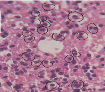
Bone marrow biopsy showed- bony trabeculae with cellular marrow composed of sheets of plasma cells (>80%)
Plasmacytosis With Grade Iii Fibrosis.
Fig 4: Bone marrow biopsy
Fig 5: Bone marrow biopsy (encircled cells are plasma cells) .
Criteria for Non secretory Multiple myeloma– 1) No M protein in serum and/or urine with
immunofixation.
2) Bone marrow clonal plasmacytosis >10%. 3) Myeloma related end organ damage
(anaemia, renal failure, osteolytic bone lesions).
The patient met all three criterias supporting non secretory multiple myeloma.
Final diagnosis- Non Secretory Multiple Myeloma.
Discussion
Multiple Myeloma
Multiple Myeloma represents a malignant proliferation of plasma cells derived from a single clone. The tumor, its products, and the host response to it result in a number of organ dysfunctions and symptoms, including bone pain, renal failure, anemia, hypercalcemia and manifestations of hyperviscosity.
Clinical features and complications- Bone pain
Dr Mamidi V Sandeep Kumar et al JMSCR Volume 08 Issue 01 January 2020 Page 1035
Infections
More susceptible to bacterial infections, most frequent pathogens are Streptococcus pneumoniae, Staphylococcus aureus, and Klebsiella pneumoniae in the lungs and Escherichia coli and other gram-negative organisms in the urinary tract.
Impaired Renal function
Renal failure occurs in nearly 25% of myeloma patients. Hypercalcemia is the most common cause of renal failure. When the glomeruli are involved, nonselective proteinuria is also observed.
Haematolgical
Normocytic and normochromic anemia occurs in 80% of myeloma patients. Due to replacement of normal marrow by expanding tumor cells, reduced production of erythropoietin by the kidney.
Raynaud’s phenomenon and impaired circulation may result if the ‘M’ component forms cryoglobulins, and hyperviscosity syndromes may develop depending on the physical properties of the M component (most common with IgM, IgG3, and IgA paraproteins).
Neurological Features
Hypercalcemia may produce lethargy, weakness, depression, and confusion. Hyperviscosity may lead to headache, visual disturbances, ataxia, vertigo, retinopathy, somnolence, and coma. Bony damage and collapse may lead to cord compression, radicular pain, and loss of bowel and bladder control, sensorimotor mono- and polyneuropathies.
Diagnostic Criteria for Multiple Myeloma, Myeloma Variants
Symptomatic Multiple Myeloma-
Clonal bone marrow plasma cells or biopsy-proven bony or extramedullary plasmacytoma and any one or more of the following myeloma defining events:
Evidence of one or more end-organ damage that can be attributed to the underlying plasma cell proliferative disorder, specifically:
1) Hypercalcemia: serum calcium >0.25 mmol/L (>1 mg/dL) higher than the upper limit of normal or >2.75 mmol/L (>11 mg/dL).
2) Renal insufficiency: creatinine clearance <40 mL per min or serum creatinine >177 μmol/L (>2 mg/dL).
3) Anemia: hemoglobin value of >2g/dL below the lower limit of normal, or a hemoglobin value <10g/dL.
4) Bone lesions: one or more osteolytic lesions on skeletal radiography, CT, or PET-CT.
5) Any one or more of the following biomarkers of malignancy:nClonal bone marrow plasma cell percentage ≥60%. Involved: uninvolved serum free light chain ratio≥100,
Non secretory Myeloma
1) No M protein in serum and/or urine with immunofixation.
2) Bone marrow clonal plasmacytosis ≥10% or plasmacytoma.
3) Myeloma-related organ or tissue impairment (end-organ damage, as described above)
Non Secretory Multiple Myeloma
Multiple Myeloma and Non Secretory Multiple Myeloma have essentially the same clinical and radiologic features. However, in the case of NSMM, the plasma cells fail to secrete an immunoglobulin and therefore both serum and urine electrophoresis are normal[2]. Diagnosis depends on bone marrow biopsy.
Two distinct types of Non Secretory Multiple Myeloma
Producer” type or True NSMM
Dr Mamidi V Sandeep Kumar et al JMSCR Volume 08 Issue 01 January 2020 Page 1036 or absence and alteration of intracellular light
chains.
Non Producer type NSMM
In this type plasma cells are unable to produce immunoglobulin [3].
Theories for absence of M protein in NSMM
One theory postulates that the producer type may be as a result of increased breakdown of abnormal immunoglobulin produced, while the non producer type may be as a result of problems with the assembly process of proteins, thereby leading to difficulty with immunoglobulin heavy and light chain synthesis.
Significance of bone marrow fibrosis in multiple myeloma-
Although the cause of fibrosis in myeloma is not well established, it may be related to lymphokine secretion by the malignant cells. Pich et al., observed that plasmablastic myeloma is usually associated with higher marrow plasma cell percentage, a diffuse pattern of infiltration and increased fibrosis[4]. Bartl et al. reported that increased fibrosis at the time of presentation was frequently associated with a subsequent relapse later[5].
Treatment
Patients with symptomatic and/or progressive myeloma require therapeutic intervention.
Induction therapy
1) Thalidomide, when combined with dexamethasone, achieved responses in two-thirds of newly diagnosed MM patients.
2) Subsequently, lenalidomide, an immunomodulatory derivative of thalidomide, and bortezomib, a proteasome inhibitor, have each been combined with dexamethasone.
Maintenance therapy
Lenalidomide improved progression –free survival in Multiple myeloma patients even in non transplant candidates.
Relapse
The combination of bortezomib and liposomal doxorubicin is active in relapsed myeloma.
The second-generation proteasome inhibitor carfilzomib and immunomodulatory agent pomalidomide have shown efficacy in relapsed and refractory MM, even MM refractory to lenalidomide and bortezomib.
Two antibodies are approved for treatment of relapsed MM. Daratumumab targeting CD38 and Elotuzumab targeting SLAMF7 has shown significant activity.
Supportive Management
Hypercalcaemia generally responds well to bisphosphonates, glucocorticoid therapy, hydration and rarely requires calcitonin as well. Plasmapheresis may be the treatment of choice for hyper viscosity syndromes. The anemia associated with myeloma may respond to erythropoietin.
Mortality
The median overall survival of patients with myeloma is 8+ years, with subsets of younger patients surviving >10 years. The major causes of death are progressive myeloma, renal failure, sepsis, or therapy-related myelodysplasia.
Conclusion
In conclusion non demonstration of an M protein in either serum and urine does not rule out the diagnosis of Multiple myeloma. Hence Non Secretory Multiple myeloma should be suspected in patients with such clinical features.
References
1. Longo, Dan (2012). Harrison's Principles of Internal Medicine 18th Edition. Mc Graw Hill Medical. p. 938.
Dr Mamidi V Sandeep Kumar et al JMSCR Volume 08 Issue 01 January 2020 Page 1037 Southern Medical Journal, vol. 95, no. 7,
pp. 761–764, 2002.
3. M. Cavo, P. Galieni, M. Gobbi et al., “Nonsecretory multiple myeloma. Presenting findings, clinical course and prognosis,” Acta Haematologica, vol. 74, no. 1, pp. 27–30, 1985.
4. Pich A, Chiusa L, Marmont F, Navone R. Risk groups of myeloma patients by histologic pattern and proliferative activity. Am J Surg Pathol 1997; 21: 339– 47.