R E S E A R C H
Open Access
Combinations of bio-active dietary
constituents affect human white adipocyte
function in-vitro
Ines Warnke
1*, Johan W. E. Jocken
2, Rotraut Schoop
1, Christine Toepfer
1, Regina Goralczyk
1and
Joseph Schwager
1Abstract
Background:Specific bio-active dietary compounds modulate numerous metabolic processes in adipose tissue (AT), including pre-adipocyte proliferation and differentiation. AT dysfunction, rather than an increased fat massper se, is strongly associated with the development of insulin resistance and is characterized by impaired adipogenesis, hypertrophic adipocytes, inflammation, and impairments in substrate metabolism. A better understanding of mechanisms underlying AT dysfunction may provide new strategies for the treatment of obesity-associated metabolic diseases. Here we evaluated the role of (all-E)-lycopene (Lyc), eicosapentaenoic acid (EPA) ortrans -resveratrol (Res) and combinations thereof on human white adipocyte function.
Methods:In-vitro differentiating human pre-adipocytes were treated with EPA, Lyc and Res or their combinations for 14 days. The effects on intracellular lipid droplet (LD) accumulation, secreted anti- and pro-inflammatory cyto-/ adipokines (e.g. adiponectin, IL-6, IL-8/CXCL-8 and MCP-1/CCL2) and on gene expression of markers of adipocyte differentiation and substrate metabolism (e.g. PPAR-gamma, C/EBP-alpha, GLUT-4, FAS, ATGL, HSL, and PLIN-1) were measured by fluorescent microscopy (Cellomics™), multi-parametric LiquiChip® technology and quantitative RT-PCR, respectively.
Results:Treatment of differentiating adipocytes for 14 days with the combination of Lyc/Res and EPA/Res resulted in significantly inhibited LD formation (~ -25 and -20%, respectively) compared to the effects of the single
compounds. These morphological changes were accompanied by increased mRNA levels of the adipogenic marker PPAR-gamma and the lipase ATGL and by decreased expression levels of lipogenic markers (LPL, FAS, GLUT-4) and the LD-covering protein PLIN-1. In addition, a blunted adipocyte secretion of pro-inflammatory cytokines (IL-6 and MCP-1) and adiponectin was observed following treatment with these compounds.
Conclusion:The combination of the dietary bio-actives Lyc and EPA with Res might influence adipocyte function by affecting the balance between adipogenic, lipogenic and lipolytic gene expression, resulting in a reduced LD storage and a less inflammatory secretion profile. Taken together, our results indicate that combinations of dietary compounds may be beneficial for the prevention and treatment of metabolic disorders via effects on human white adipocyte function.
Keywords:Primary human adipocyte function, Lipid accumulation, Adipokine secretion, Adipogenic/ lipogenic genes, Lipolytic genes, Lycopene, Resveratrol, EPA, Combinations of bio-actives
* Correspondence:[email protected]
1DSM Nutritional Products Ltd., Department of Human Nutrition and Health,
CH-4002 Basel, Switzerland
Full list of author information is available at the end of the article
Background
Over the last decade research focusing on adipose tissue (AT) biology and function enormously advanced due to the sustained increase in obesity prevalence [1]. AT dys-function, characterized by impaired adipogenesis, hyper-trophic adipocytes, inflammation, and impairments in lipid and glucose metabolism, rather than an increased body fat massper se, is strongly associated with the devel-opment of insulin resistance [2]. A better understanding of mechanisms causing or maintaining AT dysfunction may provide novel and improved strategies for the treat-ment of obesity-associated metabolic diseases.
The major role of AT is storage and release of fatty acids (FAs) depending on energy intake and expenditure. FAs are stored in the form of triacylglycerides (TAGs) in intra-cellular lipid droplets (LDs) and released by lipolysis, the hydrolysis of TAGs into free FAs and glycerol via the ac-tion of intracellular lipases (including hormone sensitive lipase (HSL) and adipose triglyceride lipase (ATGL)). This storage and removal capacity (lipid turn-over) of AT is regulated by a tight alignment between adipogenic differ-entiation, lipogenesis and lipolysis [3, 4], which has been shown to be impaired in obesity and may modulate whole-body insulin sensitivity [5, 6]. The differentiation of pre-adipocytes into mature adipocytes is controlled by a complex transcriptional cascade involving peroxisome proliferator-activated receptor gamma (PPAR-gamma) and CCAAT/enhancer binding protein alpha (C/EBP-alpha) (reviewed in [7]). Furthermore, lipid-storing adipo-cytes are enclosed by adipose-derived stromal cells includ-ing pre-adipocytes, endothelial and hematopoietic cells, and immune cells (e.g. macrophages [8]). Hence, AT is not only an energy storage tissue but also an active endo-crine organ, producing and secreting an abundance of specific mediators (for review see [9]). Pro-inflammatory cytokines and chemokines are increasingly secreted by AT cells of obese individuals [10], resulting in a state of ‘ low-grade inflammation’[11], which affects local adipose me-tabolism, systemic inflammation and insulin sensitivity.
Overall dietary quality and specifically diets high in bio-active constituents may have beneficial clinical effects on metabolic processes, by altering AT function. A number of natural compounds, such as plant-derived polyphenols, carotenoids and polyunsaturated fatty acids (PUFAs), or their metabolic derivatives have been tested for their impact on adipocyte differentiation and metabolism in several in-vitro and in-vivo murine models [12–14]. How-ever, human data are scarce and mainly the action of indi-vidual compounds have been tested [15, 16]. Several studies using pre-adipocytes, mostly from murine and less frequent from human origin, have demonstrated that the polyphenol Res and the n-3 PUFA EPA are potent modu-lators of adipocyte function (for review see [17, 18], re-spectively). However, distinct biological activities of
lycopene or its metabolites in human adipocyte function remain to be elucidated. Here, we investigated the effects of individual and combinations of bio-active dietary con-stituents including (all-E)-lycopene (Lyc), eicosapenta-enoic acid (EPA) and trans-resveratrol (Res) on lipid accumulation, adipogenic, lipogenic and lipolytic gene ex-pression and cyto-/adipokine secretion in in-vitro differ-entiating (14 days) primary human white adipocytes.
Methods
Cell culture
All cell culture reagents were obtained from Life Technolo-gies (Karlsbad, CA, US). Unless otherwise stated, chemicals were purchased from Sigma-Aldrich (St. Louis, MO, US). Individual subcutaneous primary human pre-adipocytes (HPAd 1375 and 1377) were obtained from Cell Applica-tion, Inc. (San Diego, CA, US) whereas a multi-donor vial, termed super lot (SL0035), was purchased from Zen-Bio, Inc. (North Carolina, US). Available donor characteristics are indicated in Additional file 1: Table S1. Pre-adipocytes were maintained in growth medium (GM): DMEM/Ham’s F-12 (1:1, v/v) complemented with 10% FCS, 1% pen/strep (v/v), 1% HEPES pH 7.4, 0.2% amphotericin B, and 2.5 ng/ ml recombinant basic fibroblast growth factor (bFGF). GM was changed every 2–3 days. Cells were passaged when reaching ~80% confluence and used for experiments be-tween passage 3 and 7. For experiments, 6000 cells/cm2 were incubated (37 °C, 5–8% CO2, relative humidity of
85%) on collagen-I-coated 24-well plates in GM for ~5 days. For adipogenesis confluent HPAd were cultured for 14 days in differentiation medium (DM): DMEM/Ham’s F-12 sup-plemented with 5% FCS, 1% pen/strep, 1.5% HEPES pH 7.4, 0.2% amphotericin B, 17μM calcium-pantothenate, 33 μM biotin, 0.5 μM recombinant human insulin and 100μM rosiglitazone. After 3 days of differentiation 0.5μM dexamethasone and 250 μM isobutylmethylxanthine (IBMX) were omitted from the medium.
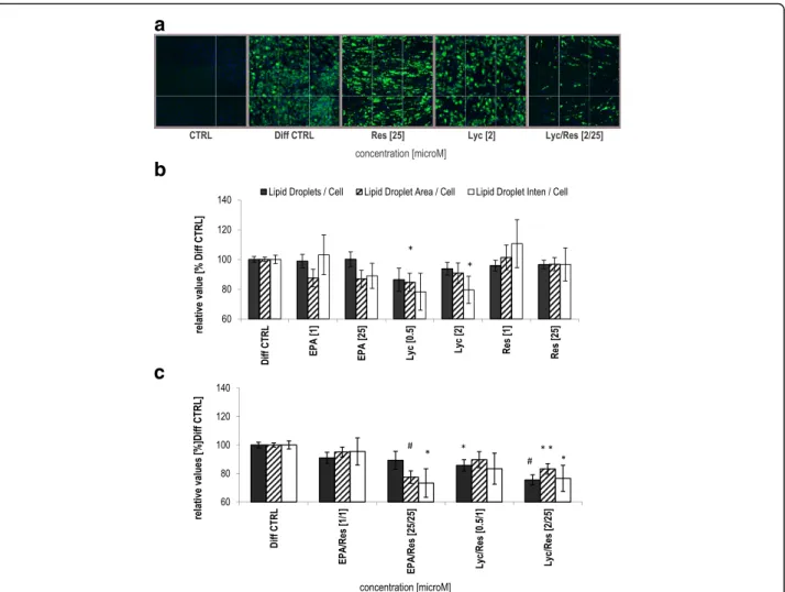
Lipid droplet quantification (ArrayScan)
Cellular LDs were quantified adapting the Cellomics™assay described previously [19] applying the Thermo Scientific™ ArrayScan™ VTI High Content Reader (Thermo Fisher Scientific, Waltham, MA, US). Briefly, differentiated adipo-cytes (day 14) were fixed, stained with the fluorescent dyes Hoechst 33342 (nuclei) and BODIPY® 493/503 (LDs; Life Technologies) followed by quantification of accumulated LDs with the SpotDetector® V2 algorithm. Adjusted proto-col parameters are listed in Additional file 3: Table S2. For analysis 100 fields per well were scanned and data of each channel were reported on a“per field”basis. The nuclei re-lated features Spot Count (= LD-number), Spot Total Area (= LD-area) and Spot Total Intensity (= LD-intensity), de-scribing the differentiation status of treated adipocytes, were calculated as percent of Diff CTRL per plate.
Gene expression analysis (TaqMan™)
Total RNA was isolated from cells at day 8 of treatment (RNeasy® 96 Kits; Qiagen, Hilden, Germany). Primers and probes were designed using the Primer Express software (Applied Biosystems, Foster City, CA, US) and synthesised by Sigma Genosys (St. Louis, MO, US) (Additional file 4: Table S3). Quantitative TaqMan™RT-PCR was performed on first strand cDNA (Omniscript® RT Kit; Qiagen) as de-tailed previously [19] utilizing an ABI-PRISM® 7900 HT Sequence Detection System. mRNA abundance was calcu-lated using the comparative CT method: ΔCT= CT [gene
of interest] – CT [endogenous control] and ΔΔCT=ΔCT
[Diff CTRL cells] -ΔCT[treated cells]. The fold expression
for the gene of interest was expressed as 2−ΔΔCT.
Adipokine and cytokine secretion (Luminex)
Supernatants of differentiating adipocytes were collected at day 8 (after 5 days conditioning, day 4–8) of the treat-ment period and stored at -80 °C till analysis. MILLI-PLEX MAP Human Adipocyte Panel (Cat#HADCYT-61 K) kits were purchased from Millipore (Billerica, Massachusetts, USA) and used according to the manu-facture’s protocol on the LiquiChip® Workstation IS 200 (Luminex technology; Qiagen, Hilden, Germany). De-tected molecules were: adiponectin (Adipo), hepatic growth factor (HGF), interleukin (IL)-6, IL-8/CXCL8, monocyte chemoattractant protein (MCP)-1/CCL2 and plasminogen activator inhibitor (PAI)-1 (active, serpin E1). Measurements were run in triplicates and final con-centrations were normalized by nucleus count per well. Data evaluation was performed with the LiquiChip® Ana-lyser software from Qiagen.
Statistical analysis
In brief, data points from repetitive experiments con-ducted with pre-adipocytes from the same donor were
set relative to the corresponding Diff CTRL mean (=100%) and averaged. Subsequently all relative values from the different donors and super lot (Additional file 1) were used for calculating the overall mean ± SEM. Statistical significance of the mean differences between treatment and Diff CRTL was tested by a linear mixed model or Student's t-test. P values <0.05 were consid-ered significant. ArrayScan™ results and cytokine con-centrations are shown as relative mean ± SEM. Gene expression data are expressed as fold change (FC) ± error (based on SEM). For details of the statistical analysis ap-plied to the three data sets see Additional file 5.
Results
Combinations of bio-active dietary constituents inhibited lipid accumulation
To determine effects of bio-active dietary constituents on lipid accumulation in adipocytes, pre-adipocytes were dif-ferentiated for 14 days in the presence or absence of the compounds. Subsequently, LD accretion in the mature adi-pocytes was assessed by fluorescent microscopy (Fig. 1a). Lyc alone decreased the LD-area by 15% at 0.5 μM (p= 0.021) and the LD-intensity by 20% at 2 μM (p< 0.05), while Res and EPA alone did not affect lipid accumulation (Fig. 1b) compared to Diff CTRL. However, the combin-ation Lyc/Res substantially reduced the adipocyte lipid content (represented by shown LD-parameters). Lyc/Res at 2/25 μM, significantly inhibited LD-number, -area and -intensity by 25% (p< 0.001), 17% (p< 0.01) and 23% (p= 0.044), respectively (Fig. 1a + c, right), in comparison with the Diff CTRL. In addition, combined treatment with EPA/ Res at 25/25μM reduced LD-area and -intensity by 22% (p < 0.001) and 26% (p= 0.011), respectively, whereas the LD-number was slightly attenuated (Fig. 1c). Together, these data indicate that the combination of Lyc/Res and EPA/Res significantly reduced the adipocyte lipid content compared to Diff CTRL.
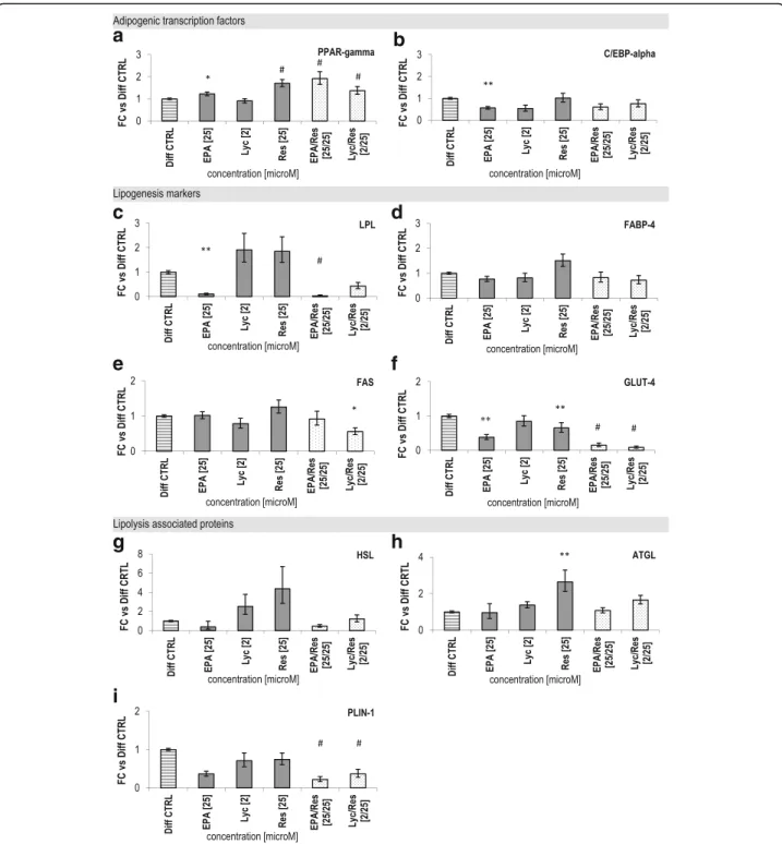
Dietary bio-actives affected adipogenic, lipogenic and lipolytic gene expression
0.15-fold, respectively; Fig. 2c + f,p< 0.001). Both combinations did not affect FABP-4 (cytosolic fatty acid binding protein 4) expression (Fig. 2d). Res alone at 25μM moderately en-hanced the expression of PPAR-gamma to 1.7-fold (p< 0.001) and reduced GLUT-4 expression 0.7-fold (p= 0.007) compared to Diff CTRL. The expression of C/EBP-alpha, LPL and GLUT-4 was diminished to 0.5-fold, 0.1-fold and 0.4-0.1-fold, respectively, when pre-adipocytes were differentiated in the presence of 25μM EPA (Fig. 2b, c + f, p< 0.01). In contrast, Lyc alone (at 2 μM) did not affect adipogenic and lipogenic gene expression (Fig. 2a-f ).
The mRNA levels of the major lipases HSL and ATGL were neither significantly affected by Lyc and EPA nor by the combinations, although Res alone increased ATGL expression 2.6-fold (p= 0.006; Fig. 2g + h) in com-parison to Diff CTRL. However, combining these
bio-active compounds significantly diminished the expres-sion of the LD-covering protein PLIN-1 (perilipin 1; Fig. 2i, EPA/Res: 0.2-fold and Lyc/Res: 0.4-fold, p< 0.001). A comparable effect on the expression of lipo-genic and lipolytic genes was observed at day 14 for both combinations (Additional file 6: Figure S2). Altogether, these results demonstrate that combining of Lyc/Res and EPA/Res affects the balance between adipogenic, lipogenic and lipolytic gene expression in white human adipocytes.
Bio-active compounds attenuated secretion of pro-inflammatory cytokines
Finally, we analysed the influence of dietary bio-actives on cyto-/adipokine secretion of differentiating human adipocytes. EPA alone and the combination EPA/Res
a
b
c
strongly reduced the secretion of the insulin sensitizer Adipo by ~90% whereas Res and Lyc/Res blunted it by ~60% (Fig. 3a) compared to Diff CTRL. The combina-tions EPA/Res and Lyc/Res and EPA suppressed inflammatory IL-6 secretion by 70%. Release of the
pro-inflammatory cytokine MCP-1 was significantly reduced by more than 30% by all compounds and combinations except Res. Both combinations caused a similar secre-tion pattern for the above mensecre-tioned cyto-/adipokines at day 14 of differentiation (Additional file 6: Figure S2).
a
c
d
e
f
g
i
h
b
Further, the secretion of angiogenic HGF was markedly diminished by Lyc/Res (70%, p< 0.01), whereas PAI-1 levels were not modified by the compounds (Fig. 3e + f ) in comparison to the Diff CTRL.
Together, our data indicate that EPA and the combina-tions Lyc/Res and EPA/Res are able to suppress the se-cretion of pro-inflammatory markers, but concurrently attenuate the anti-inflammatory and insulin sensitizing adipocyte-specific hormone adiponectin.
Discussion
Weight gain is accompanied by increased lipid storage in AT, altered gene expression and production of pro-and anti-inflammatory mediators [20]. In the present study, we examined the effect of the three abundantly
consumed bio-actives Lyc, Res and EPA on human white adipocyte function and whether combining Res with EPA or Lyc has amplifying effects. Our in-vitro data demonstrate that combining Lyc with Res caused a sig-nificant reduction in LD-number, -area and -intensity in human adipocytes. This reduced lipid accumulation was accompanied by increased expression of adipogenic and reduced expression of lipogenic and lipolytic markers, and by an attenuated pro-inflammatory secretion profile (i.e. IL-6 and MCP-1).
Single bio-active dietary constituents are known to in-fluence pre-adipocyte differentiation, proliferation and adipocyte function directly or indirectly [15–18]. Sur-prisingly, in our study Res and EPA treatment alone showed no significant effect on the LD accumulation in
a
b
c
d
e
f
human adipocytes. This is contrary to the general re-ported findings in in-vitro and animal studies, which suggest that Res and EPA are capable of diminishing lipid storage in rodent adipocytes [21–24]. However, in human adipocytes lipid accumulation was either pro-moted or unaffected by EPA [25, 26], what seems in line with our observation after 14 days treatment. For Res, both enhancing and suppressive effects on lipid accumu-lation were described in murine 3T3-L1 cells and follow-ing a high fat diet (HFD) in mice [27, 28]. In human Simpson-Golabi-Behmel syndrome (SGBS) adipocytes anti-adipogenic effects of Res were reported for concen-trations > 30 μM [29, 30], suggesting that Res at 25 μM (this study) might be at switch between its pro- and anti-adipogenic actions. The limited number of human clinical trials with Res or EPA supplementation showed controversial results regarding their effects on adipo-cyte size, function [31, 32] and adipose inflammation [33, 34], possible due to variation in population, dose and duration of supplementation. Together, these data indicate that further research is necessary to elucidate the exact effect of Res and EPA on adipogenic potential of human (pre-) adipocytes, under the consideration that the usage of primary white adipocytes can affect the experimental outcomes due to intrinsic donor characteristics [35].
Supplementation with the carotenoid Lyc can increase its levels in human AT [36] and 3T3-L1 cells [37] and it is well studied for its anti-oxidative effects [38]. In addition, our results show for the first time that Lyc alone moderately but significantly reduces lipid accumu-lation in human adipocytes. This effect was also ob-served in C3H10 T1/2 adipocytes [19]. Conversely, Lyc or its metabolite apo-10’-lycopenoic acid (APO10LA) did not modulate AT mass in rodents [39] (following oral administration for 6 weeks) [40]. In contrast to the minor effect of single compounds, our data clearly dem-onstrate that the combination Lyc/Res has a stronger ef-fect on LD accumulation, suggesting that Res enhances Lyc’s modest anti-adipogenic effect.
Although not a direct measure of lipid turn-over within adipocytes, investigating mRNA levels of genes involved in related pathways could provide possible underlying mechanisms for the amplifying effects ob-served with combinations of bio-actives. Res’s anti-adipogenic properties may be attributed to its effects on both master regulators of adipogenesis, PPAR-gamma and C/EBP-alpha, which are repressed via activation of AMP-activated protein kinase (AMPK) [41]. Interest-ingly, in the present study Res at 25 μM up-regulated PPAR-gamma expression and showed no effect on C/ EBP-alpha mRNA in differentiating human adipocytes. This seems contrary to the reported down-regulation of PPAR-gamma and lipogenic markers in 3T3-L1 [41] and
SGBS cells [29] at concentrations >25 μM and the ac-companying reduction of fat pads in mice following 10 weeks of HFD feeding [42]. We hypothesize that in the present study the concentration of Res (25μM) is in-sufficient to counteract the strong adipogenic effects of the PPAR-gamma agonist rosiglitazone [43], contained in the media at 100 μM during the complete differenti-ation period. This is further supported by results in differ-entiating SGBS cells, demonstrating no effect of 20 μM Res on PPAR-gamma expression even in the presence of a low rosiglitazone concentration (2μM) [29].
PUFAs can act as PPAR agonists and display PPAR-independent gene control function [44]. Regarding EPA, a well-known n-3 PUFA, a reduction in stored LDs and a concomitant modification of adipogenic markers, e.g. a decrease of PPAR-gamma expression, was described in murine fat cells [19, 21]. However in line with our re-sults, an upregulation of PPAR-gamma expression is de-scribed in isolated human adipocytes [45], suggesting species-specific effects of EPA on adipogenesis. Finally, Lyc is not presented as an adipogenic modulator [16], however some conflicting data exist regarding its impact on adipose gene expression. In accordance with our hu-man data, in murine adipocytes, Lyc treatment did not affect adipogenic genes [39] but moderately decreased lipid deposition [19], whereas Lyc administration for 6 weeks decreased PPAR-gamma mRNA in AT of rats without changes in total fat mass [46].
In addition to adipogenesis, the balance between lipo-genesis and lipolysis largely influences total lipid amount in human adipocytes. The combination of Lyc/Res and EPA/Res strongly suppressed the lipogenic markers GLUT-4, LPL and FAS in our human fat cells. However, for the single compounds conflicting effects on these markers were reported previously. Supporting our re-sults, a reduction of GLUT-4 mRNA and unaffected levels of FAS mRNA were shown in 3T3-L1 cells after Res (20μM) [47] and EPA (100 μM) [48] treatment, re-spectively. Whereas, opposing to our human data, GLUT-4 and LPL mRNA expression was up-regulated after treating murine adipocytes with Lyc (2 μM) [19] and EPA (100μM) [21], respectively.
no impact on PLIN-1 expression [51]. On the other hand, the combinations significantly suppressed PLIN-1 mRNA in our human adipocyte model, suggesting interactions between bio-active compounds which influence PLIN-1 expression. However, it needs to be investigated whether this translates into functional changes in the lipolytic po-tential of the adipocytes.
Together, the pronounced down-regulation of lipo-genic (GLUT-4, LPL, FAS) and lipolytic genes (PLIN-1) by the combination of bio-active compounds (EPA and Lyc with Res) cannot be justified by additive effects of the single compounds, suggesting strong amplifying/syn-ergistic interactions. Therefore, combining bio-active constituents facilitates the interplay of different direct and indirect transcriptional controlling mechanisms, which warrants further investigations.
Finally, we showed that the reduction in lipid accumula-tion following EPA/Res or Lyc/Res treatment was accom-panied by a diminished release of the pro-inflammatory cytokines IL-6 and MCP-1. In line, a previous study, in dif-ferent adipocyte models and in murine AT explants, dem-onstrated that pre-incubation with the anti-inflammatory nutrient Lyc (24 h, 2μM) reduced the secretion of the pro-inflammatory cytokines IL-6 and MCP-1 induced by tumour necrosis factor (TNF)-alpha stimulation or feeding, respectively [52]. Additionally, a study with HFD-induced obese rats showed that Lyc supplementation in-creased plasma adiponectin levels and dein-creased the mRNA of MCP-1 and IL-6 in AT and their circulating pro-tein levels [40]. Without stimulation of the inflammatory pathways (this study), Lyc only suppressed the secretion of the macrophage attracting chemokine MCP-1 [53] from human adipocytes. Furthermore, several studies indicate that treatment of mature SGBS and 3T3-L1 adipocytes with Res (100μM, 48 h) [30, 54] and EPA (200μM, 48 h) [55, 56], respectively, and supplementation of HFD-fed mice with both bio-actives (Res, 10 weeks [42]; EPA, 5 or 11 weeks [55, 56]) can reduce the expression and secretion of pro-inflammatory cytokines such as MCP-1, IL-6 and IL-8 in AT.
In addition, we observed a decrease of Adipo secretion with Res, EPA and the combinations. However, several groups reported an increase of the anti-inflammatory and insulin sensitizing adipokine adiponectin and a decrease of leptin expression and secretion after treatment of mature 3 T3-L1- and SGBS-adipocytes with Res (up to 200μM) or its metabolites for 24 or 48 h, respectively [30, 57]. Simi-larly, EPA treatment (100μM, 24 or 48 h) was also able to increase Adipo secretion from mature primary human adi-pocytes [26, 58]. Nevertheless, our findings are in line with recent data from Lorente-Cebrian et al. [59], who reported that 200μM EPA (96 h) significantly decreased adiponectin gene expression and protein secretion in freshly isolated rat adipocytes [59]. Furthermore, we noticed a strong
attenuation of the angiogenic protein HGF by the combin-ation Lyc/Res. HGF is secreted from AT and its expression is elevated in the obese state allowing for remodelling of AT when expanding [60] and exhibits anti-inflammatory characteristics on the cross-talk between macrophages and adipocytes in mice [61].
Altogether our data suggest that treatment with dietary bio-actives during adipogenic differentiation, alters the cyto-/adipokine release towards a less pro-inflammatory secretion profile, indicated by an attenuated IL-6 and MCP-1 release. The combinations EPA/Res and Lyc/Res partially augmented the effects of the single compounds on the secretion of adiponectin, IL-6, MCP-1 and HGF, supporting the concept of strong amplifying/synergistic interactions between nutrients [62].
Conclusions
Here we show for the first time, that the combination of the dietary bio-actives Lyc and EPA with Res might influ-ence adipocyte function synergistically by affecting the bal-ance between adipogenic, lipogenic and lipolytic gene expression, resulting in a reduced lipid accumulation and improved inflammatory profile. Our data suggest that plying combinations of bio-actives is a more favourable ap-proach to tackle AT dysfunction because anti-lipogenic and anti-inflammatory effects can be magnified compared to the single dietary constituents [63–66]. Given that obesity is recognized as a state of chronic‘low-grade inflammation’ [10, 11], reducing the secretion of pro- and stimulating anti-inflammatory adipokines with combinations of dietary bio-actives could contribute to manage and treat obesity-related metabolic complications.
Additional files
Additional file 1: Table S1.Relevant donor characteristics from used human pre-adipocytes (HPAd). (DOCX 32 kb)
Additional file 2: Figure S1.Lipid droplet number, lipogenic gene expression markers and adipokine secretion of adipocytes treated with different doses of bio-active compounds. Assessment of different doses of Lyc, Res and EPA alone and of the combinations EPA/Res and Lyc/Res on distinct adipocyte parameters reflecting adipocyte function. Depicted are the effects on the number of lipid droplets (Lipid Droplets/Cell in % of Diff CTRL; A-B), the expression of the lipogenic genes LPL and GLUT-4 (FC ± error compared to Diff CTRL set as 1; C-D) and the secretion of the adipokines adiponectin and IL-6 (overall mean ± SEM; E-F) after 8 (C-F) or 14 (A-B) days treatment of HPAd (donors≥2, n≥2). These data indicate that not all parameters follow a linear dose-response relationship. (*)p< 0.05, (**)p< 0.01, (#)p< 0.001 (versus Diff CTRL, linear mixed model). (PDF 44 kb)
Additional file 3: Table S2.Altered Cellomics™parameters for the analysis of HPAd (20x objective). (DOCX 33 kb)
Additional file 4: Table S3.Sequences of human primers and probes for adipocyte specific genes used for quantitative RT-PCR. (DOCX 34 kb)
Additional file 5:Detailed Statistical Analysis. (DOCX 27 kb)
differentiated human adipocytes. Shown are the effects of the combinations EPA/Res and Lyc/Res on gene expression and adipokine secretion after 14 days treatment of HPAd. mRNA levels of lipogenesis markers LPL (A), GLUT-4 (B) and FAS (C) and the lipolytic marker PLIN-1 (D) were determined by quantitative RT-PCR and the accumulation of the adipokines adiponectin (E), IL-6 (F), MCP-1 (G) and PAI-1 (H) was measured in media after 5 days conditioning (day 10–14) on the LiquiChip® workstation. Data are shown as crude fold change (FC) ± error (based on SEM, A-D) compared to Diff CTRL set as 1 and as overall mean ± SEM (E-H) for one experimental series (all donors included: HPAd 1375, 1377 and super lot SL0035). (*)p< 0.05, (**)p< 0.01, (#)p< 0.001 (versus Diff CTRL, Student’st-test)
Abbreviations
Adipo:Adiponectin; AT: Adipose tissue; ATGL: Adipose triglyceride lipase; bFGF: basic fibroblast growth factor; C/EBP-alpha: CCAAT/enhancer binding protein alpha; DM: Differentiation medium; EPA: Eicosapentaenoic acid; FA: Fatty acid; FABP-4: Cytosolic fatty acid binding protein 4; FAS: Fatty acid synthase; FC: Fold change; FCS: Fetal calf serum; GLUT-4/SLC2A4: Glucose transporter type 4; GM: Growth medium; HFD: High fat diet; HGF: Hepatic growth factor; HPAd: Human pre-adipocytes; HSL: Hormone sensitive lipase; IL-6: Interleukin-6; IL-8: Interleukin-8; LD: Lipid droplet; LPL: Lipoprotein lipase; Lyc: (all-E)-lycopene; MCP-1/CCL2: Monocyte chemoattractant protein-1; n: number of experimental series; PAI-1: Plasminogen activator inhibitor; PBS: Phosphate-buffered saline; PLIN-1: Perilipin 1; PPAR-gamma: Peroxisome Proliferator-Activated Receptor Gamma; PUFA: Polyunsaturated fatty acid; Res:trans-resveratrol; SEM: Standard error of the mean; SGBS: Simpson-Golabi-Behmel syndrome; TAGs: Triacylglycerides.
Acknowledgements
We would like to thank Nathalie Richard and Albine Bompard for helping to perform the LiquiChip® experiments, Nicole Seifert for cell culture support, Eliane Wandeler and Ann Sion for assistance with the ArrayScan® reader and protocol establishment and Giorgio La Fata and Timo Lischke for critical review of the manuscript.
Funding Not applicable.
Availability of data and material
Datasets generated or analysed during this study are included in this published article [and its Additional files] and raw data of the current study are available from the corresponding author on reasonable request.
Authors’contributions
Conceived and designed the experiments: IW and JS. Performed the experiments: IW and CT. Analysed the data: IW, CT and RS. Wrote the paper: IW, JJ and JS. All authors read and approved the final manuscript.
Competing interests
The authors declare that they have no conflict of interest. IW, RS, CT, RG and JS are/were employed at DSM, a manufacturer of vitamins and nutrients.
Consent for publication Not applicable.
Ethics approval and consent to participate Not applicable.
Author details
1DSM Nutritional Products Ltd., Department of Human Nutrition and Health,
CH-4002 Basel, Switzerland.2Department of Human Biology, NUTRIM School
of Nutrition and Translational Research in Metabolism, Maastricht University Medical Center+, Maastricht, The Netherlands.
Received: 3 August 2016 Accepted: 9 November 2016
References
1. Vandevijvere S, Chow CC, Hall KD, Umali E, Swinburn BA. Increased food energy supply as a major driver of the obesity epidemic: a global analysis. Bull World Health Organ. 2015;93(7):446–56.
2. Guilherme A, Virbasius JV, Puri V, Czech MP. Adipocyte dysfunctions linking obesity to insulin resistance and type 2 diabetes. Nat Rev Mol Cell Biol. 2008;9(5):367–77.
3. Arner E, Westermark PO, Spalding KL, Britton T, Ryden M, Frisen J, Bernard S, Arner P. Adipocyte turnover: relevance to human adipose tissue
morphology. Diabetes. 2010;59(1):105–9.
4. Arner P, Bernard S, Salehpour M, Possnert G, Liebl J, Steier P, Buchholz BA, Eriksson M, Arner E, Hauner H, et al. Dynamics of human adipose lipid turnover in health and metabolic disease. Nature. 2011;478(7367):110–3. 5. Hammarstedt A, Graham TE, Kahn BB. Adipose tissue dysregulation and
reduced insulin sensitivity in non-obese individuals with enlarged abdominal adipose cells. Diabetol Metabol Syndr. 2012;4:42-42. 6. Arner P, Langin D. Lipolysis in lipid turnover, cancer cachexia, and
obesity-induced insulin resistance. Trends Endocrinol Metab. 2014;25(5):255–62. 7. Rosen ED, MacDougald OA. Adipocyte differentiation from the inside out.
Nat Rev Mol Cell Biol. 2006;7(12):885–96.
8. Weisberg SP, McCann D, Desai M, Rosenbaum M, Leibel RL, Ferrante Jr AW. Obesity is associated with macrophage accumulation in adipose tissue. J Clin Invest. 2003;112(12):1796–808.
9. Galic S, Oakhill JS, Steinberg GR. Adipose tissue as an endocrine organ. Mol Cell Endocrinol. 2010;316(2):129–39.
10. Kim CS, Park HS, Kawada T, Kim JH, Lim D, Hubbard NE, Kwon BS, Erickson KL, Yu R. Circulating levels of MCP-1 and IL-8 are elevated in human obese subjects and associated with obesity-related parameters. Int J Obes. 2006;30(9):1347–55.
11. Calder PC, Ahluwalia N, Brouns F, Buetler T, Clement K, Cunningham K, Esposito K, Jonsson LS, Kolb H, Lansink M, et al. Dietary factors and low-grade inflammation in relation to overweight and obesity. Br J Nutr. 2011;106 Suppl 3:S5–S78.
12. Hsu CL, Yen GC. Phenolic compounds: evidence for inhibitory effects against obesity and their underlying molecular signaling mechanisms. Mol Nutr Food Res. 2008;52(1):53–61.
13. Madsen L, Petersen RK, Kristiansen K. Regulation of adipocyte differentiation and function by polyunsaturated fatty acids. Biochim Biophys Acta. 2005;1740(2):266–86.
14. Kawada T, Kamei Y, Fujita A, Hida Y, Takahashi N, Sugimoto E, Fushiki T. Carotenoids and retinoids as suppressors on adipocyte differentiation via nuclear receptors. BioFactors. 2000;13(1-4):103–9.
15. Wang S, Moustaid-Moussa N, Chen L, Mo H, Shastri A, Su R, Bapat P, Kwun I, Shen CL. Novel insights of dietary polyphenols and obesity. J Nutr Biochem. 2014;25(1):1–18.
16. Luisa Bonet M, Canas JA, Ribot J, Palou A. Carotenoids and their conversion products in the control of adipocyte function, adiposity and obesity. Arch Biochem Biophys. 2015;572:112–25.
17. Aguirre L, Fernández-Quintela A, Arias N, Portillo M. Resveratrol: Anti-Obesity Mechanisms of Action. Molecules. 2014;19(11):18632.
18. Martínez-Fernández L, Laiglesia LM, Huerta AE, Martínez JA, Moreno-Aliaga MJ. Omega-3 fatty acids and adipose tissue function in obesity and metabolic syndrome. Prostaglandins Other Lipid Mediat. 2015;121(Part A): 24–41.
19. Warnke I, Goralczyk R, Fuhrer E, Schwager J. Dietary constituents reduce lipid accumulation in murine C3H10 T1/2 adipocytes: A novel fluorescent method to quantify fat droplets. Nutr Metab. 2011;8(1):30.
20. Alligier M, Meugnier E, Debard C, Lambert-Porcheron S, Chanseaume E, Sothier M, Loizon E, Hssain AA, Brozek J, Scoazec JY, et al. Subcutaneous adipose tissue remodeling during the initial phase of weight gain induced by overfeeding in humans. J Clin Endocrinol Metab. 2012;97(2):E183–92. 21. Manickam E, Sinclair AJ, Cameron-Smith D. Suppressive actions of
eicosapentaenoic acid on lipid droplet formation in 3T3-L1 adipocytes. Lipids Health Dis. 2010;9(1):57.
22. Rayalam S, Yang JY, Ambati S, Della-Fera MA, Baile CA. Resveratrol induces apoptosis and inhibits adipogenesis in 3T3-L1 adipocytes. Phytother Res. 2008;22(10):1367–71.
24. Alberdi G, Rodríguez VM, Miranda J, Macarulla MT, Arias N, Andrés-Lacueva C, Portillo MP. Changes in white adipose tissue metabolism induced by resveratrol in rats. Nutr Metab. 2011;8:29-29.
25. Hanada H, Morikawa K, Hirota K, Nonaka M, Umehara Y. Induction of apoptosis and lipogenesis in human preadipocyte cell line by n-3 PUFAs. Cell Biol Int. 2011;35(1):51–9.
26. Romacho T, Glosse P, Richter I, Elsen M, Schoemaker MH, van Tol EA, Eckel J. Nutritional ingredients modulate adipokine secretion and inflammation in human primary adipocytes. Nutrients. 2015;7(2):865–86.
27. Hu P, Zhao L, Chen J. Physiologically achievable doses of resveratrol enhance 3T3-L1 adipocyte differentiation. Eur J Nutr. 2015;54(4):569–79. 28. Chang CC, Lin KY, Peng KY, Day YJ, Hung LM. Resveratrol exerts anti-obesity
effects in high-fat diet obese mice and displays differential dosage effects on cytotoxicity, differentiation, and lipolysis in 3T3-L1 cells. Endocr J. 2016; 63(2):169–178.
29. Fischer-Posovszky P, Kukulus V, Tews D, Unterkircher T, Debatin KM, Fulda S, Wabitsch M. Resveratrol regulates human adipocyte number and function in a Sirt1-dependent manner. Am J Clin Nutr. 2010;92(1):5–15.
30. Rosenow A, Noben JP, Jocken J, Kallendrusch S, Fischer-Posovszky P, Mariman EC, Renes J. Resveratrol-induced changes of the human adipocyte secretion profile. J Proteome Res. 2012;11(9):4733–43.
31. Yoshino J, Conte C, Fontana L, Mittendorfer B, Imai S, Schechtman KB, Gu C, Kunz I, Rossi Fanelli F, Patterson BW, et al. Resveratrol supplementation does not improve metabolic function in nonobese women with normal glucose tolerance. Cell Metab. 2012;16(5):658–64.
32. Konings E, Timmers S, Boekschoten MV, Goossens GH, Jocken JW, Afman LA, Muller M, Schrauwen P, Mariman EC, Blaak EE. The effects of 30 days resveratrol supplementation on adipose tissue morphology and gene expression patterns in obese men. Int J Obes. 2014;38(3):470–3. 33. Skarke C, Alamuddin N, Lawson JA, Li X, Ferguson JF, Reilly MP, FitzGerald
GA. Bioactive products formed in humans from fish oils. J Lipid Res. 2015; 56(9):1808–20.
34. Ferguson JF, Xue C, Hu Y, Li M, Reilly MP. Adipose tissue RNASeq reveals novel gene-nutrient interactions following n-3 PUFA supplementation and evoked inflammation in humans. J Nutr Biochem. 2016;30:126–32. 35. Lo Surdo J, Bauer SR. Quantitative approaches to detect donor and passage
differences in adipogenic potential and clonogenicity in human bone marrow-derived mesenchymal stem cells. Tissue Eng Part C, Methods. 2012;18(11):877–89.
36. Walfisch Y, Walfisch S, Agbaria R, Levy J, Sharoni Y. Lycopene in serum, skin and adipose tissues after tomato-oleoresin supplementation in patients undergoing haemorrhoidectomy or peri-anal fistulotomy. Br J Nutr. 2003;90(4):759–66.
37. Moussa M, Gouranton E, Gleize B, Yazidi CE, Niot I, Besnard P, Borel P, Landrier JF. CD36 is involved in lycopene and lutein uptake by adipocytes and adipose tissue cultures. Mol Nutr Food Res. 2011;55(4):578–84. 38. Basu A, Imrhan V. Tomatoes versus lycopene in oxidative stress and
carcinogenesis: conclusions from clinical trials. Eur J Clin Nutr. 2007;61(3):295–303.
39. Gouranton E, Aydemir G, Reynaud E, Marcotorchino J, Malezet C, Caris-Veyrat C, Blomhoff R, Landrier JF, Ruhl R. Apo-10'-lycopenoic acid impacts adipose tissue biology via the retinoic acid receptors. Biochim Biophys Acta. 2011;1811(12):1105–14.
40. Luvizotto RAM, Nascimento AF, Imaizumi E, Pierine DT, Conde SJ, Correa CR, Yeum K-J, Ferreira ALA. Lycopene supplementation modulates plasma concentrations and epididymal adipose tissue mRNA of leptin, resistin and IL-6 in diet-induced obese rats. Br J Nutr. 2013;110(10):1803–9.
41. Chen S, Li Z, Li W, Shan Z, Zhu W. Resveratrol inhibits cell differentiation in 3T3-L1 adipocytes via activation of AMPK. Can J Physiol Pharmacol. 2011;89(11):793–9. 42. Kim S, Jin Y, Choi Y, Park T. Resveratrol exerts anti-obesity effects via
mechanisms involving down-regulation of adipogenic and inflammatory processes in mice. Biochem Pharmacol. 2011;81(11):1343–51.
43. Lehmann JM, Moore LB, Smith-Oliver TA, Wilkison WO, Willson TM, Kliewer SA. An antidiabetic thiazolidinedione is a high affinity ligand for peroxisome proliferator-activated receptor gamma (PPAR gamma). J Biol Chem. 1995; 270(22):12953–6.
44. Sessler AM, Ntambi JM. Polyunsaturated fatty acid regulation of gene expression. J Nutr. 1998;128(6):923–6.
45. Chambrier C, Bastard JP, Rieusset J, Chevillotte E, Bonnefont-Rousselot D, Therond P, Hainque B, Riou JP, Laville M, Vidal H. Eicosapentaenoic acid
induces mRNA expression of peroxisome proliferator-activated receptor gamma. Obes Res. 2002;10(6):518–25.
46. Luvizotto RA, Nascimento AF, Miranda NC, Wang XD, Ferreira AL. Lycopene-rich tomato oleoresin modulates plasma adiponectin concentration and mRNA levels of adiponectin, SIRT1, and FoxO1 in adipose tissue of obese rats. Hum Exp Toxicol. 2015;34(6):612–9.
47. Mercader J, Palou A, Bonet ML. Resveratrol enhances fatty acid oxidation capacity and reduces resistin and Retinol-Binding Protein 4 expression in white adipocytes. J Nutr Biochem. 2011;22(9):828–34.
48. Wójcik C, Lohe K, Kuang C, Xiao Y, Jouni Z, Poels E. Modulation of adipocyte differentiation by omega-3 polyunsaturated fatty acids involves the ubiquitin-proteasome system. J Cell Mol Med. 2014;18(4):590–9. 49. Fruhbeck G, Mendez-Gimenez L, Fernandez-Formoso JA, Fernandez S,
Rodriguez A. Regulation of adipocyte lipolysis. Nutr Res Rev. 2014;27(1):63–93. 50. Lasa A, Schweiger M, Kotzbeck P, Churruca I, Simon E, Zechner R, Portillo
MP. Resveratrol regulates lipolysis via adipose triglyceride lipase. J Nutr Biochem. 2012;23(4):379–84.
51. Polus A, Kiec-Wilk B, Razny U, Gielicz A, Schmitz G, Dembinska-Kiec A. Influence of dietary fatty acids on differentiation of human stromal vascular fraction preadipocytes. Biochim Biophys Acta. 2015;1851(9):1146–55. 52. Gouranton E, Thabuis C, Riollet C, Malezet-Desmoulins C, El Yazidi C, Amiot
MJ, Borel P, Landrier JF. Lycopene inhibits proinflammatory cytokine and chemokine expression in adipose tissue. J Nutr Biochem. 2011;22(7):642–8. 53. Kanda H, Tateya S, Tamori Y, Kotani K, Hiasa K, Kitazawa R, Kitazawa S,
Miyachi H, Maeda S, Egashira K, et al. MCP-1 contributes to macrophage infiltration into adipose tissue, insulin resistance, and hepatic steatosis in obesity. J Clin Invest. 2006;116(6):1494–505.
54. Zagotta I, Dimova EY, Debatin KM, Wabitsch M, Kietzmann T, Fischer-Posovszky P. Obesity and inflammation: reduced cytokine expression due to resveratrol in a human in vitro model of inflamed adipose tissue. Front Pharmacol. 2015;6:79.
55. Siriwardhana N, Kalupahana NS, Fletcher S, Xin W, Claycombe KJ, Quignard-Boulange A, Zhao L, Saxton AM, Moustaid-Moussa N. n-3 and n-6 polyunsaturated fatty acids differentially regulate adipose angiotensinogen and other inflammatory adipokines in part via NF-kappaB-dependent mechanisms. J Nutr Biochem. 2012;23(12):1661–7.
56. Kalupahana NS, Claycombe K, Newman SJ, Stewart T, Siriwardhana N, Matthan N, Lichtenstein AH, Moustaid-Moussa N. Eicosapentaenoic acid prevents and reverses insulin resistance in high-fat diet-induced obese mice via modulation of adipose tissue inflammation. J Nutr. 2010;140(11):1915–22. 57. Eseberri I, Lasa A, Churruca I, Portillo MP. Resveratrol metabolites modify
adipokine expression and secretion in 3T3-L1 pre-adipocytes and mature adipocytes. PLoS One. 2013;8(5):e63918.
58. Tishinsky JM, Ma DW, Robinson LE. Eicosapentaenoic acid and rosiglitazone increase adiponectin in an additive and PPARgamma-dependent manner in human adipocytes. Obesity. 2011;19(2):262–8.
59. Lorente-Cebrián S, Pérez-Matute P, Martínez JA, Marti A, Moreno-Aliaga MJ. Effects of eicosapentaenoic acid (EPA) on adiponectin gene expression and secretion in primary cultured rat adipocytes. J Physiol Biochem. 2006;62(2):61–9. 60. Cao Y. Angiogenesis modulates adipogenesis and obesity. J Clin Invest.
2007;117(9):2362–8.
61. Kusunoki H, Taniyama Y, Otsu R, Rakugi H, Morishita R. Anti-inflammatory effects of hepatocyte growth factor on the vicious cycle of macrophages and adipocytes. Hypertens Res. 2014;37(6):500–6.
62. Most J, Goossens GH, Jocken JW, Blaak EE. Short-term supplementation with a specific combination of dietary polyphenols increases energy expenditure and alters substrate metabolism in overweight subjects. Int J Obes. 2014; 38(5):698–706.
63. Rayalam S, Della-Fera MA, Yang JY, Park HJ, Ambati S, Baile CA. Resveratrol potentiates genistein’s antiadipogenic and proapoptotic effects in 3T3-L1 adipocytes. J Nutr. 2007;137(12):2668–73.
64. Park HJ, Yang JY, Ambati S, Della-Fera MA, Hausman DB, Rayalam S, Baile CA. Combined effects of genistein, quercetin, and resveratrol in human and 3T3-L1 adipocytes. J Med Food. 2008;11(4):773–83.
65. Hadad N, Levy R. The synergistic anti-inflammatory effects of lycopene, lutein,β-carotene, and carnosic acid combinations via redox-based inhibition of NF-κB signaling. Free Radic Biol Med. 2012;53(7):1381–91. 66. Björk C, Wilhelm U, Mandrup S, Larsen BD, Bordoni A, Hedén P, Rydén M,