Open Access
Review
Potential involvement of the immune system in the development of
endometriosis
Cleophas M Kyama
1,2, Sophie Debrock
1, Jason M Mwenda
2and
Thomas M D'Hooghe*
1,2Address: 1Leuven University Fertility Centre, Department of Obstetrics & Gynaecology, University Hospital Gasthuisberg, Leuven, Belgium and 2Division of Reproductive Biology, Institute of Primate Research, Karen, Nairobi, Kenya
Email: Cleophas M Kyama - [email protected]; Sophie Debrock - [email protected]; Jason M Mwenda - [email protected]; Thomas M D'Hooghe* - [email protected] * Corresponding author
Abstract
This article presents an overview of immunological factors and their role in the development of endometriosis, with emphasis on inflammatory cytokines, growth and adhesion factors. Although retrograde menstruation is a common phenomenon among women of reproductive age, not all women who have retrograde menstruation develop endometriosis. The development of endometriosis is hypothesised to be a complex process, which may be facilitated by several factors, including the quantity and quality of endometrial cells in peritoneal fluid (PF), increased inflammatory activity in PF, increased endometrial-peritoneal adhesion and angiogenesis, reduced immune surveillance and clearance of endometrial cells, and increased production of autoantibodies against endometrial cells. Potential biomarkers like cytokines and autoantibodies upregulated during development of endometriosis may be useful in the development of a non-surgical diagnostic tool. Although endometriosis can be treated using hormonal suppression, there is need for non-hormonal drugs, which can inhibit the development of endometriosis and alleviate pain or infertility without inhibition of ovulation. New molecules that modulate immune function in endometriosis should be the targets for future research.
Introduction
Endometriosis is a gynaecological disorder characterised by the presence and growth of endometrial tissues in the ectopic site. The endometrial deposits are mostly found in the pelvis (ovaries, peritoneum, uterosacral ligaments, pouch of Douglas and rectovaginal septum). The preva-lence of endometriosis among asymptomatic women ranges from 2–22%, while in women with dysmenor-rhoea, the incidence of endometriosis is 40% to 60% [1]. It is the most common cause of pelvic pain and occurs in 13%-33% of women with infertility [2]. Although endometriosis stands as one of the most investigated
dis-orders of gynaecology [3], our current understanding of aetiology and pathophysiology of the disease remains elu-sive. Retrograde menstruation is a widely accepted and proposed mechanism that may explain mostly the pres-ence of endometrial cells in ectopic sites [4]. However, it does not account for the fact that these misplaced cells survive in women with endometriosis and not in healthy women. An immunological/inflammatory aetiology has been conjectured, as demonstrated by increased concen-trations of activated macrophages, cytokines, T cells and B cells [2]. The endometrial fragments desquamated during menstruation and deposited into peritoneal cavity,
Published: 02 December 2003
Reproductive Biology and Endocrinology 2003, 1:123
Received: 26 August 2003 Accepted: 02 December 2003
This article is available from: http://www.rbej.com/content/1/1/123
implant, proliferate and develop into endometriotic lesions. The development of endometriosis may be influ-enced by quantity and quality of endometrial cells in the PF, and by immune factors, including increased inflam-matory activity in PF, impaired immune recognition and clearance of ectopic endometrial cells, and formation of autoantibodies. These factors will be discussed in this review.
Retrograde Menstruation
Several theories exist that attempt to explain the mecha-nisms involved in development of endometriosis, but Sampson's theory [4] of retrograde menstruation has gained most supportive evidence. Retrograde menstrua-tion is the reflux of menses through fallopian tube to ectopic site especially the peritoneal cavity. Viable endometrial cells have been found in the peritoneal cavity during nonmenstrual phases of the cycle [5] and during menses (reviewed by D'Hooghe et al. [2]). Although retro-grade menstruation occurs in 70–90% of women [6] and 83% in baboons [7], endometriosis is diagnosed in at least 10% of the former and 25% of the latter. If only a small percentage of all women who have reverse menstru-ation actually develop the disease, the puzzling enigma is why the pelvic environment becomes receptive to allow implantation and proliferation of endometrial cells and development of endometriosis? Other factors must be involved allowing retrogradely displaced endometrial tis-sue to implant and develop into endometriotic lesions. It has been hypothesized that the quantity of endometrial cells deposited into the peritoneal cavity during menstru-ation could be higher among women who develop endometriosis, as suggested by a positive correlation between the number of menstrual cycles and the preva-lence, cumulative incidence and progression of spontane-ous endometriosis in baboons (reviewed by D'Hooghe and Debrock [8]). Similarly, it is well known that women with short cycles and long duration of menstrual flow are more likely to develop endometriosis [9]. Furthermore, outflow obstruction of menstrual effluent, resulting in excessive retrograde menstruation has been associated with endometriosis both in humans [9,10] and in baboons [11]. It may be possible that both dysmenor-rhoea (painful menstruation) and endometriosis are manifestations of outflow obstruction [9].
Role of increased inflammatory activity in the
development of endometriosis
Peritoneal fluid (PF) in women with endometriosis is marked by increased inflammation, including increased volume of PF, increased concentration of white blood cells and macrophages, and increased activation status of these macrophages (reviewed by D'Hooghe and Hill [12]). These activated peripheral mononuclear cells as well as endometriotic cells in situ are hypothesised to
secrete various cytokines with pleiotropic biological activ-ities. Cytokines are low molecular weight proteins or glyc-oproteins typically synthesised by peritoneal macrophages, lymphocytes, ectopic endometrial implants or mesothelial cells of the peritoneum [13,14]. Usually, inflammatory cytokines and growth factors are secreted and culminate in recruitment of numerous cell types to the peritoneal cavity [15]. Aberrant expression of several cytokines by activated macrophages, such as interleukin (IL)-1, IL-6, IL-8 and TNF-α in peritoneal fluid of women with endometriosis compared to controls [16] may con-tribute to a peritoneal microenvironment, which favours the implantation of endometrial cells and the establish-ment of endometriosis [17]. Indeed, as reviewed in the next section, cytokines like IL-8 and TNF-alpha are known to promote endometrial cell proliferation, endometrial adhesion and angiogenesis. Not only peritoneal macro-phages, but also endometriotic lesions and mesothelial cells of peritoneal origin may secrete cytokines such as Tumor Necrosis Factor-alpha (TNF-α) and Interleukin-1 (IL-1) in women with endometriosis. These cytokines in turn modulate the stimulation of other cytokines and chemokines such as Interleukin-8 (IL-8) and RANTES (Regulated upon activation, normal T-cell expressed and secreted). RANTES is a potent attractant and activator of macrophages, T-lymphocytes and eosinophils [18,19], while IL-8 promotes angiogenesis [20]. In one study [21], a positive correlation was found between the rAFS stages of endometriosis and the concentration of TNF-α in PF. The concentration of TNF-α in PF was significantly higher in patients with stage III/IV disease (168 pg/ml) than in women with stage 1/II disease (60.2 pg/ml) or control patients (3.3 pg/ml) [21]. TNF-α and IL-8 concentrations in peritoneal fluid have also been reported to correlate with the size and number of active lesions [22]. The increased concentration of TNF-α reflects enhanced secre-tory activity of the peritoneal macrophages and not just the mere increase in the number of peritoneal macro-phages [20].
Are changes in immunological/inflammatory
mediators a cause or consequence of
endometriosis?
Does endometriosis lead to inflammation caused by an inappropriate and exaggerated immune response to ectopic endometrial debris? Or is endometriosis caused by peritoneal inflammation? Obviously, these cause-effect relationships cannot be studied in women for ethi-cal reasons. In baboons, current evidence suggests that peritoneal inflammation is a consequence, not a cause of endometriosis.
inflammation (increased PF volume and increased PF concentration of white blood cells and inflammatory cytokines) [23,24]. This peritoneal inflammatory effect is observed within one month after intrapelvic injection of endometrium (24), but disappears after 2 to 3 months later [25]. Secondly, it has been reported that the WBC concentration and proportion of macrophages and cyto-toxic T cells is increased in the PF of baboons with spon-taneous endometriosis [25,26]. Thirdly, the percentage of CD4+ and IL2R+ cells has been shown to be increased in the peripheral blood of baboons with stage II to IV endometriosis, (both spontaneous long term endometri-osis and induced) when compared to those with recent spontaneous endometriosis (Stage I) or a normal pelvis. Even if peritoneal inflammation is a consequence rather than a cause of endometriosis [Fig. 1], the coexistence of endometriosis and peritoneal inflammation may offer new anti-inflammatory therapeutic options in the treat-ment of endometriosis. In an earlier study in baboons, a high dose of immunosuppression with azathioprin and methylprednisolone during 3 months did not affect the incidence of spontaneous endometriosis, the extent of induced endometriosis, and had a marginal stimulatory effect on the progression of spontaneous endometriosis [27], suggesting that overall immunosuppression does not have much effect on the incidence, prevalence or degree of endometriosis. Similarly, there is no evidence that the prevalence of endometriosis is higher in women using long term immunosuppression. However, more specific anti-inflammatory agents may affect the develop-ment of endometriosis. For instance drugs suppressing macrophage activation, such as verapamil (calcium chan-nel blocking agent) and pentoxifylline, have been tested in hamster and mice respectively [28,29]. Other drugs have also been tested in rodents and nonhuman primates, as is reported in the last section of this paper.
Role of the immune system in
endometrial-peritoneal adhesion
Quality of viable endometrial cells
It has been hypothesised that the quality of endometrial cells in PF of women with endometriosis is different from women with normal pelvis. Viable endometrial cells from human endometriotic biopsies but not from human endometrial biopsies are invasive in an in vitro collagen invasion assay, probably because they have a higher pro-portion of potentially invasive E-cadherin-negative epi-thelial cells [30]. Inflammatory cytokines (TNF-alpha, IL-8 and IL-6) produced by endometrial cells probably con-tribute to this adhesion process [31-33]. IL-8 has been shown to stimulate the adhesion of endometrial cells to fibronectin [31]. TNF-α has been reported to also pro-mote endometrial stromal cell proliferation in vitro [32] and endometrial stromal cell adhesion to extracellular
matrix components [33]. TNF-α may induce IL-8 gene and IL-8 protein expression in a dose-dependent manner, and the stimulating effect of TNF-α on endometrial stromal cell proliferation can be reversed by adding IL8 anti-bodies [30].
Does endometriosis then only occur among women with a high degree of endometrial-peritoneal adhesion? This is unknown at present, since it is impossible to study this process in women in vivo. Debrock and colleagues [34] reported a 80–100% success rate of endometrial-perito-neal adhesion in cultured explants after 48 hours, regard-less of the presence or absence of endometriosis. Witz et al
[35] showed endometrial adhesion occurs within 1 hour and transmesothelial invasion occurs within 18 hours. However, all these assays are merely descriptive and there is a need to develop a quantitative in vitro assay to measure endometrial-peritoneal adhesion.
Endometrial quality can also be affected by local estrogen production in eutopic/ectopic endometrium. Indeed, the expression of uncontrolled aromatase mRNA in endome-triotic lesions [36] suggests that a local estrogenic milieu is important in the development of endometriosis. It is possible that persistent expression of aromatase and 17β -hydroxysteroid dehydrogenase in endometriotic lesions may also be driven by a T-like autoantibody response [Table. 1]. Indeed, autoantibodies recognising T-like anti-gens have been reported to be upregulated in endometri-osis and may trigger the synthesis of cytokines such as IL-1, TNF-α and IL-6, which in turn may induce the expres-sion of aromatase and 17β-hydroxysteroid dehydrogenase in endometriotic lesions [37].
Increased Angiogenesis
A simplified view of the role of immune system in the development and maintenance of endometriosis
Figure 1
A simplified view of the role of immune system in the development and maintenance of endometriosis. It illustrates the sequence of events leading to survival of endometrial cells in peritoneum, adherence, implantation, invasion and progression to disease.
Retrograde menstruation
Viable endometrial cells in
peritoneal cavity
Endometrial-Peritoneal adhesion
Ectopic implantation
and
invasion
Growth and maintenance of
endometriosis
Increased Quantity of PF Endometrial cells and/or Decreased immune surveillance
x Defective NK cells
x Secretion of sICAM-1
x Abnormal apoptosis
x Reduced T cell cytotoxicity
Specific Quality of PF endometrial cells and/or Pelvic Inflammation
x Increased number and activation of macrophages
x Increased PF levels of IL-8, TNF-v, IL-6
x Upregulation of MMPs
x IL-1, TNF-v
x Suppression of TIMPs
x Increased angiogenesis
x Increased secretion of VEGF
x Increased expression of IL-8, RANTES
x TNF-v
x Increased DCs which presents released autoantigens to autoreactive T- Cells
x Reduced activity of NK cells to DCs presenting autoantigens
x Increased autoantibodies
x Active Hormonal cycles
Upregulation of matrix metalloproteinases (MMPs) and potential involvement of auto-antibodies
After attachment, endometrial cells invade the extracellu-lar matrix, a process influenced by matrix metalloprotein-ases (MMPs). MMPs are a group of enzymes important for the control of extracellular matrix turnover [43]. MMPs are upregulated by TNF-α and IL-1, which could contrib-ute to the invasiveness of endometrial fragments in women with endometriosis [44]. TNF-α may also contrib-ute to the decreased expression of endogenous tissue inhibitors of MMPs (TIMPS) under in vitro conditions [45]. Both the downregulation of TIMP and increased expression of MMPs may support the invasive growth of endometriotic explants (Fig. 1). It has been postulated that the hemopexin domain expressed by most MMPs is involved in this process of MMP upregulation. Hemo-pexin is a Thomsen-Friendenriech antigen bearing serum protein that binds to jacalin. A hemopexin domain with high sequence homology to plasma hemopexin is expressed by MMPs except MMP-7 and can be recognised by T-like autoantibodies in women with endometriosis [37]. It has been postulated that the binding of T-like autoantibodies to the hemopexin domain may lead to dysregulation of the expression of MMPs and TIMPs in ectopic lesions, leading to increased invasiveness of these lesions in women with endometriosis (37). However, the role of autoantibodies in the development of endometri-osis remains an area of great interest and ignorance, as reviewed before [46], even though new reports have sug-gested that in women with endometriosis B-cell activity is altered accompanied by increased incidence of autoanti-bodies [47,48].
Altered immune function in women with
endometriosis
Defective natural killer cells activity?
Retrograde menstruation occurs to some degree in all women. Therefore, it has been hypothesised that the clear-ance of endometrial cells within the pelvic cavity could be decreased among women with endometriosis. A defective cellular immunity (Fig. 1), especially impaired natural
killer (NK) cell function may contribute to the survival and ectopic implantation of sloughed endometrial cells. Wilson and colleagues [49] reported decreased NK cells cytotoxicity in women with endometriosis. D'Hooghe and colleagues [50] documented no difference in lym-phocyte-mediated cytotoxicity and NK cell activity between baboons with and without endometriosis. A dys-function of two subclasses of NK cells may nurture autoimmunity associated with endometriosis [51]. One subset, NK T cells, is characterised by the capacity to kill cell target and secrete cytokines, such as IL4 and IL-10, which are important in the regulation of autoimmunity [52]. Another subset of NK cells, CD16/CD56 NK cells kills autologous dentritic cells (DCs) presenting self-anti-gens to autoreactive T cells. The inability of NK cells in eliminating autologous DCs expressing endometrial self-antigens, may allow their presentation to autoreactive T cells and the production of autoantibodies [51]. The fail-ure of NK cells to scavenge autologous endometrial cells may allow development of endometriosis. It has been hypothesized that natural killer cells in the endometrium can attack the implantation site of the embryo as it tries to attach to the uterine wall and that autoantibodies can play a role in this process [53]. However, a recent review has shown that there is no proven relationship between abor-tion, recurrent abortion and endometriosis [54], and at present there is no proof that endometriosis-associated subfertility is caused by impaired embryo implantation.
Decreased T Lymphocyte Cytotoxicity
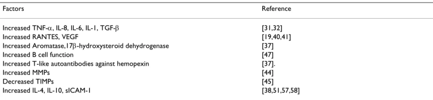
CD4 T cells are divided into type1 (Th1) helper T cells which secrete interleukin (IL)-2, IL-12 and interferon γ and type2 (Th2) helper T cells, which secrete IL-4, 5, 6 10, 13 [38]. Cell-mediated immunity, including T-cell-medi-ated cytotoxicity is activT-cell-medi-ated or suppressed by cytokines produced by Th1 and Th2 cells, respectively. Under nor-mal conditions, there is a tightly regulatory control mech-anism between Th1 and Th2 cells. For instance, Th1 cells secrete IL-12, which activates cytotoxic NK cell activity, whereas Th2 cells may reduce NK cell activity by produc-ing IL-10 [38]. In women with endometriosis, Th2 helper Table 1: Aberrantly regulated immune/inflammatory factors associated with the development of endometriosis
Factors Reference
Increased TNF-α, IL-8, IL-6, IL-1, TGF-β [31,32]
Increased RANTES, VEGF [19,40,41]
Increased Aromatase,17β-hydroxysteroid dehydrogenase [37]
Increased B cell function [47]
Increased T-like autoantibodies against hemopexin [37].
Increased MMPs [44]
Decreased TIMPs [45]
cells from PF are reported to aberrantly suppress cell-mediated immunity by upregulating IL-4 and 10 secre-tions in PF from women with endometriosis [38,55]. As a result, decreased T cell cytotoxicity may allow implanta-tion of endometrial cells in peritoneum.
Impaired immune-surveillance and abnormal apoptosis
The failure of immune cells to transmit death signals to endometrial cells, and/or the ability of endometrial frag-ments to avoid cell death may be associated with the development of endometriosis. Indeed, in women with endometriosis, it has been hypothesed that endometrial cells in the peritoneal fluid avoid immunosurveillance and implant into peritoneum [56]. It has been speculated that lymphocytes can adhere to endometrial cells through the Lymphocyte Function-Associated Antigen-1(LFA-1) – Intercellular adhesion molecule-1 (ICAM-1) dependent pathway and present them as a target to NK cells. Soluble forms of ICAM-1 (s-ICAM-1) secreted by PF endometrial cells/endometriotic lesions can also bind to LFA-1 pre-senting lymphocytes and could prevent the recognition of endometrial cells by these lymphocytes and prevent sub-sequent NK cell-mediated cytotoxicity [57,58]. Further-more, IL-6 secreted by endometriotic cells in concert with interferon-γ may upregulate sICAM-1 production by mac-rophages of patients with endometriosis [59]. As a result, increased secretion of sICAM-1 may allow endometrial fragments to evade immunosurveilance, survive and implant.
Another major pathway in programmed cell death, Fas-Fas Ligand (Fas-FasL) system, could also be abnormal in women with endometriosis [60,61]. It has been specu-lated that the expression of FasL by viable endometrial cells induces apoptosis of T cells through ligation of Fas, allowing endometrial fragments to escape cell death, implant and develop to endometriotic lesions [62]. Inter-estingly, Garcia-Velasco et al [62] showed that macro-phage-conditioned media might stimulate Fas-Fas ligand (FasL) expression by endometrial cells.
Relevance in clinical research
Biomarkers to predict endometriosis non-surgically
Presently, the diagnosis of endometriosis can be made only by laparoscopy and biopsy of suspicious lesions with subsequent histological confirmation of endometrial tis-sue and there is no non-invasive way to diagnose this con-dition. Laparascopy is minimally invasive procedure, but requires general anaesthesia and surgical skills with potential complications and procedural costs. Hence, a non-surgical diagnostic tool would be of paramount ben-efit to both physicians and patients.
Efforts to evaluate the diagnostic value of endometrial markers for endometriosis have been hampered by the
lack of easy, reliable and quantitative techniques to assess the expression levels of these markers in sample material. Emerging proteomic techniques offer new approaches to identifying biomarkers for the early detection and follow-up of endometriosis.
Aromatase P450 mRNA has been identified as a candidate diagnostic marker but low sensitivity and specificity impair its application in clinical practice (Reviewed by Brosens et al [63]). In a recent study [64], the measure-ment of serum IL-6 levels and PF TNF-α levels could dis-criminate between patients with endometriosis and those without the disease. Endometriosis could be diagnosed if TNF-alpha levels in PF were higher than 15 pg/ml (100% sensitivity and 89% specificity) and if IL-6 levels in serum were above 2 pg/ml (90% sensitivity and 67% specificity) [64]. Potentially, the quantitation of autoantibodies against endometrial cells could also provide a novel method for the non-invasive diagnosis for endometriosis [65]. However, more studies are needed to confirm that this approach may be clinically useful. The role of Trans-vaginal Ultrasonography (TVU) and Magnetic resonance Imaging (MRI) in diagnosis and follow up of small endometriotic lesions is limited at present, but represents an interesting area of research [63].
New treatment options and future management of endometriosis
Overall, medical treatment of endometriosis is limited by cost, side effects and recurrence of endometriosis after the cessation of treatment. Therefore, there is need for new drugs that treat endometriosis-associated pain and infer-tility without inhibition of ovulation. Future potential tar-gets in the treatment or management of endometriosis may be inflammatory cytokines, MMPs, adhesion and growth factors [80,38]. Pentoxifylline has been shown to reduce endometriotic implant growth without inducing hypoestrogenism in both humans and hamsters [38]. In a recent study, Hornung and colleagues [81] demonstrated that Thiazolidinedione (TZD) significantly reduced leu-kocyte infiltration in the mouse model with endometrio-sis. Inhibition of TNF-alpha activity has also been a new target in the prevention and treatment of endometriosis. Experimental endometriosis in rats was treated with recombinant human tumour necrosis factor-binding pro-tein-1 (r-hTBP-1), a soluble form of tumour necrosis fac-tor-α receptor type-1 [82]. It was demonstrated that r-hTBP-1 could reduce the size of endometriotic-like perito-neal lesions by 64% [82]. Similarly, a study carried out in baboons showed that r-hTBP-1 effectively inhibited the development of endometriosis and endometriosis-related adhesions [83]. The potential of etanercept, a soluble TNF receptor (TNFR) fusion protein, for the treatment of endometriosis is also being considered [84].
Conclusion
Understanding the involvement of the immune system in the development of endometriosis may help to under-stand the pathogenesis and spontaneous evolution of this condition. At present, most evidence suggests that pelvic inflammation and other immunological changes are a consequence of endometriosis. The development of a non-invasive diagnostic tools based on cytokines and autoantibodies could be of great benefit in the clinical management of endometriosis. Therapeutic strategies to eliminate the inflammatory reaction associated with endometriosis could lead to new treatment options for endometriosis.
References
1. Farquhar CM: Extracts from the "clinical evidence". Endometriosis.BMJ 2000, 320:1449-1452.
2. D'Hooghe TM, Debrock S, Hill JA, Meuleman C: Endometriosis and subfertility: is the relationship resolved?Semin Reprod Med
2003, 21:243-254.
3. Guarnaccia M, Olive DL: The structure and future of endome-triosis research.Obstet Gynecol Clin North Am 1997, 24:455-465. 4. Sampson JA: Peritoneal endometriosis due to menstrual
dis-semination of endometrial tissue into the peritoneal cavity.
Am J Obstet Gynecol 1927, 14:422-469.
5. Bartosik D, Jacobs SL, Kelly LJ: Endometrial tissue in peritoneal fluid.Fertil Steril 1986, 46:796-800.
6. Blumenkrantz MJ, Gallaagher N, Bashore RA, Tenckhoff H: Retro-grade menstruation in women undergoing chronic perito-neal dialysis.Obstet Gynecol 1981, 142:890-895.
7. D'Hooghe TM, Bambra CS, Raeymaekers BM, Koninckx PR:
Increased incidence and recurrence of retrograde
menstru-ation in baboons with spontaneous endometriosis. Hum Reprod 1996, 11:2022-2025.
8. D'Hooghe TM, Debrock S: Endometriosis, retrograde menstru-ation and peritoneal inflammmenstru-ation in women and baboons.
Human Reproduction Update 2002, 8:84-88.
9. Cramer DW, Missmer SA: The epedimiology of endometriosis.
Ann N Acad Sci 2002, 955:11-22.
10. Pinsonneault O, Goldstein DP: Obstructing malformations of the uterus and vagina.Fertil Steril 1985, 44:241-247.
11. D'Hooghe TM, Bambra CS, Suleman MA, Dunselman GA, Evers HL, Koninckx PR: Development of a model of retrograde menstru-ation in baboons (Papio anubis.Fertil Steril 1994, 62:635-638. 12. D'Hooghe TM, Hill JA: Immunobiology of endometriosis. In:
Immunology of reproduction Edited by: Bronston R, Anderson DJ. Cam-bridge, MA: Blackwell Scientific; 1996:322-356.
13. Betjes MG, Tuk CW, Struijk DG, Krediet RT, Arisz L, Hart M, Beelen RH: Interleukin-8 production by human peritoneal mesothe-lial cells in response to tumor necrosis factor-alpha, inter-leukin-1, and medium conditioned by macrophages cocultured with Staphylococcus epidermidis.J Infect Dis 1993,
168:1202-1210.
14. Akoum A, Lemay A, Paradis I, Rheault N, Maheux R: Secretion of interleukin-6 by human endometriotic cells and regulation by proinflammatory cytokines and sex steroids.Hum Reprod
1996, 11:2269-2275.
15. D'Hooghe TM, Debrock S, Meuleman C, Hill JA, Mwenda JM: Future directions in endometriosis research.Obstet Gynecol Clin North Am 2003, 30:221-244.
16. Wieser F, Fabjani G, Tempfer C, Schneeberger C, Zeillinger R, Huber JC, Wenzl R: Tumor necrosis factor-alpha promotor polymor-phisms and endometriosis.J Soc Gynecol Investig 2002, 9:313-318. 17. Pizzo A, Salmeri FM, Ardita FV, Sofo V, Tripepi M, Marsico S: Behav-iour of cytokine levels in serum and peritoneal fluid of women with endometriosis.Gynecol Obstet Invest 2002, 54:82-87. 18. Altman GB, Gown AM, Luchtel DL, Baker C: RANTES production by cultured primate endometrial epithelial cells.Am J Reprod Immunol 1999, 42:168-174.
19. Hornung D, Fujii E, Lim KH, Vigne JL, McMaster MT, Taylor RN: His-tocompatibility leukocyte antigen-G is not expressed by endometriosis or endometrial tissue. Fertil Steril 2001,
75:814-817.
20. Bullimore DW: Endometriosis is sustained by tumour necrosis factor-alpha.Med Hypotheses 2003, 60:84-88.
21. Rana N, Braun DP, House R, Gebel H, Rotman C, Dmowski WP:
Basal and stimulated secretion of cytokines by peritoneal macrophages in women with endometriosis.Fertil Steril 1996,
65:925-930.
22. Harada T, Enatsu A, Mitsunari M, Nagano Y, Ito M, Tsudo T, Tan-aguchi F, Iwabe T, Takinawa M, Terakawa N: Role of cytokines in progression of endometriosis.Gynecol Obstet Invest 1999, Suppl 47:34-39.
23. D'Hooghe TM, Bambra CS, Raeymaekers BM, Hill JA: Pelvic inflam-mation induced by diagnostic laparoscopy in baboons.Fertil Steril 1999, 72:1134-1141.
24. D'Hooghe TM, Bambra CS, Ling X, Peixe K, Hill JA: The effect of menstruation and intrapelvic injection of endometrium on peritoneal fluid parameters in the baboon.Am J Obstet Gynecol
2001, 184:917-925.
25. D'Hooghe TM, Hill JA, Oosterlynck DJ, Koninckx PR, Bambra CS:
Effect on endometriosis and the menstrual cycle on white blood cell subpopulations in the peripheral blood and perito-neal of baboons.Hum Reprod 1996, 11:1736-1740.
26. D'Hooghe TM: Clinical relevance of the baboon as a model for the study of endometriosis.Fertil Steril 1997, 68:613-625. 27. D'Hooghe TM, Bambra CS, Raeymaekers BM, De Jonge I, Hill JA,
Koninckx PR: The effects of immunosuppression on develop-ment and progression of endometriosis in baboons (Papio anubis).Fertil Steril 1995, 6:172-178.
28. Steinleitner A, Lambert H, Suarez M, Serpa N, Robin B, Canter B:
Periovulatory calcium channel blockade enhances reproduc-tive performance in an animal model for endometriosis-associated subfertility.Am J Obstet Gynecol 1991, 164:949-952. 29. Steinleitner A, Lambert H, Roy S: Immunomodulation with
treat-ment of endometriosis-associated infertility.Fertil Steril 1991,
55:26-31.
30. Gaetje R, Kotzian S, Herrmann G, Baumann R, Starzinski-Powitz A:
Nonmalignant epithelial cells, potentially invasive in human endometriosis, lack the tumor suppressor molecule E-cad-herin.Am J Pathol 1997, 150:461-467.
31. Berkkanoglu M, Arici A: Immunology and endometriosis.Am J Reprod Immunol 2003, 50:48-59.
32. Iwabe T, Harada T, Tsudo T, Nagano Y, Yoshida S, Tanikawa M, Ter-akawa N: Tumor necrosis factor-alpha promotes proliferation of endometriotic stromal cells by inducing interleukin-8 gene and protein expression. J Clin Endocrinol Metab 2000,
85:824-829.
33. Witz CA, Monotoya-Rodriguez IA, Schenken RS: Whole explants of peritoneum and endometrium: a novel model of the early endometriosis lesion.Fertil Steril 1999, 71:56-60.
34. Debrock S, Vander Perre S, Meuleman C, Moerman P, Hill JA, D'Hooghe TM: In-vitro adhesion of endometrium to autolo-gous peritoneal membranes: effect of the cycle phase and the stage of endometriosis.Hum Reprod 2002, 17:2523-2528. 35. Witz CA, Thomas MR, Montoya-Rodriguez IA, Nair AS, Centonze
VE, Schenken RS: Short-term culture of peritoneum explants confirms attachment of endometrium to intact peritoneal mesothelium.Fertil Steril 2001, 75:385-390.
36. Nothnick WB, D'Hooghe TM: Medical Management of Endome-triosis: Novel Targets and Approaches towards the Develop-ment of Future TreatDevelop-ment Regimes.Gynecol Obstet Invest 2003,
55:189-198.
37. Yeaman GR, Collins JE, Lang GA: Autoantibody responses to car-bohydrate epitopes in endometriosis.Ann N Y Acad Sci 2002,
955:174-182.
38. Harada T, Iwabe T, Tarakawa N: Role of cytokines in endometriosis.Fertil Steril 2001, 76:1-10.
39. Nothnick WB: Treating endometriosis as an autoimmune disease.Fertil Steril 2001, 7:223-231.
40. McLaren J, Prentice A, Charnock-Jones DS, Smith SK: Vascular endothelial growth factor (VEGF) concentrations are ele-vated in peritoneal fluid of women with endometriosis.Hum Reprod 1996, 11:220-223.
41. McLaren J: Vascular endothelial growth factor and endometri-otic angiogenesis.Hum Reprod Update 2000, 6:45-55.
42. Kats R, Collette T, Metz CN, Akoum A: Marked elevation of mac-rophage migration inhibitory factor in the peritoneal fluid of women with endometriosis.Fertil Steril 2002, 78:69-76. 43. Bruner KL, Eisenberg E, Gorstein F, Osteen KG: Progesterone and
transforming growth factor-beta coordinately regulate sup-pression of endometrial matrix metalloproteinases in a model of experimental endometriosis. Steroids 1999,
64:648-653.
44. Sillem M, Prifti S, Koch A, Neher M, Jauckus J, Runnebaum B: Regu-lation of matrix metalloproteinases and their inhibitors in uterine endometrial cells of patients with and without endometriosis.Eur J Obstet Gynecol Reprod Biol 2001, 95:167-174. 45. Gottschalk C, Malberg K, Arndt M, Schmitt J, Roessner A, Schultze D,
Kleinstein J, Ansorge S: Matrix metalloproteinases and TACE play a role in the pathogenesis of endometriosis.Adv Exp Med Biol 2000, 477:483-486.
46. D'Hooghe TM, Hill JA: Is endometriosis an autoimmunological disease? In: Immunology of reproduction Edited by: Kurpisz M, Fernan-dez N. Oxford University Press; 1995:133-162.
47. Chishima F, Hayakawa S, Hirata Y, Nagai N, Kanaeda T, Tsubata K, Satoh K: Peritoneal and peripheral B-1-cell populations in patients with endometriosis. J Obstet Gynaecol Res 2000,
26:141-149.
48. Startseva NV: Clinical immunological aspects of genital endometriosis.Akush Ginekol (Mosk) 1980, 3:23-26.
49. Wilson TJ, Hertzog PJ, Angus D, Munnery L, Wood EC, Kola I:
Decreased natural killer cell activity in endometriosis patients: relationship to disease pathogenesis.Fertil Steril 1994,
62:1086-1088.
50. D'Hooghe TM, Scheerlinck JP, Koninckx PR, Hill JA, Bambra CS:
Anti-endometrial lymphocytotoxicity and natural killer cell activity in baboons (Papio anubis and Papio cynocephalus) with endometriosis.Hum Reprod 1995, 10:558-562.
51. Matarese G, De Placido G, Nikas Y, Alviggi C: Pathogenesis of endometriosis: natural immunity dysfunction or autoim-mune disease?Trends Mol Med 2003, 9:223-228.
52. Wilson MT, Van Kaer L: Natural killer T cells as targets for ther-apeutic intervention in autoimmune diseases.Curr Pharm Des
2003, 9:201-220.
53. Dmowski WP, Rana N, Michalowska J, Friberg J, Papierniak C, el-Roeiy A: The effect of endometriosis, its stage and activity, and of autoantibodies on in vitro fertilization and embryo transfer success rates.Fertil Steril 1995, 63:555-562.
54. Vercammen EE, D'Hooghe TM: Endometriosis and recurrent pregnancy loss.Semin Reprod Med 2000, 18:363-368.
55. Hsu CC, Yang BC, Wu MH, Huang KE: Enhanced interleukin-4 expression in patients with endometriosis. Fertil Steril 1997,
67:1059-1064.
56. Somigliana E, Vigano P, Gaffuri B, Guarneri D, Busacca M, Vignali M:
Human endometrial stromal cells as a source of soluble intercellular adhesion molecule (ICAM)-1 molecules.Hum Reprod 1996, 11:1190-1194.
57. Vigano P, Gaffuri B, Somigliana E, Busacca M, Di Blasio AM, Vignali M:
Expression of intercellular adhesion molecule (ICAM)-1 mRNA and protein is enhanced in endometriosis versus endometrial stromal cells in culture.Mol Hum Reprod 1998,
4:1150-1156.
58. Becker JC, Termeer C, Schmidt RE, Brocker EB: Soluble intercel-lular adhesion molecule-1 inhibits MHC-restricted specific T cell/tumor interaction.J Immunol 1993, 151:7224-7232. 59. Fukaya T, Sugawara J, Yoshida H, Murakami T, Yajima A:
Intercellu-lar adhesion molecule-1 and hepatocyte growth factor in human endometriosis: original investigation and a review of literature.Gynecol Obstet Invest 1999, 47:11-16.
60. Lebovic DI, Mueller M, Taylor R: Immunobiology of endometriosis.Fetil Steril 2001, 75:1-10.
61. Garcia-Velasco JA, Arici A: Apoptosis and the pathogenesis of endometriosis.Semin Reprod Med 2003, 21:165-172.
62. Garcia-Velasco JA, Arici A, Zreik T, Noftolin F, Mor G: Macrophage derived growth factors modulate Fas ligand expression in cultured endometrial stromal cells: a role in endometriosis.
Mol Hum Reprod 1999, 5:642-650.
63. Brosens J, Timmerman D, Starzinski-Powitz A, Brosens I: Noninva-sive diagnosis of endometriosis: the role of imaging and markers.Obstet Gynecol Clin North Am 2003, 30:95-114.
64. Bedaiwy MA, Falcone T, Sharma RK, Goldberg JM, Attaran M, Nelson DR, Agarwal A: Prediction of endometriosis with serum and peritoneal fluid markers: a prospective controlled trial.Hum Reprod 2002, 17:426-431.
65. Inagaki J, Sugiura-Ogasawara M, Nomizu M, Nakatsuka M, Ikuta K, Suzuki N, Kaihara K, Kobayashi K, Yasuda T, Shoenfeld Y, Aoki K, Matsuura E: An association of IgG anti-laminin-1 autoantibod-ies with endometriosis in infertile patients.Hum Reprod 2003,
18:544-549.
66. D'Hooghe TM, Hill JA: Chapter 25. Endometriosis. In: Novak's Gynecology 13th edition. Edited by: Williams and Wilkins, JS Berek. Phil-adelphia, USA; 2002:931-972.
67. El-Roeiy A, Dmowski WP, Gleicher N, Radwanska E, Harlow L, Binor Z et al.: Danazol but not gonadotropin-releasing hormone agonists suppresses autoantibodies in endometriosis. Fertil Steril 1988, 50:864-871.
68. Ota H, Maki M, Shidara Y, Kodama H, Takahashi H, Hayakawa M et al.: Effects of danazol at the immunologic level in patients with adenomyosis, with special reference to autoantibodies: a multi-center cooperative study.Am J Obstet Gynecol 1992,
167:481-486.
69. Takahashi K, Yoshino K, Kusakari M, Katoh S, Shibukawa T, Kitao M:
Prognostic potential of serum CA125 levels in danazol-treated patients with external endometriosis: a preliminary study.Int J Fertil 1990, 35:226-229.
70. Franssen AMHW, van der Heijden PFM, Thomas CMG, Doesburg WH, Willemsen WNP, Rolland R: On the origin and significance of serum CA125 concentrations in 97 patients with endome-triosis before, during, and after buserelin acetate, nafarelin, or danazol.Fertil Steril 1992, 57:974-979.
71. Bischof P, Galfetti MA, Seydoux J, von Hospenthal JU, Campana A:
Peripheral CA125 levels in patients with uterine fibroids.
Publish with BioMed Central and every scientist can read your work free of charge
"BioMed Central will be the most significant development for disseminating the results of biomedical researc h in our lifetime."
Sir Paul Nurse, Cancer Research UK
Your research papers will be:
available free of charge to the entire biomedical community
peer reviewed and published immediately upon acceptance
cited in PubMed and archived on PubMed Central
yours — you keep the copyright
Submit your manuscript here:
http://www.biomedcentral.com/info/publishing_adv.asp
BioMedcentral
72. Acien P, Shaw RW, Irvine L, Burford G, Gardner R: CA125 levels in endometriosis patients before, during and after treatment with danazol or LHRH agonists. Eur J Obstet Gynecol 1989,
32:241-246.
73. Hill JA, Barbieri RL, Anderson DJ: Immunosuppressive effects of danazol in vitro.Fertil Steril 1987, 48:414-418.
74. Mori H, Nakagawa M, Itoh N, Wada K, Tamaya T: Danazol sup-presses the production of interleukin-1b and tumor necrosis factor by human monocytes. Am J Reprod Immunol 1990,
24:45-50.
75. Braun DP, Gebel H, Rotman C, Rana N, Dmowski WP: The devel-opment of cytotoxicity in peritoneal macrophages from women with endometriosis.Fertil Steril 1992, 1203:1203-1210. 76. Gelfand JA, Sherins RJ, Alling DW, Frank MM: Treatment of
hered-itary angioedema with danazol. N Engl J Med 1976,
295:1444-1448.
77. Ahn YS, Harrington WJ, Mylvaganam R, Ayub J, Pall LM: Danazol therapy for autoimmune hemolytic anemia.Ann Intern Med
1985, 102:298-301.
78. Agnello V, Pariser K, Gell J, Gelfand J, Turksoy RN: Preliminary observations on danazol therapy of systemic lupus ery-thematosus: effect on DNA antibodies, thrombocytopenia and complement.J Rheumatol 1983, 10:682-687.
79. Mylvaganam R, Ahn YS, Harrington WJ, Kim CI: Immune modula-tion by danazol in autoimmune thrombocytopenia.Clin Immu-nol Immunopathol 1987, 42:281-287.
80. D'Hooghe TM: Immunomodulators and aromatase inhibitors: are they the next generation of treatment for endometriosis?Curr Opin Obstet Gynecol 2003, 15:243-249. 81. Hornung D, Chao VA, Vigne JL, Wallwiener D, Taylor RN:
Thiazo-lidinedione inhibition of peritoneal inflammation. Gynecol Obstet Invest 2003, 55:20-24.
82. D'Antonio M, Martelli F, Peano S, Papoian R, Borrelli F: Ability of recombinant human TNF binding protein-1 (r-hTBP-1) to inhibit the development of experimentally-induced endome-triosis in rats.J Reprod Immunol 2000, 48:81-98.
83. D'Hooghe TM, Nugent N, Cuneo S, Chai D, Deer F, Debrock S, Mwenda J: Recombinant human TNF binding protein (r-hTBP-1) inhibits the development of endometriosis in baboons: a prospective, randomised, placebo- and drug con-trolled study. Presented at the Annual Meeting of the Amer-ican Society for Reproductive Medicine, Orlando, USA, October 22nd–24th 2001 [abstract].Fertil Steril 2001, 76:O-1-S-1. 84. Pugsley MK: Etanercept. Immunex.Curr Opin Investig Drugs 2001,