© 2014, IJCSMC All Rights Reserved
95
Available Online atwww.ijcsmc.com
International Journal of Computer Science and Mobile Computing
A Monthly Journal of Computer Science and Information Technology
ISSN 2320–088X
IJCSMC, Vol. 3, Issue. 6, June 2014, pg.95 – 108
RESEARCH ARTICLE
MODIFIED MUTLIRESOLUTION
MEDICAL IMAGE SEGMENTATION
BASED ON WAVELET TRANSFORM
Dr. K.R.Ananth
1, Dr. P.Navaneetham
21-Associate Professor, 2- Professor & Head, Department of MCA Velalar College of Engineering and Technology, Erode, TamilNadu, India
E-mail: [email protected], [email protected]
ABSTRACT: Wavelet Transform is used to analyze image. The watershed transformations a useful morphological segmentation tool is used for a variety of grey-scale images. An efficient method of segmentation is presented in this proposed method by modified multiresolution, which combines the pyramidal image segmentation with hierarchical watershed segmentation algorithm. The segmentation procedure consists of pyramid representation, image segmentation, region merging and region projection. Each layer is split into a number of regions by rooting labeling technique and boundary is extracted by threshold. Morphological operation is used to smooth the image.
Keyword: Wavelet Transform, Multi resolution, Image segmentation
1.
INTRODUCTION
Image processing plays a vital role for the diagnosis and analysis of various medical imaging modalities such as X-rays, PET, CT and MRI. Qualification by manual tracing of outlines of structures is tedious and time consuming. As a result a segmentation technique that is robust and is characterized by a high degree of accuracy is proposed. Various categories of segmentation are clustering, edge detection and region extraction.
The wavelet transform has good localization property in both time and frequency domain. Many researchers have used the wavelet analysis to detect and characterize the irregular parts of signals [7. The capability of neural networks can be further extended by embedding the wavelets inside the network in place of sig-moidal activation functions.
© 2014, IJCSMC All Rights Reserved
96
and noise problem are reduced as the watershed operations are carried out on the low pass filtered low- resolution image from wavelet transform.
This proposed modified multiresolution using transform aspires in improving the segmentation process, which is based on modified Multi-resolution analysis for a pyramidal hierarchical image. The over segmentation and noise problem are reduced as the watershed operation is carried out on the low-pass filtered low-resolution images from wavelet transform. Morphological operation is introduced into image processing.
2.
SEGMENTATION METHOD
Original image is smoothened by using morphological filter to reduce noise particles; pyramid representation creates multiresolution images using wavelet transform which has been modified by the parametric inverse transformation. The images at different layers represent various image resolutions. Images are first segmented into a number of regions at each layer of the pyramid with watershed transformation. Starting from top layer regions are merged with merging parameter. The resulting image is projected onto the next layer by inverse wavelet transform until L equals 0.
3.
Wavelet Transform
Since wavelets are localized in both the space and frequency domains, they have a multiresolution property. This makes it suitable for extracting multiresolution from low-frequency backgrounds and high-frequency noise[8]. In particular, the wavelet transforms decomposes the signal into signal bands of different frequency ranges. It can help to identify useful information relevant to MCCs and discard the signal bands which make little contribution to detection.
The wavelet used in this study is Daubechies orthogonal wavelet of length four. The ROIs are decomposed up to four levels using the wavelet transform.
4.
THE PROPOSED METHOD
The objective of our research is to design a boundary-based object recognition approach that utilizes the modified multiresolution to extract distinctive wavelet features at the optimum scale–translation grids of the boundary curvature representation and perform the classification automatically. The extracted wavelet features are the outputs of the wavelet nodes that correspond to the Wavelet transform coefficients yielded by convolving the normalized curvature function with wavelet basis functions at the optimum scale– translation grids.
The wavelet transform is a powerful tool [3], [4]. Unlike the Fourier sinusoids, which provide a sharp frequency characterization of a given signal but which are unable to identify transient events, wavelets achieve a balance between localization in space or time, and localization in the frequency domain. This balance is intrinsic to multiresolution, which allows the analysis to deal with image features at any scale.
As the discrete wavelet transform corresponds to basis decomposition, it provides a non-redundant and unique representation of the signal. These fundamental properties are key to the efficient decomposition of the non-stationary processes typical of fMRI experimental settings. Consequently, wavelets have received a large recognition in biomedical signal and image processing; several overviews are available [9], including work that is tailored to fMRI [10].
The discrete wavelet transform (DWT) of a one-dimensional signal v(x)—typically represented by its samples v[n], n Z—is a decomposition into a sum of basis functions. These functions are shifted and dilated versions of a (bandpass) wavelet function (x) and shifted versions of a (lowpass) scaling function
(x).
In 2-D, for example, this construction leads to 1 scaling function ((x1) (x2)) and 3 wavelets ((x1)(x2)), ((x1)(x2)) , ((x1)(x2)) . It leads to 1 scaling function and 7 wavelets in 3-D.
A.
Multi-scale morphological filtering method
© 2014, IJCSMC All Rights Reserved
97
gray- level images by a determined composition of opening and closing with a given structuring element. The main shortcoming of conventional opening and closing is that it does not allow a perfect preservation of the edge information. This operator emphasizes only on the size of the feature but ignores their shape completely. However it is possible to design morphological filters by reconstruction for both shapes and sizes of the feature.
The elementary geodesic dilation and erosion is defined as the minimum between dilated by as SE, S of size one. Hence reconstructing dilation and erosion of arbitrary size is obtained through iteration. Based on the operation opening and closing by reconstruction of opening and closing may be defined by the equation (1.1)
Mi*S =δS (Mi*S) … (1.1)
Because reconstructing opening and closing void drawback of conventional operation this morphological operation helps segmentation obtain more semantic region partitioning in the later stages. Before removing noise particles, it is a necessity to estimate their scale according to the analysis.
B.
Pyramidal representation
In recent years, the wavelet transform has become an active research area in the signal and image analysis communities. Unlike Fourier descriptors that use global sinusoids as the basis functions, the wavelet description is more efficient in representing and detecting local features of an object boundary due to the time frequency localization of wavelet bases [8]. With the detection of local features, an object can easily be recognized.
The modified Multiresolution methods attempt to obtain a global view of an image by examining it at various resolutions by modifying the levels building the pyramid representation of an image when compared to the existing method. Several types of multiresolution image decomposition include Gaussian Pyramids, Laplacian Pyramids and wavelets. Both Gaussian and Laplacian Pyramid generally entail some loss of information. Unlike these methods, the wavelet transform provides a complete image representation and performs decomposition according to both scale orientation. To create a multiresolution image, Haar wavelet transform is used.
The wavelet transform of a signal f (x) is performed by convolution of the signal with a family of basis function given in the equation (1.2) as
(F (x), Ψ2(x)) = ∫ f (x) Ψ2 (Xi) dx … (1.2)
Where Ψ2 (x) is the basis function s and l are referred as dilation and translation parameters, respectively.
The image can be decomposed into its wavelet coefficients using Mallat pyramid algorithm. By using Haar wavelets, the original image is first passed through low-pass filters to generate LL, LH, HL and HH sub bands. The decompositions are repeated on the LL Sub band to obtain the next four-sub bands. For J scale transforms; the original image can be represented in the equation (1.3) by
(W2i, ( WH2i, WV2i, WD2i))1 ≤ I ≤ J … (1.3)
where the size of the wavelet representation is the same as the original signal. This representation is composed of a coarse signal at resolution 2i and a set of detail signal at resolution 2i-2j.
C. Image segmentation and segmentation Projection
After creating the pyramid image using wavelet transform, the different resolution images is segmented through the application of watershed algorithm. Generally the blurred images represented in each layer of the pyramid are used for segmentation. Segmentation is applied in two stages. First, the scale with which morphological operation is defined at every layer .the n the classical the watershed algorithm is used to generate an object contours.
© 2014, IJCSMC All Rights Reserved
98
5.
Steps followed in applying watershed algorithm
Step 1: Read the image.
Step 2: Maximize the contrast.
Step 3: Subtract images.
Step 4: Convert object of interest.
Step 5: Detect intensity valley.
Step 6: Apply watershed segmentation.
Read Image:
Read in the given carotid artery image, which is the medical image got from ultrasonic scanning.
Maximize Contrast:
Many objects of different sizes touch each other. To minimize the number of valleys found by the watershed transform, maximize the contrast of the objects of interest. A common technique for contrast enhancement is the combined use of the top-hat and bottom-hat transforms.
The top-hat transform is defined as the difference between the original image and its opening. The opening of an image is the collection of foreground parts of an image that fit a particular structuring element.
The bottom-hat transform is defined as the difference between the closing of the original image and the original image. The closing of an image is the collection of background parts of an image that fit a particular structuring element. The common structuring elements are squares, rectangles, disks, diamonds, balls, and lines. Since the objects of interest in the image look like disks, structuring element, which is disk shape, is used. This disk size is the average object radius in the image.
Subtract Images:
The top-hat image contains the "peaks" of objects that fit the structuring element. In contrast, the bottom-hat image shows the gaps between the objects of interest. To maximize the contrast between the objects and the gaps that separate them from each other, the "bottom-hat" image is subtracted from the "original + top-hat" image.
Convert Objects of Interest:
Watershed transform detects intensity "valleys‖ in the image. The enhanced image is converted
into the objects of interest with high intensity valleys.
Detect Intensity Valleys:
All the intensity valleys are below a particular threshold .The output of the imextendedmin function is a binary image. The location rather than the size of the regions is important. The imimposemin function modifies the image to contain only those valleys found by the imextendedmin function. The imimposemin function will also change a valley's pixel values to zero (deepest possible valley for uint8 images). All regions containing an imposed minimum will be detected by the watershed transform.
Watershed Segmentation:
Watershed segmentation of the imposed minima image is accomplished with the watershed function. The watershed function returns a label matrix containing non-negative numbers that correspond to watershed regions. Pixels that do not fall into any watershed region are given a pixel value of zero.
However the image is degraded by noise so causes over-segmentation. Therefore over-segmentation images are further merged in some regions. The region to be merged is based on homogeneity and similarity criteria based on the wavelet coefficients. Each of the region having mean, second and third order central moments values of the wavelet coefficients are calculated.
Merging over-segmentation regions:
© 2014, IJCSMC All Rights Reserved
99
watershed transform, the same aim or the same background is divided into several parts. The conditions for combining rules are:
a) The regions that will be combined are neighbors.
b) The characteristics of the region, which will be combined, should be similar. c) the big region after combining is useful.
To merge the region which are neighbors and similar can be according to the similitude. If similitude is higher than threshold, then the two neighbor fields can be seen as the same region that can be combined. After combination, the background and the aim are separated. Usually, similitude can be defined according to gray, texture, and so on.
Boundary gray mean can be observed by the dissimilarity within the each CB of given medical image. Suppose BPi is set of boundary points of catchment basin I (CBj), Ni stands for the number of i,
then the boundary gray mean of the CBj, The definition of mean (i) is given by
mean (i) = f(pj) / i … (1.4)
Normally, the gray mean of the whole CB is applied as the standard for joining neighbor areas, but because the main dissimilarity among each CB of medical images after the watershed transforms lies in boundary, the method is to calculate gray mean of boundary points. It not only reduces the work of calculation but also neglects the side factors which affect judgment and decision; it also stresses the primary factors.
In medical image for thee effect of noise many small CB are produced. these small CB are combined to form the large image.
Once the merged image M is generated at L layer it must be projected onto next layer in order to finish the full resolution image segmentation. Direct projection of the segmented image offers very poor results. At each projection pyramid level, the region frontiers become less smooth ,showing a heavy blocking effects appearance .To overcome the problem inverse wavelet transform to implement projection from low to high resolution layer step by step.
During projection from I th layer to I-1 th layer, a parent-child spatial relationship between the image elements of two successive layers is defined. This relationship is evaluated by means of a similarity measure. The children of a layer can belong to different parents in the upper layer. Similarity between a child image element and its possible parents describes the similarities. By feature of image elements similarity is defined, for example by comparing the contrast or texture properties of a child and its possible parents.
Figure: Block diagram representation of image segmentation
Ultrasound image / CT /
© 2014, IJCSMC All Rights Reserved
100
6.
RESULTS AND DISCUSSION
Initially the ultrasound image is read from a ultrasound scanner and this is shown in figure 4.1. The input image is the common carotid artery. The aim of this project is to extract the boundary of common carotid artery. The input common carotid artery image is then subjected to second level decomposition using wavelet transformation. This is shown in figure 4.2.
One level decomposition Produces HH, HL, LH, LL images .On two level Decomposition HHHH, HHHL, LLHL, LLLL is got. This LLLL image on watershed transform overcomes over segmentation. It is proved from the obtained results that the second level decomposition is more accurate than first level decomposition.
Such an image is subjected to top hat and bottom hat method. This is shown in figure 4.3 and 4.4. Then the top hat image is combined with original image from that bottom hat image is subtracted This is shown in figure 4.5. Such a subtracted image is enhanced .That image is subjected to watershed segmentation. This is shown in figure 4.5.By setting threshold values the boundary is extracted this is shown in figure 4.7 and the boundary is superimposed on the original image as shown in figure 4.8.The Figure 4.9 shows the output got using Haar wavelets for various levels of decomposition (ie) First, second and third Level Decomposition. Similarly the figures 4.10 and 4.11 shows the outputs got using DB4 and DB10 at various decomposition levels. Figure 4.12 shows the extracted boundaries of 28 frames. Figure 4.13 shows the similar output got using watershed segmentation for the longitudinal section of carotid image. Figure 4.14 gives the graphical representation of frames Vs distance
7.
EXPERIMENTAL EVALUTION
In this section, to test the applicability of the proposed Method based recognition method in different domains; we report the results of three object recognition experiments conducted on three different data sets. The results illustrate the stability and efficiency of our classification technique when compared with other object classification solutions, in particular, the approaches using the Wavelet transform preceded by a wavelet filter bank.
Figure 4.1: ULTRASOUND IMAGE OF COMMON CAROTID ARTERY (TRANSVERSE VIEW)
© 2014, IJCSMC All Rights Reserved
101
Figure 4.2 TWO LEVEL IMAGE DECOMPOSITION
Figure4.3 TOP HAT METHOD
Figure.4.4BOTTOM HAT METHOD
F
igure4.5 ORIGINAL IMAGE + TOP-HAT-BOTTOM HAT METHOD© 2014, IJCSMC All Rights Reserved
102
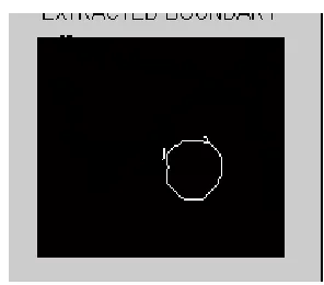
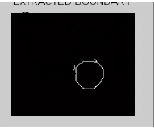
Figure4.7 BOUNDARY EXTRACTION
Figure 4.8SUPERIMPOSED IMAGE
Figure 4.9:OUTPUTS FOR VARIOUS LEVELS OF DECOMPOSITION USING HAARONE LEVEL DECOMPOSITION WITH HAAR
© 2014, IJCSMC All Rights Reserved
103
THREE LEVEL DECOMPOSITION WITH HAAR
Figure 4.10:OUTPUTS FOR VARIOUS LEVELS OF DECOMPOSITION USING DB4 ONE LEVEL WAVELET DECOMPOSITION WITH DB4
TWO LEVEL WAVELET DECOMPOSITION WITH DB4
© 2014, IJCSMC All Rights Reserved
104
Figure 4.11:OUTPUTS FOR VARIOUS LEVELS OF DECOMPOSITION USING DB10 ONE LEVEL WAVELET DECOMPOSITION WITH DB10
TWO LEVEL WAVELET DECOMPOSITION WITH DB10
© 2014, IJCSMC All Rights Reserved
105
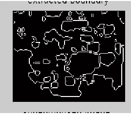
Figure 4.12: EXTRACTED BOUNDARIES OF 28 FRAMES
© 2014, IJCSMC All Rights Reserved
106
Figure 4.13: ULTRASOUND IMAGE OF COMMON CAROTID ARTERY (LONGITUDINAL VIEW)
INPUT IMAGE
TOP HAT-METHOD
BOTTOM HAT METHOD
ORIGINAL+TOP-BOTTOMHAT
© 2014, IJCSMC All Rights Reserved
107
SUPER IMPOSED IMAGE
The above outputs are similar results obtained using watershed segmentation with the longitudinal section of the carotid artery image. The boundary is extracted successfully using DB10 and Second level of decomposition.
FIGURE 4.12: GRAPHICAL OUTPUT OF FRAMES (1 TO 28):
8.
CONCLUSION
The proposed a method has clearly detected the boundary of common carotid artery. Earlier method of boundary extraction concentrated on wavelet transform (or) watershed transform but those methods had a drawback of over-segmentation due to noise. So a method of applying watershed transform for the proposed modified multiresolution method, after wavelet decomposition is proposed and the results are proved to be successful. In case of transversal view of carotid artery the shape is disk shaped so the structuring element used is disk, but in case of longitudinal view the structuring element used is line. The outputs of both transversal section and longitudinal section are obtained.
© 2014, IJCSMC All Rights Reserved
108
REFERENCES
1) Bhandarkar,S.M.,Hui,Z.,‖Image segmentation using evolutionary computation‖,IEEE Trans,Comput 3 (1-21),1999.
2) Rezaee,M.R.,vander zwet, P.M.J .Lelieveldt, B.P.E, van der eest. ,R.J.,Reibert,J.H.C, ‖A Multiresolution in segmentation technique based on pyramidal segmentation and fuzzy clustering‖,IEEE Trans, Image Process,2012.
3) Liu.J.,Yang, Y.H., ―Multiresolution color image segmentation‖, IEEE Trans,2011 4) Canny.J,‖A Computational approach to edge detection‖,IEEE Trans ,2006
5) Paul T. Jackway ,‖ Gradient watersheds in morphological scale-spac‖,IEEE Trans ,Image Processing 2012
6) V.Grau, A.U.J.Mewes and M. Alcaniz, ‖improved watershed transform for medical image segmentation using watershed transformation‖ April 2004.
7) S. Mallat and W. L. Hwang, ―Singularity detection and processing with wavelets,‖ IEEE Trans. Inf. Theory, vol. 38, no. 2, pp. 617–643, Mar.2012.
8) S. L. Phung and A. Bouzerdoum, ―A pyramidal neural network for visual pattern recognition,‖ IEEE Trans. Neural Network., vol. 18, no. 2,pp. 329–343, Mar. 2011.
9) Michael Wirth, Matteo Fraschini, Jennifer Lyon. ―Contrast enhancement of micro calcifications in mammograms using morphological enhancement and non-flat structuring elements‖, IEEE Symposium on CBMS’04, pp.1063-7125, 2004.