DOI: 10.1534/genetics.107.078386
SPD-3 Is Required for Spindle Alignment in
Caenorhabditis elegans
Embryos and Localizes to Mitochondria
Maria V. Dinkelmann,*
,†Haining Zhang,*
,†Ahna R. Skop* and John G. White
†,‡,1*Laboratory of Genetics,†Laboratory of Molecular Biology and‡Department of Anatomy, University of Wisconsin, Madison, Wisconsin 53706
Manuscript received July 3, 2007 Accepted for publication September 20, 2007
ABSTRACT
During the development of multicellular organisms, cellular diversity is often achieved through asymmetric cell divisions that produce two daughter cells having different developmental potentials. Prior to an asymmetric cell division, cellular components segregate to opposite ends of the cell defining an axis of polarity. The mitotic spindle rotationally aligns along this axis of polarity, thereby ensuring that the cleavage plane is positioned such that segregated components end up in individual daughter cells. Here we report our characterization of a novel gene required for spindle alignment inCaenorhabditis elegans. During the first mitosis inspd-3(oj35)embryos the spindle failed to align along the anterior/posterior axis, leading to abnormal cleavage configurations.spd-3(oj35)embryos had additional defects reminiscent of dynein/dynactin loss-of-function possibly caused by the mislocalization of dynactin. Surprisingly, we found that SPD-3TGFP localized to mitochondria. Consistent with this localization,spd-3(oj35)worms exhibited
slow growth and increased ATP concentrations, which are phenotypes similar to those described for other mitochondrial mutants in C. elegans. To our knowledge, SPD-3 is the first example of a link between mitochondria and spindle alignment inC. elegans.
A
SYMMETRIC cell divisions are an essential feature of metazoan development as they provide cell diversity within a multicellular organism. During deter-minative asymmetric cell divisions, specific cellular components are first polarized to opposite ends of the cell. The cleavage plane is then positioned such that it bisects the polarization axis, thereby giving rise to daughter cells with different developmental potentials (Hyman and White1987; Whiteand Strome1996).These actions ensure that the polarized components are segregated to different daughter cells, where they act to determine the differentiated state.
The single-cell embryo ofCaenorhabditis elegans under-goes an asymmetric cell division. Upon fertilization, a centrosome enters the oocyte along with the paternal pronucleus and splits into two. Initially, the daughter centrosomes are aligned perpendicular to the long axis of the embryo. The axis of polarity is determined by the site of fertilization: the sperm entry site designates the posterior of the embryo. This is achieved by an in-teraction of the sperm centrosomes with the cell cortex, which causes cortical material to flow away from the centrosomes (Hird and White1993; Goldstein and
Hird 1996; Golden 2000; Munroet al. 2004). As
po-larity is being established, the pronuclei migrate toward each other, meeting at70% egg length (slightly toward the posterior). When the pronuclei meet, the entire pronuclear/centrosome complex rotates 90°, placing the centrosomes, and consequently the developing mi-totic spindle, along the anterior/posterior (A/P) axis. Because the cleavage plane is always set up to bisect the mitotic spindle (Rappaport1986), the process of
ro-tational alignment ensures that polarized components are segregated to unique daughter cells, thereby mak-ing the division determinative.
Microtubules are required for rotational alignment. Exposure to the microtubule depolymerizing drug nocodazole or mutations in genes such as zyg-9 and
mbk-2result in short astral microtubules that are unable to reach the cortex (Hymanand White1987; Matthews
et al. 1998; Pang et al. 2004). Consequently, the
pro-nuclear/centrosome complex fails to rotate, resulting in an improperly aligned mitotic spindle. Because the resulting cleavage plane does not coordinate with the polarity axis, cell division with a misaligned mitotic spindle often does not segregate polarized determi-nants into appropriate daughter cells and the embryos exhibit gross developmental defects.
The microtubule-associated minus end-directed motor dynein, together with its activator dynactin, is required for rotational alignment (Skopand White1998; Gonczy
et al.1999). Cells deficient in dynein or dynactin function are defective in several additional early developmental Sequence data from this article have been deposited with the EMBL/
GenBank Data Libraries under accession no. NM-068635.
1Corresponding author:Laboratory of Molecular Biology, 1525 Linden Dr., Madison, WI 53706. E-mail: [email protected]
processes including meiosis, pronuclear migration, cen-trosome separation, and chromosome segregation (Skop
and White 1998; Gonczy et al.1999; Yoderand Han
2001). Generally, it is believed that spindle alignment occurs by the capture of astral microtubule plus ends at the cell cortex by localized or locally activated dynein/ dynactin complexes, followed by shortening of the captured microtubules (Whiteand Strome1996; Tsou
et al.2002). Spindle rotation is thought to occur as a result of shortening more captured microtubules from one centrosome over the other. Several models exist that predict mechanisms for this break in the symmetry of pulling forces (Cowanand Hyman2004).
While the search and capture model is attractive, the actual mechanism that determines spindle orientation remains poorly understood. Recent evidence has impli-cated several ‘‘housekeeping’’ genes as having specific and essential roles during spindle alignment. Pro-teins involved in diverse processes such as secretion and nuclear pore formation are required for proper spindle orientation (H. Zhang and J. G. White,
per-sonal communication; Skopet al.2001; Schetteret al.
2006). Here we present evidence of another unexpected player in the process of spindle rotational alignment: mitochondria.
MATERIALS AND METHODS
C.elegansstrains and alleles:The Bristol strain N2 was used as the standard wild-type strain. Culturing, handling, and genetic manipulation of C. elegans were performed using standard procedures (Brenner1974). Temperature-sensitive strains were maintained at 16°and L4 hermaphrodites were shifted to 25°for 24 hr prior to analysis. The following strains were used: WH113 (spd-3(oj35)), MT5734 (nDf41 IV/ nT1½unc(n754), let(IV;V)), MT5241 (unc-5(e53), unc-44(e362)), DR1213 (unc-44(e362), unc-24(e138)), CB4856 (Hawaiian map-ping strain), WH204 (unc-119(ed3);ojIs1[b-tubulinTGFP unc-119(1)]), WH258 (unc-119(ed3); ojIs5[dnc-2TGFP unc-119(1)]),
WH400 (spd-3(oj35); ojIs5[dnc-2TGFP unc-119(1)]), WH342
(unc-119(ed3); ojIs31[spd-3TGFP unc-119(1)]), MQ130 ( clk-1(qm30)), TR1450 (unc-54(r293)), and VC1332 (H34C03.1 (ok1817) IV/nT1[qIs51](IV;V)) (InternationalC. elegansGene Knockout Consortium).
RNA-mediated interference: For spd-3, H34C03.2, Y57A10A.26, and Y48C3A.3 RNA-mediated interference (RNAi) by injection, dsRNA was synthesizedin vitrousing T3 and T7 (or just T7)in vitrotranscription kits (Ambion, Austin, TX) with cDNA clones (yk262b3 or yk1669c08 for spd-3, yk1310d6 for H34C03.2, yk111g2 for Y57A10A.26, and OST104E5-1 for Y48C3A.3) or genomic DNA ½H34C03.2 primers (T7 site in boldface type): TAATACGACTCACTA TACGCGTCTATAAATCCCCGAAAGT and TAATACGACTC
ACTATATGTCACCAGGCCGCATCAT.
All dsRNA was injected at final concentrations of 1–4mg/ml into young hermaphrodite adults (Fireet al.1998). Worms were allowed to recover for 24 hr before embryos were collected.dnc-1,smg-1,smg-2,atp-2,cco-1, andcyc-1RNAi were administered by feeding. Feeding vectors were obtained from the Ahringer library (Kamathet al.2003). The dnc-1RNAi feeding vector used to test the DNC-1 antibody specificity was constructed by cloning the full-length yk11c8 cDNA into the
L4440 feeding vector, followed by transformation into Escher-ichia coli HT115(DE3) bacteria. The E. coli HT115(DE3) bacteria containing the feeding vectors were cultured and used to seed RNAi plates (NGM 1 1mIPTG 1 25mg/ml carbenicillin). L4 hermaphrodites were allowed to feed for
$24 hr before analysis (Timmonsand Fire1998). As a control forsmg-2RNAi efficiency,unc-54(r293)worms were fed RNAi in parallel with spd-3(oj35) worms. smg-2 RNAi has been pre-viously reported to suppress the paralysis phenotype seen in unc-54(r293), which was consistent with our studies (Hodgkin et al.1989).
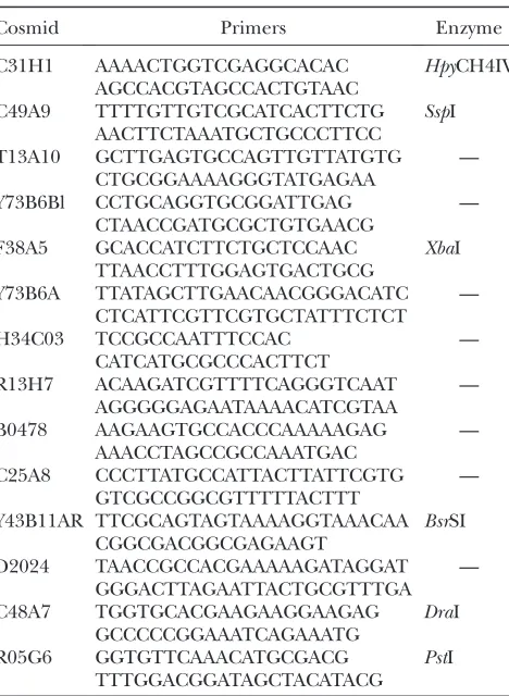
Mapping/cloning: Three-point mapping was done at 16° by crossing spd-3(oj35) males to either MT5241 or DR1213 hermaphrodites. Several heterozygous F1 progeny were sin-gled and allowed to self-fertilize. The resulting F2 progeny were screened for recombinants containing one marker but losing the other. Recombinant worms were singled and homozygosed. After lines were established, several worms from each line were tested for thespd-3(oj35)allele by shifting to the restrictive temperature and screening for dead embryos. SNP mapping was done by crossing CB4856 Hawaiian males to eitherunc-5(e53), spd-3(oj35)hermaphrodites orspd-3(oj35), unc-24(e138) hermaphrodites (Jakubowski and Kornfeld 1999). Several heterozygous F1progeny were singled and al-lowed to self-fertilize. The resulting F2progeny were screened for unc worms that had lost the spd-3(oj35) allele as distin-guished by fertility at 25°. Worms were then genotyped at the appropriate SNPs (Table 1) to determine the region of recombination.
For rescue experiments, cosmid DNA containing the putative wild-type spd-3 gene was injected individually into spd-3(oj35)young adult worms at a concentration of 100mg/ml along with pTG96, a GFP coinjection marker (Yochemet al. 1998) at a concentration of 1 mg/ml (Figure 1A). Progeny were screened for the uptake of a GFP-expressing transgene using a fluorescence dissecting microscope; green worms were singled and allowed to self-fertilize. Once lines were estab-lished, L1 worms were shifted to 25°to test for rescue of the spd-3(oj35)postembryonic phenotype of sterility.
The PCR product used for rescue was generated using the primers 59 TCACATGAAAGCAATAAAACCAAT 39 and 59 TCGGAAAATAAATTGGAAAAGGAG 39.
Thespd-3complementation test was carried out by crossing spd-3(oj35)to VC1332 (H34C03.1(ok1817) IV/nT1[qIs51](IV;V)), which carries a deletion including the first three exons of the gene (WormBase). Thespd-3(oj35)/H34C03.1(ok1817)L4 worms were shifted to 25°overnight to screen for embryonic lethality.
SPD-3 domain searches were done using the following websites: http://smart.embl-heidelberg.de/, http://www.ch. embnet.org/software/COILS_form.html, and http://www.ch. embnet.org/software/TMPRED_form.html.
Measuring embryonic lethality:Dead egg counts were done by placing an L4 worm at restrictive temperature and shifting to a new plate every 24 hr. Dead eggs and larvae were scored 24 hr after the shift.
Live imaging: For colocalization experiments, NGM agar plates were seeded with OP50 culture containing 2mm Mito-Tracker CMXRos (Invitrogen, San Diego) (Chen et al.2000). SPD-3TGFP (or wild-type control) L4 worms were allowed to
feed on the plates for 24 hr prior to imaging. Mitochondria in oj35 embryos were visualized with rhodamine 6G (Sigma, St. Louis) as described (Badrinathand White2003).
Nomarski imaging was done using either a Nikon diaphot 300 inverted microscope or a Nikon optiphot-2 upright microscope. Data were collected using 4-D Grabber software as described previously (Skopand White1998). Fluorescent imaging was done using either a multiphoton microscope as described previously (Wokosinet al.2003; M. Z. Nazir, K. W. Eliceiri, A. Ahmed, E. Hathaway, A. Hashmi, V. Agarwal, Y. Rao, S. Kumar, T. Lukas, K. M. Riching, C. T. Rueden, Y. Wangand J. G. White, unpublished results) or a spinning-disk confocal (QLC100; Visitech International) and collected with Open Lab 4.0.3. Images were analyzed using ImageJ software.
Quantification of the average fluorescence intensity in DNC-2TGFP and DNC-2TGFP;spd-3(oj35)embryos:Images
of DNC-2TGFP and DNC-2TGFP;spd-3(oj35) embryos were
taken in parallel under the same conditions using a multipho-ton microscope. A single focal plane (adjusted manually) was observed over time. We used the elliptical tool in ImageJ to select the embryonic region during metaphase and then calculated the average intensity of this region. Student’st-test with two-tailed equal variance was used to determine if the
difference between DNC-2TGFP and DNC-2TGFP;spd-3(oj35)
was statistically significant.
Immunohistochemistry: The following peptide sequence was used to generate rabbit polyclonal DNC-1 antibody: Ac-DPNEPQFTAPDPRRQSLC-amide. Protein production, anti-body production, and affinity purification were performed by Quality Controlled Biochemicals (Hopkinton, MA). Slides for indirect immunofluorescence were prepared by placing 30–40 adult worms in M9 buffer on a subbed slide and slicing the worms open to release the embryos. After placing a coverslip over the embryos, excess M9 was wicked out using filter paper. The slides were then placed on a metal block on dry ice for 30 min, after which the coverslip was cracked off using a flat-edge razor to crack the eggshells of the embryos. The embryos were then fixed in 100% methanol for 10 min at room temperature and blocked with PBS10.5% BSA10.5% Tween 20 (PBSBT) for 30 min at room temperature. The slides were incubated with primary antibodies at 4° overnight. The slides were washed with PBS10.5% Tween 20 (PBST) 3310 min. The slides were then incubated with Alexa ½488-conjugated sec-ondary antibodies (Molecular Probes, Eugene, OR) at 1:200 for 1–2 hr at room temperature. After washing with PBST, the slides were mounted with 8ml VectaShield and a glass coverslip and sealed with nail polish. Slides were viewed using a Bio-Rad (Hercules, CA) 1024 confocal microscope. The following primary antibodies were used: mouse a-tubulin (n357) at 1:100 (Amersham Pharmacia Biotech, Piscataway, NJ) and rabbita-DNC-1 at 1:400.
SPD-3TGFP construct:SPD-3TGFP vector was constructed
using full-length genomicspd-3DNA that was cloned into pFJ1 vector. pFJ1 contains an N-terminal GFP tag and pie-1 regulatory elements, as well asunc-119(1). An altered version of pFJ1, which contains a GFP tag at the C terminus of the insert, was also used. The GFP vectors were introduced into unc-119(ed3) worms by biolistic bombardment to create in-tegrated lines (Praitiset al.2001).
Suppressor screen:Two L4spd-3(oj35)worms were placed in each well of a 12-well plate at 25°and fedE. coliHT115(DE3) bacteria containing dsRNA from the Ahringer library (Kamath et al. 2003). After 3 days, plates were screened for larvae (significantly higher in number than controls). Candidates were retested two times for consistency.
Pull-down assay:Worm lysate was obtained by passing wild-type worms grown in liquid culture through a French press two times and removing cellular debris by high-speed centrifuga-tion. SPD-3TGST was constructed by cloning an 882-bp
fragment of spd-3cDNA (encoding the last 294 amino acids of the predicted 3 protein) into the pGEX3X vector. SPD-3TGST was produced by transforming the vector intoE. coli
BL21 Rosetta cells and inducing protein production with IPTG. Cells were lysed by sonication, and protein was purified with glutathione Sepharose 4B beads (Amersham). Purified protein was concentrated using centriprep columns (Amicon, Danvers, MA) and dialyzed into 13PBS using PD-10 desalting columns (Amersham). Final concentrations were estimated by comparison to BSA standards on a Coomassie-stained SDS– PAGE gel.
Approximately 500 mg of purified protein were coupled to 500ml of glutathione Sepharose 4B beads with 1.5 mm dithiobis(succinimidyl)propionate (Pierce, Rockford, IL). Worm lysate was precleared by incubating with beads bound with GST alone for 1 hr at 4°. Precleared lysate was then transferred to beads bound with SPD-3TGST and incubated
for 1 hr at 4°. All beads were washed three times with 5% 1m Tris, pH 8.013% 5mNaCl11% NP40 (IP buffer) and then three times with IP buffer minus NP40. Binding partners were eluted with 0.5% SDS in 13PBS (SDS elution buffer), and proteins were precipitated by adding 3–4% trichloroacetic acid TABLE 1
spd-3SNP map
Cosmid Primers Enzyme
C31H1 AAAACTGGTCGAGGCACAC HpyCH4IV AGCCACGTAGCCACTGTAAC
C49A9 TTTTGTTGTCGCATCACTTCTG SspI AACTTCTAAATGCTGCCCTTCC
T13A10 GCTTGAGTGCCAGTTGTTATGTG — CTGCGGAAAAGGGTATGAGAA
Y73B6Bl CCTGCAGGTGCGGATTGAG — CTAACCGATGCGCTGTGAACG
F38A5 GCACCATCTTCTGCTCCAAC XbaI TTAACCTTTGGAGTGACTGCG
Y73B6A TTATAGCTTGAACAACGGGACATC — CTCATTCGTTCGTGCTATTTCTCT
H34C03 TCCGCCAATTTCCAC —
CATCATGCGCCCACTTCT
R13H7 ACAAGATCGTTTTCAGGGTCAAT — AGGGGGAGAATAAAACATCGTAA
B0478 AAGAAGTGCCACCCAAAAAGAG — AAACCTAGCCGCCAAATGAC
C25A8 CCCTTATGCCATTACTTATTCGTG — GTCGCCGGCGTTTTTACTTT
Y43B11AR TTCGCAGTAGTAAAAGGTAAACAA BsrSI CGGCGACGGCGAGAAGT
D2024 TAACCGCCACGAAAAAGATAGGAT — GGGACTTAGAATTACTGCGTTTGA C48A7 TGGTGCACGAAGAAGGAAGAG DraI
GCCCCCGGAAATCAGAAATG
R05G6 GGTGTTCAAACATGCGACG PstI TTTGGACGGATAGCTACATACG
and leaving them on ice in a 4°incubator overnight. Tubes were centrifuged at maximum speed for 30 min at 4°. The protein pellet was washed with acetone two times, dried completely, resuspended in SDS sample buffer (Bio-Rad), and run on a 10% SDS polyacrylamide gel.
Coomassie bands exclusive to SPD-3TGST were excised,
trypsin digested, and analyzed at the University of Wisconsin Molecular Interaction Facility using Mascot after MALDI-Mass Spectrometry. The reportedC. eleganshomolog to MTX2 is ZC97.1 (Armstronget al.1999). However, Y57A10A.26 has stronger sequence conservation than ZC97.1 (e-value: 6.9e-12 over 90% of the protein sequence vs.9.1e-4 over 85.5% of sequence).
Growth rate experiments:Growth rates were measured by placing a single½wild type,clk-1(qm30), orspd-3(oj35)L4 worm on a 35-mm plate at 16° and recording the number of days until the appearance of F1L4 worms.
Mitochondrial inhibitor drugs:Fifty microliters of a 40-mm antimycin A (Sigma) solution in ethanol were spread on a 35-mm NGM plate seeded with OP50. Fifty microliters of a 20-mm sodium azide (Sigma) solution in water were spread on a
35-mm NGM plate seeded with OP50. Water and ethanol alone were used for controls. Plates were allowed to dry overnight, and growth-rate experiments were performed as described above.
ATP concentration measurements: Mixed-stage wild-type andspd-3(oj35)hermaphrodites were shifted to 25°for 24 hr and then harvested, washed twice with M9 buffer, resuspended in cell lysis buffer, and frozen in liquid nitrogen. After samples were thawed, ATP concentration was determined using a bioluminescence assay kit (Roche, Indianapolis) and total protein concentration was measured using a BCA protein assay kit (Pierce).
RESULTS
Cloning spd-3: spd-3(oj35) was isolated in an EMS-mutagenesis screen in search of cell division mutants (O’Connellet al. 1998). It is a temperature-sensitive,
maternal-effect mutant (O’Connell et al. 1998).
at the fourth larval stage produce embryos manifesting thespd-3(oj35)mutant phenotype (hereafter referred to asoj35embryos).spd-3animals shifted to the restrictive temperature just prior to hatching develop into sterile adults (O’Connell et al. 1998). spd-3 was initially
mapped to the left arm of chromosome IV between 0 and 6.3 MU using linkage analysis, three-point map-ping, and deficiency mapping (O’Connellet al.1998).
To further narrow the spd-3 region, we used a combination of classic three-point mapping and SNP mapping. We mappedspd-3to a small region covered by five overlapping cosmids (Figure 1A). We next showed that cosmid H34C03 partially rescues the oj35 pheno-type. This cosmid contains six predicted open reading frames. Subsequently, we narrowed the rescuing frag-ment to a PCR product containing only two genes that compose the H34C03 operon. Operons are defined as a set of two or more genes sharing a single promoter; genes in C. elegans operons may or may not be functionally related to each other (Blumenthal and
Gleason 2003). The H34C03 operon consists of only
two genes: a ubiquitin carboxy-terminal hydrolase and a novel gene, neither of which show any obvious pheno-types with RNAi (data not shown). Upon sequencing the entire operon in the oj35 mutant, we found a single mutation in the downstream novel gene H34C03.1 (Figure 1A). The oj35 allele contained a cytosine-to-thymine transition resulting in a leucine-to-phenylala-nine change at amino acid 130. We subsequently showed that an H34C03.1TGFP transgene under pie-1
control elements rescued defects in the oj35embryos
(Table 2). In addition, an amino-terminal deletion (ok1817) in H34C03.1 failed to complement the oj35
allele, suggesting that this is a loss-of-function mutation (Figure 1A). Taken together, we concluded thatspd-3is equivalent to H34C03.1.
The predicted SPD-3 protein does not contain any recognizable functional domains. However, there are two putative transmembrane domains in the amino terminus and several predicted coiled coils (Figure 1B). The oj35 mutation destroys a weakly predicted coil (Figure 1C, arrow). While there are unambiguous orthologs in both C. briggsae andC. remanei, there are no obvious homologs outside of nematodes.
spd-3(oj35)is defective in meiosis, pronuclear migra-tion, and mitotic spindle alignment:Multiple defects in
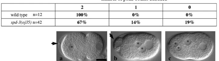
oj35early embryos were observed using noninvasive, live Nomarski imaging. The earliest defect that we observed in oj35 embryos was the failure to faithfully extrude polar bodies.C. elegansembryos normally undergo two rounds of meiosis shortly after fertilization. The meiotic spindle aligns perpendicular with the anterior cortex, resulting in the extrusion of two compact polar bodies (Figure 2a, arrow). In oj35 embryos, the number of polar bodies was variable, with some embryos com-pletely failing to extrude extra maternal pronuclei (Figure 2c), some having abnormally large polar bodies (Figure 2b, arrow), and some with normal extrusion. The failure to extrude polar bodies, as well as the presence of large polar bodies, suggests possible defects in the alignment of the meiotic spindle.
Pronuclear migration was often delayed in oj35
embryos. In wild-type embryos, the pronuclei form shortly after the completion of meiosis. The paternal pronucleus is located in the posterior, while the mater-nal pronucleus is most often located in the anterior. Shortly following the formation of the pronuclear envelopes, the maternal pronucleus in wild-type em-bryos migrated toward the paternal pronucleus at an average rate of 0.126 60.039mm/sec (n ¼10). How-ever, in oj35embryos, the maternal pronuclear migra-tion rate was 0.067460.0371mm/sec (n¼12), almost 50% slower than that in wild type. Often, the paternal pronuclear envelope broke down near the posterior cortex before the pronuclei met.
TABLE 2
SPD-3:GFP rescuesoj35embryonic defects
Genotype % dead eggs
% transverse spindle in P0
SPD-3TGFP 1.060.89 (n¼6) 0 (n¼4) unc-5(e53), spd-3(oj35) 99.361.7 (n¼6) 100 (n¼4) SPD-3TGFP;
unc-5(e53), spd-3(oj35)
4.063.2 (n¼6) 0 (n¼6)
All experiments were done at restrictive temperature.
The most apparent and detrimental defect observed inoj35embryos was the failure to properly orient the first mitotic spindle. Following fertilization in a wild-type embryo, the pronuclear/centrosomal complex rotates 90°to align the mitotic spindle along the A/P axis,i.e., the axis of polarity (Figure 3, a–e). This rota-tion failed inoj35embryos, resulting in a mitotic spindle aligned transversely to the A/P axis (Figure 3, g–k). The misaligned spindle gave rise to a mispositioned cleavage plane that did not coordinate with the polarity axis, resulting in a failure of the asymmetric division. It is worth noting that even in instances when the pronuclei did meet prior to nuclear envelope breakdown, the spindle was still misaligned, suggesting that the rotation failure was not simply a result of delayed pronuclear migration.
It has been shown that astral microtubule interactions with the cell cortex are imperative for spindle rotation to occur. Loss-of-function alleles of genes required for microtubule growth such as zyg-9 and mbk-2 result in short spindles that fail to rotate, presumably due to the inability of the short astral microtubules to interact with the cortex (Matthewset al. 1998; Pang et al. 2004).
Exposure to microtubule depolymerizing drugs such as nocodazole also results in this phenotype (Hymanand
White1987). Given these observations, we examined
the microtubules in fixedoj35embryos by immunoflu-orescent staining. Surprisingly, we found that astral microtubules inoj35embryos were robust and sufficient in length to interact with the cortex (Figure 3l). Indeed, the microtubules in the mutant embryos appeared excessive in length, compared to wild type. In wild-type embryos, several microtubules appear to have been captured and presumably stabilized upon reaching the cortex (Figure 3f). However, in oj35 embryos, the microtubules appear to have contacted the cortex and then continued to grow along it (Figure 3l).
spd-3(oj35) embryos exhibit defects in dynactin localization: Interestingly, a similar spindle alignment
defect is observed with loss-of-function of dynactin com-ponents (Skopand White1998; Gonczy et al.1999).
The dynein/dynactin complex has been well character-ized for its role in spindle alignment in various systems, including theC. elegansearly embryo (Carminatiand
Stearns 1997; Mcgrail and Hays 1997; Skop and
White1998; Gonczyet al.1999; O’Connelland Wang
2000). Therefore, we next examined whether the dynac-tin complex was localized properly inoj35embryos.
We examined the localization of DNC-1 (p150glued) in oj35embryos using an antibody to DNC-1. In wild-type embryos, DNC-1 localizes to the centrosomes, along spindle microtubules, and to the plus ends of astral microtubules (Figure 4). Surprisingly, we found that although the microtubule localization of DNC-1 appeared normal inoj35embryos, the majority of the protein appeared to localize into posterior structures resembling P granules. In addition, the centrosome labeling appeared brighter than that in wild type, a phenotype reminiscent of defects in dynein heavy chain function (Schmidtet al. 2005).
During mitosis, DNC-2TGFP (p50, dynamitin)
nor-mally localizes cytoplasmically with an enrichment in the pericentriolar region and along microtubules (Fig-ure 4). This microtubule localization is particularly apparent on the mitotic spindle where there is a distinct absence of signal on the chromosomes. Inoj35embryos, DNC-2TGFP exhibited a slight enrichment in the
pericentriolar region and the mitotic spindle and also a higher, delocalized cytoplasmic signal (Figure 4). Quantification of DNC2TGFP fluorescence intensity
showed a consistent increase in the oj35 background (Figure 4 graph). In addition, the contrasting lack of staining at the centrioles and the chromosomes is not as obvious in theoj35embryos compared to wild type.
SPD-3 localizes to mitochondria: To determine the subcellular localization of SPD-3, we generated trans-genic worms expressing SPD-3TGFP under the control
expression in the early embryo. The transgene consists of full-length genomicspd-3DNA tagged with GFP at the N or the C terminus and rescued the embryonic lethality and P0spindle orientation defects in theoj35mutants
(Table 2). Several lines were produced with a C-terminal GFP tag and one with an N-terminal GFP tag, which all showed identical localization patterns.
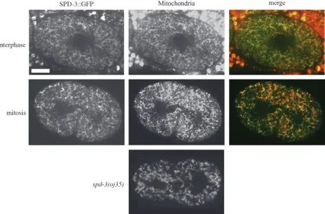
Surprisingly, we found that SPD-3TGFP localized to
mitochondria (Figure 5). In the earlyC. elegansembryo, mitochondria are diffusely distributed throughout the cytoplasm with no distinct localization pattern. In most organisms during interphase, mitochondria are local-ized throughout the cytoplasm. However, in some organisms such as yeast and the nematode Acrobe-loides, mitochondria cluster around the centrosomes during mitosis, presumably to ensure proper segrega-tion into separate daughter cells (Badrinath and
White 2003; Yaffe et al. 2003). This centrosome
clustering is consistent with a possible dependence on dynein. However, inC. elegansembryos, mitochondria do not undergo any obvious rearrangements during mitosis (Figure 5) (Badrinath and White 2003),
making SPD-3TGFP localization even more surprising.
It is worth noting that mitochondrial morphology and localization as assessed with mitochondrial vital dyes were not affected inoj35embryos, at least at levels that we could detect in these studies (Figure 5).
We identified the protein product ofC. elegansgene Y57A10A.26 as a potential interactor with SPD-3 in a GST pull-down assay. This gene has strong homology to murine metaxin 2, which has been shown in mice to
localize to the mitochondrial outer membrane and is involved in transporting proteins into the mitochon-drion (Armstronget al.1999). Although Y57A10A.26
alone did not exhibit any RNAi phenotype (data not shown), knocking down both Y57A10A.26 and its homo-log Y48C3A.3 (36% identities, 55% positives) by RNAi gave 20% embryonic lethality after 31 hr, quickly followed by sterility (n¼30). Because the worms exhibit sterility so soon after the RNAi takes effect, we were unable to thoroughly analyze embryonic defects. However, it is worth noting that in one instance we did witness an embryo phenocopy of theoj35spindle alignment defect. The identification of the mitochondrial protein metaxin as a potential binding partner for SPD-3 and the suggestion that loss-of-function of the two metaxin genes leads to sterility, a postembryonic defect seen in
oj35 worms (O’Connell et al. 1998), provide further
evidence that SPD-3 does localize to mitochondria and suggest that SPD-3 either may be part of a protein import complex along with the metaxin 2-like protein or may itself be imported by this complex.
spd-3(oj35)worms exhibit metabolic defects:A com-mon phenotype inC. elegansmutants with perturbations in metabolism is reduced growth rates. We therefore measured the growth rates ofoj35worms at permissive temperature by recording the generation time of the worms. We found thatoj35worms had reduced growth rates compared to wild type, similar to that of strains with disruption in mitochondrial function such as clk-1(qm30)(Table 3) (Lakowskiand Hekimi1996; Dillin
et al.2002).
Figure4.—spd-3(oj35)embryos have defects in dynactin localization. In wild-type embryos, DNC-1 localizes to the centrosomes, the mitotic spindle, and the plus ends of astral microtubules (n¼12).oj35embryos exhibit an additional, anomalous localization of DNC-1 to posterior structures resembling P granules (n¼12). DNC-1 signal is absent indnc-1RNAi embryos. DNC-2TGFP in
wild-type embryos is present throughout the cytoplasm, with enrichments in the pericentriolar region and along the mitotic spin-dle (n¼5). This enrichment is decreased inoj35embryos, while the cytoplasmic signal is significantly increased (n¼6). DNC-2TGFP images portrayed are snapshots taken from live imaging, while tubulin (n357) and DNC-1 antibodies were used with fixed
We wondered whether metabolic defects in worms might affect dynein motor function due to alterations in energy production. We addressed this possibility by comparing the concentration of free ATP between wild-type and oj35 worms. Surprisingly, oj35 worms
exhibited almost 40% greater ATP concentration com-pared to wild type.spd-3(oj35)worms contained 0.1486 0.016mmATP/mg total protein (n¼3) while wild-type
worms contained 0.093 6 0.003 mm ATP/mg total
protein (n ¼3). Functional disruptions in other pro-teins required for normal growth patterns including CLK-1 (required for ubiquinone biosynthesis) and DAF-2 (involved in insulin growth factor signaling) also result in increased ATP concentrations, suggesting that SPD-3 may be involved in these pathways (Braeckman et al.
1999; Dillinet al.2002).
To address whether disruptions in metabolism could lead to dynein loss-of-function phenotypes, we disrup-ted the electron transport chain using various mito-chondrial inhibitor drugs including antimycin A, a complex III inhibitor, and sodium azide, a complex IV inhibitor commonly used as a chemical surrogate for hypoxia (Scott et al.2002). While exposure to these
drugs led to slow growth (Table 3), there were no obvious defects in the early embryos analyzed after 24 hr of drug exposure in the mother. In addition, disruption of various other mitochondrial genes through muta-tions ½clk-1(qm30) (n ¼ 23) or RNAi ½atp-2 (ATP synthase) (n ¼ 13), cyc-1 (cytochrome C) (n ¼ 19), and cco-1 (cytochrome C oxidase) (n ¼ 20) did not Figure5.—SPD-3TGFP colocalizes with mitochondria. SPD-3TGFP has a similar localization pattern throughout the cell cycle (top, interphase; bottom, mitosis) and colocalizes with MitoTracker CMXRosetta. Bar, 10mm. There are no gross abnormalities in mitochondrial morphology or localization inoj35embryos as visualized with Rhodamine 6G (Sigma) (n¼3).
TABLE 3
Generation rates
Genotype Temperature
Generation rate (days)
Wild type (n¼30) 16° 5.0360.18 spd-3(oj35)(n¼29) 16° 7.6260.68 clk-1(qm30)(n¼19) 16° 8.6360.68
Drug exposure Temperature
Generation rate (days)
Water (control) (n¼5) 20° 4.060 Sodium azide
(20 mm) (n¼3)
20° 12.6762.3
Ethanol (control) (n¼5) 20° 4.060 Antimycin A
(40mm) (n¼4)
20° 5.560.58
resemble phenotypes seen in embryos with dynein loss-of-function (Sonnichsen et al. 2005), suggesting that
the purported dynein loss-of-function is specific to the loss of SPD-3 function.
spd-3genetically interacts withsmg-1:In an attempt to identify genetic interactors, we performed a suppressor screen using RNAi. RNAi has proven to be a convenient method to significantly knock down gene expression in
C. elegansand can be administered to worms through feeding (Timmons and Fire 1998). Using a feeding
library designed in the Ahringer lab (Kamath et al.
2003), we screened for genes on chromosomes I and II that suppressed oj35 phenotypes when depleted by RNAi. Normally, oj35 larvae placed at the restrictive temperature produce almost all dead eggs. We searched for genetic suppressors by screening for decreased levels of lethality after RNAi treatment.
We identifiedsmg-1as a suppressor ofoj35. oj35worms exposed tosmg-1 RNAi at restrictive temperature pro-duce an average of 9.8364.25 (n¼9) larvae per plate compared to untreated mutant worms with an average of 0.5 6 0.84 (n ¼ 8) larvae per plate. Further, we showed that smg-1RNAi rescued the spindle rotation defect in six of sevenoj35single-cell embryos observed. SMG-1 is a kinase that has been well studied in worms for its role in nonsense-mediated mRNA decay (NMD) (Grimsonet al.2004). However, there is evidence that
smggene products may also be involved in other types of regulation of mRNA expression levels (Mango 2001;
Mitrovichand Anderson 2005). Theoj35allele
con-tains a missense, rather than a nonsense, mutation. There-fore, if smg-1 RNAi is suppressing the oj35 phenotype through regulation of gene expression levels, it must be through a mechanism other than NMD. Supporting the hypothesis thatspd-3expression is regulated by the
smggene products, gene chip data suggest thatspd-3(1) RNA levels are elevated 2.2760.64-fold insmgmutant backgrounds compared to wild type (D. Markwardt
and P. Anderson, personal communication).
We reasoned that if suppression of theoj35phenotype was the result of changes in expression levels by the disruption of the SMG complex, then loss-of-function of othersmg genes would lead to the same suppression. However, smg-2 RNAi does not suppress the oj35
phenotype (n ¼ 10), but does suppress the paralysis phenotype seen inunc-54(r293)worms (n ¼10) done in parallel as an RNAi control. This suggests thatsmg-1
RNAi suppression of the oj35 phenotype may not be through regulation of gene expression levels by the SMG complex, but rather through an alternative, spe-cific function of SMG-1.
DISCUSSION
SPD-3 and dynein:We have shown thatoj35embryos have defects in meiosis, maternal pronuclear migration, and mitotic spindle alignment leading to embryonic
lethality. Interestingly, these defects are similar to those seen in weak loss-of-function of the dynein motor protein or components of its activating protein com-plex, dynactin (Skop and White1998; Gonczy et al.
1999; Yoder and Han 2001; Schmidt et al.2005). In
addition, we showed that SPD-3 is required for proper localization or expression of the dynactin components DNC-2 (p50 dynamitin) and DNC-1 (p150glued). In-terestingly, while dynein and dynactin localize to micro-tubules, the nuclear envelope, the cell cortex, and centrosomes (Skop and White 1998; Gonczy et al.
1999), SPD-3 was found to localize to mitochondria. Furthermore, oj35 worms have additional metabolic defects including altered ATP levels and slow growth, which are common phenotypes seen inC. elegansworms with disruptions in mitochondrial function (Lakowski
and Hekimi 1996; Dillin et al. 2002), making oj35
worms distinct from typical dynein/dynactin loss-of-function mutants.
SPD-3 and the cytoskeleton: It has been shown in several systems that dynein/dynactin is required for proper subcellular localization of several organelles in-cluding mitochondria (Burkhardtet al.1997; Helfand
et al.2002; Varadiet al.2004). While there appears to be
no specific localization of mitochondria in theC. elegans
early embryo, it is possible that localization in the early embryo is not random, but too subtle to detect in these studies. Therefore, SPD-3 may be required for proper dynein function in spindle alignment as well as proper subcellular localization of organelles such as mitochon-dria. While we cannot rule out this possibility, we deem this unlikely as mitochondrial localization and morphol-ogy do not appear to be perturbed in theoj35 mutant embryos.
SPD-3 and mitochondria: Surprisingly, we found SPD-3TGFP localized at mitochondria. Several pieces
of evidence shed clues as to a potential function of SPD-3 in relation to metabolism: the slow growth exhibited byoj35worms introduces the possibility that SPD-3 may be required for optimal mitochondrial function, as this is a common phenotype seen when the electron trans-port chain is compromised (Lakowski and Hekimi
1996; Dillinet al.2002).
In addition, we have identified a potential interaction between SPD-3 and Y57A10A.26, a protein with homol-ogy to murine metaxin 2. Metaxin 2 has been shown in mice to localize to the mitochondrial outer mem-brane and is involved in mitochondrial protein im-port (Armstronget al. 1999). Decreased function of
patterns and increased ATP levels (Braeckman et al.
1999; Dillin et al. 2002). CLK-1 is required for the
biosynthesis of ubiquinone, a carrier in the electron transport chain ( Jonassenet al.1998), while DAF-2 is an
insulin growth factor receptor that is thought to affect longevity by ultimately regulating gene expression through a phosphatidylinositol 3-kinase signaling path-way (Kenyon et al. 1993; Kimura et al. 1997). This
suggests the possibility that SPD-3 may also be involved in biosynthesis or signaling pathways.
Interestingly, it has been shown that disrupting the function of the mitochondrial ATP synthase,stunted, in Drosophila embryos results in defects in spindle align-ment in the syncytial blastoderm (Kiddet al.2005). The
authors show that ATP levels in the cortices ofstunted
mutant embryos are decreased compared to wild type. They suggest a model in which the reduced energy level is selectively affecting molecular motors such as dynein. While we have shown that ATP levels inoj35
worms are higher than those in wild type, it remains possible that elevated ATP levels are affecting dynein regulation, as it has been shown previously that dynein activity can be dependent on ATP and ADP concen-trations (Shiroguchiand Toyoshima2001).
However, the fact that we are unable to mimic theoj35
phenotype with mitochondrial inhibitor drugs, or by the disruption of other mitochondrial genes, suggests that the metabolic perturbation inoj35worms is not directly leading to the embryonic defects reminiscent of dynein loss-of-function. In fact, disrupting mitochondrial func-tion with drugs or RNAi of various genes required in the electron transport chain resulted in almost no observ-able defects in early embryonic development, implying that the early embryo is surprisingly tolerant to pertur-bations in the electron transport chain. This observation is consistent with previous studies that suggest that the embryo does not require large amounts of ATP for early development and the small percentage of ATP pro-duced by mitochondria-independent processes such as glycolysis may be sufficient for early developmental progression (Fenget al.2001).
Partial suppression of the oj35 phenotype by smg-1
RNAi: smg-1 loss-of-function via RNAi partially sup-presses the embryonic lethality and spindle alignment defects caused by theoj35mutant allele. Becauseoj35
does not contain a nonsense mutation, the suppression must be through a mechanism other than NMD. Gene-chip data provide evidence that the SMG protein complex is regulatingspd-3(1) expression levels. When the SMG complex is compromised through mutations,
spd-3(1) expression is increased more than twofold. However, the fact that depletion of another member of the SMG complex bysmg-2RNAi does not suppress theoj35 phenotype suggests that smg-1RNAi suppres-sion of theoj35phenotype may not be through regu-lation of gene expression levels by the SMG complex, but through an alternative, specific function of the
SMG-1 kinase. In addition to its role in NMD, SMG-1 orthologs in other systems have been implicated in signaling pathways regulating rearrangements of the actin cytoskeleton and stress response (Schmidtet al.
1996; Brumbaughet al.2004). Specifically, the human
SMG-1 ortholog has recently been identified as a phosphoinositide 3-kinase, which is activated by geno-toxic stress (Abraham2004; Brumbaughet al. 2004).
Given that SPD-3TGFP localizes to mitochondria, it is
quite possible that SMG-1 is necessary to regulate ex-pression levels of genes such as spd-3 in response to oxidative stress. However, an alternative explanation is that SMG-1 and SPD-3 are both involved in a stress-response signaling pathway, and it is the depletion of SMG-1 kinase activity in relation to this process that is suppressing theoj35phenotype. Given that phosphor-ylation of various subunits of the dynein complex has been shown to regulate motor activity as well as membrane localization (Dillman and Pfister 1994;
Linet al.1994; Pfisteret al.1996), SPD-3 may play an
additional role in regulating dynein activity, expression levels, and localization. The fact that dynactin compo-nents are mislocalized inoj35embryos is consistent with this hypothesis. These observations lead us to consider the intriguing possibility that SPD-3 may be acting in multiple signaling cascades required forC. elegansearly embryonic development.
While the specific function of SPD-3 remains elusive, we believe SPD-3 is the first example of a mitochondrial localized gene product required for spindle alignment in theC. elegansembryo. Further studies are needed to clarify the precise mechanism through which SPD-3 is affecting both dynactin localization and metabolism and elucidate the role mitochondria are playing in early
C. elegansdevelopment.
We are grateful to Dave Markwardt and Phil Anderson for communications and sharing unpublished data regarding smg-1 regulation, as well as Grzegorz Sabat at the University of Wisconsin Molecular Interaction Facility for analysis of the pull-down assay. We thank the labs of Judith Kimble, Sean Carroll, and Ann Palmenburg for equipment use. This work was supported by grants from the National Institutes of Health (R01 GM052454-09)
LITERATURE CITED
Abraham, R. T., 2004 The ATM-related kinase, hSMG-1, bridges
ge-nome and RNA surveillance pathways. DNA Repair3:919–925. Armstrong, L. C., A. J. Saenzand P. Bornstein, 1999 Metaxin 1
interacts with metaxin 2, a novel related protein associated with the mammalian mitochondrial outer membrane. J. Cell Biochem.
74:11–22.
Badrinath, A. S., and J. G. White, 2003 Contrasting patterns of
mi-tochondrial redistribution in the early lineages of Caenorhabdi-tis elegans and Acrobeloides sp. PS1146. Dev. Biol.258:70–75. Blumenthal, T., and K. S. Gleason, 2003 Caenorhabditis elegans
operons: form and function. Nat. Rev. Genet.4:112–120. Braeckman, B. P., K. Houthoofd, A. DeVreeseand J. R. Vanfleteren,
1999 Apparent uncoupling of energy production and consump-tion in long-lived Clk mutants of Caenorhabditis elegans. Curr. Biol.
9:493–496.
Brenner, S., 1974 The genetics ofCaenorhabditis elegans.Genetics
Brumbaugh, K. M., D. M. Otterness, C. Geisen, V. Oliveira, J.
Brognard et al., 2004 The mRNA surveillance protein
hSMG-1 functions in genotoxic stress response pathways in mam-malian cells. Mol. Cell14:585–598.
Burkhardt, J. K., C. J. Echeverri, T. Nilsson and R. B. Vallee,
1997 Overexpression of the dynamitin (p50) subunit of the dy-nactin complex disrupts dynein-dependent maintenance of membrane organelle distribution. J. Cell Biol.139:469–484. Carminati, J. L., and T. Stearns, 1997 Microtubules orient the
mi-totic spindle in yeast through dynein-dependent interactions with the cell cortex. J. Cell Biol.138:629–641.
Chen, F., B. M. Hersh, B. Conradt, Z. Zhou, D. Riemer et al.,
2000 Translocation ofC. elegansCED-4 to nuclear membranes during cell death. Science287:1485–1489.
Cowan, C. R., and A. A. Hyman, 2004 Asymmetric cell division in C.
elegans: cortical polarity and spindle positioning. Annu. Rev. Cell. Dev. Biol.20:427–453.
Dillin, A., A. L. Hsu, N. Arantes-Oliveira, J. Lehrer-Graiwer, H.
Hsinet al., 2002 Rates of behavior and aging specified by
mito-chondrial function during development. Science 298: 2398– 2401.
Dillman, 3rd, J. F., and K. K. Pfister, 1994 Differential
phosphor-ylation in vivo of cytoplasmic dynein associated with anterog-radely moving organelles. J. Cell Biol.127:1671–1681. Feng, J., F. Bussiereand S. Hekimi, 2001 Mitochondrial electron
transport is a key determinant of life span in Caenorhabditis elegans. Dev. Cell1:633–644.
Fire, A., S. Xu, M. K. Montgomery, S. A. Kostas, S. E. Driveret al.,
1998 Potent and specific genetic interference by double-stranded RNA in Caenorhabditis elegans. Nature391:806–811. Golden, A., 2000 Cytoplasmic flow and the establishment of polarity
in C. elegans 1-cell embryos. Curr. Opin. Genet. Dev.10:414–420. Goldstein, B., and S. N. Hird, 1996 Specification of the
anteroposte-rior axis in Caenorhabditis elegans. Development122:1467–1474. Gonczy, P., S. Pichler, M. Kirkham and A. A. Hyman, 1999
Cy-toplasmic dynein is required for distinct aspects of MTOC position-ing, including centrosome separation, in the one cell stage Caenorhabditis elegans embryo. J. Cell Biol.147:135–150. Grimson, A., S. O’Connor, C. L. Newman and P. Anderson,
2004 SMG-1 is a phosphatidylinositol kinase-related protein ki-nase required for nonsense-mediated mRNA decay in Caeno-rhabditis elegans. Mol. Cell. Biol.24:7483–7490.
Helfand, B. T., A. Mikami, R. B. Valleeand R. D. Goldman,
2002 A requirement for cytoplasmic dynein and dynactin in in-termediate filament network assembly and organization. J. Cell Biol.157:795–806.
Hird, S. N., and J. G. White, 1993 Cortical and cytoplasmic flow
po-larity in early embryonic cells of Caenorhabditis elegans. J. Cell Biol.121:1343–1355.
Hodgkin, J., A. Papp, R. Pulak, V. Ambrosand P. Anderson, 1989 A
new kind of informational suppression in the nematode Caeno-rhabditis elegans.Genetics123:301–313.
Hyman, A. A., and J. G. White, 1987 Determination of cell division
axes in the early embryogenesis of Caenorhabditis elegans. J. Cell Biol.105:2123–2135.
Jakubowski, J., and K. Kornfeld, 1999 A local, high-density,
single-nucleotide polymorphism map used to clone Caenorhabditis elegans cdf-1. Genetics153:743–752.
Jonassen, T., M. Proft, F. Randez-Gil, J. R. Schultz, B. N. Marbois
et al., 1998 Yeast Clk-1 homologue (Coq7/Cat5) is a mitochon-drial protein in coenzyme Q synthesis. J. Biol. Chem.273:3351– 3357.
Kamath, R. S., A. G. Fraser, Y. Dong, G. Poulin, R. Durbinet al.,
2003 Systematic functional analysis of the Caenorhabditis ele-gans genome using RNAi. Nature421:231–237.
Kenyon, C., J. Chang, E. Gensch, A. Rudner and R. Tabtiang,
1993 A C. elegans mutant that lives twice as long as wild type. Nature366:461–464.
Kidd, T., R. Abu-Shumays, A. Katzen, J. C. Sisson, G. Jimenezet al.,
2005 The epsilon-subunit of mitochondrial ATP synthase is re-quired for normal spindle orientation during the Drosophila em-bryonic divisions. Genetics170:697–708.
Kimura, K. D., H. A. Tissenbaum, Y. Liuand G. Ruvkun, 1997 daf-2,
an insulin receptor-like gene that regulates longevity and dia-pause in Caenorhabditis elegans. Science277:942–946.
Lakowski, B., and S. Hekimi, 1996 Determination of life-span in
Caenorhabditis elegans by four clock genes. Science272:1010– 1013.
Lin, S. X., K. L. Ferroand C. A. Collins, 1994 Cytoplasmic dynein
undergoes intracellular redistribution concomitant with phos-phorylation of the heavy chain in response to serum starvation and okadaic acid. J. Cell Biol.127:1009–1019.
Mango, S. E., 2001 Stop making nonSense: the C. elegans smg
genes. Trends Genet.17:646–653.
Matthews, L. R., P. Carter, D. Thierry-Miegand K. Kemphues,
1998 ZYG-9, a Caenorhabditis elegans protein required for mi-crotubule organization and function, is a component of meiotic and mitotic spindle poles. J. Cell Biol.141:1159–1168. McGrail, M., and T. S. Hays, 1997 The microtubule motor
cyto-plasmic dynein is required for spindle orientation during germ-line cell divisions and oocyte differentiation in Drosophila. Development124:2409–2419.
Mitrovich, Q. M., and P. Anderson, 2005 mRNA surveillance
of expressed pseudogenes in C. elegans. Curr. Biol. 15:963– 967.
Munro, E., J. Nanceand J. R. Priess, 2004 Cortical flows powered
by asymmetrical contraction transport PAR proteins to establish and maintain anterior-posterior polarity in the early C. elegans embryo. Dev. Cell7:413–424.
O’Connell, C. B., and Y. L. Wang, 2000 Mammalian spindle
orien-tation and position respond to changes in cell shape in a dynein-dependent fashion. Mol. Biol. Cell11:1765–1774.
O’Connell, K. F., C. M. Leysand J. G. White, 1998 A genetic
screen for temperature-sensitive cell-division mutants of Caeno-rhabditis elegans.Genetics149:1303–1321.
Pang, K. M., T. Ishidate, K. Nakamura, M. Shirayama, C. Trzepacz
et al., 2004 The minibrain kinase homolog,mbk-2, is required for spindle positioning and asymmetric cell division in earlyC. elegansembryos. Dev. Biol.265:127–139.
Pfister, K. K., M. W. Salata, J. F. Dillman, 3rd, K. T. Vaughan,
R. B. Valleeet al., 1996 Differential expression and
phos-phorylation of the 74-kDa intermediate chains of cytoplasmic dynein in cultured neurons and glia. J. Biol. Chem.271:1687– 1694.
Praitis, V., E. Casey, D. Collarand J. Austin, 2001 Creation of
low-copy integrated transgenic lines in Caenorhabditis elegans. Genetics157:1217–1226.
Rappaport, R., 1986 Establishment of the mechanism of cytokinesis
in animal cells. Int. Rev. Cytol.105:245–281.
Schetter, A., P. Askjaer, F. Piano, I. Mattaj and K. Kemphues,
2006 Nucleoporins NPP-1, NPP-3, NPP-4, NPP-11 and NPP-13 are required for proper spindle orientation in C. elegans. Dev. Biol.289:360–371.
Schmidt, A., J. Kunzand M. N. Hall, 1996 TOR2 is required for
organization of the actin cytoskeleton in yeast. Proc. Natl. Acad. Sci. USA93:13780–13785.
Schmidt, D. J., D. J. Rose, W. M. Saxton and S. Strome,
2005 Functional analysis of cytoplasmic dynein heavy chain in Caenorhabditis elegans with fast-acting temperature-sensitive mutations. Mol. Biol. Cell16:1200–1212.
Scott, B. A., M. S. Avidanand C. M. Crowder, 2002 Regulation of
hypoxic death in C. elegans by the insulin/IGF receptor homo-log DAF-2. Science296:2388–2391.
Shiroguchi, K., and Y. Y. Toyoshima, 2001 Regulation of
mono-meric dynein activity by ATP and ADP concentrations. Cell Motil. Cytoskeleton49:189–199.
Skop, A. R., and J. G. White, 1998 The dynactin complex is required
for cleavage plane specification in early Caenorhabditis elegans embryos. Curr. Biol.8:1110–1116.
Skop, A. R., D. Bergmann, W. A. Mohler and J. G. White,
2001 Completion of cytokinesis in C. elegans requires a brefel-din A-sensitive membrane accumulation at the cleavage furrow apex. Curr. Biol.11:735–746.
Sonnichsen, B., L. B. Koski, A. Walsh, P. Marschall, B. Neumann
et al., 2005 Full-genome RNAi profiling of early embryogenesis in Caenorhabditis elegans. Nature434:462–469.
Timmons, L., and A. Fire, 1998 Specific interference by ingested
dsRNA. Nature395:854.
Tsou, M. F., A. Hayashi, L. R. DeBella, G. McGrathand L. S. Rose,
enriched in response to PAR polarity cues in C. elegans embryos. Development129:4469–4481.
Varadi, A., L. I. Johnson-Cadwell, V. Cirulli, Y. Yoon, V. J.
Allanet al., 2004 Cytoplasmic dynein regulates the subcellular
distribution of mitochondria by controlling the recruitment of the fission factor dynamin-related protein-1. J. Cell Sci. 117:
4389–4400.
White, J., and S. Strome, 1996 Cleavage plane specification in C.
elegans: how to divide the spoils. Cell84:195–198.
Wokosin, D. L., J. M. Squirrell, K. W. Eliceiriand J. G. White,
2003 An optical workstation with concurrent, independent multiphoton imaging and experimental laser microbeam capa-bilities. Rev. Sci. Instrum.74:193–201.
Yaffe, M. P., N. Stuurmanand R. D. Vale, 2003 Mitochondrial
po-sitioning in fission yeast is driven by association with dynamic mi-crotubules and mitotic spindle poles. Proc. Natl. Acad. Sci. USA
100:11424–11428.
Yochem, J., T. Guand M. Han, 1998 A new marker for mosaic
anal-ysis in Caenorhabditis elegans indicates a fusion between hyp6 and hyp7, two major components of the hypodermis. Genetics
149:1323–1334.
Yoder, J. H., and M. Han, 2001 Cytoplasmic dynein light
interme-diate chain is required for discrete aspects of mitosis in Caeno-rhabditis elegans. Mol. Biol. Cell12:2921–2933.