Effect of 2.45 GHz Pulsed Microwave
Radiation on Hippocampus Mediated
Behaviour: in a Specific Relation to the
Expression of Rac1, Cdc42 and pCREB
Manoj Kumar1, Surya Pal Singh2, Chandra Mohini Chaturvedi3
M. Sc., Department of Zoology, Banaras Hindu University, Varanasi, India1.
Ph.D., Department of Electronics Engineering, Indian Institute of Technology (Banaras Hindu University),
Varanasi, India2
Emeritus Professor, Department of Zoology, Banaras Hindu University, Varanasi, India3
ABSTRACT: Present study was aimed to investigate the effect of pulsed microwave exposure on anxiety & depression like behaviour and spatial memory function of laboratory mouse. Mice were exposed to 2.45 GHz frequency with 16 and 217 Hzpulse modulation at the rate of 1 msec, pulse width 500 µsec& 50 % duty cycle; specific absorption rate (SAR) of 0.017 W/Kg for 30 days (2 hrs daily, 10.00 to 12.00 IST ). Mice behaviour was monitored in different behavioural paradigms and hippocampal tissue was processed for expression analysis of Rac1, Cdc 42 and pCREB
(S-133)
.Our results denote that 16 and 217 Hz did not induce anxiety and depression like behaviour analysed in OFT and FST respectively. Spatial memory function was remained unaffected following pulsed 16 and 217 MW exposures (except decline in time spent in target quadrant of 217 Hz). Expression levels of the above proteins complied with the behavioural findings and remain unaltered following 16 and 217 MW exposure. Findings of the present study suggested that 2.45 GHz with the 16 and 217 Hz modulation is unable to trigger any adverse effect on behavioural function of laboratory mouse.
KEYWORDS: microwave, spatial memory, Hz, anxiety and depression like behaviour,
I. INTRODUCTION
Many investigations have led to address the possible ill biological effects of microwave (MW) on central nervous system and brain functions [1]. Hippocampus is a brain’s crucial structure involved in the behavioural functions i.e. learning, memory, stress and emotions [2-4]. Plastic nature of hippocampus confers its vulnerability towards structural and functional changes involved in behavioural regulation following MW irradiation [5]. Furthermore, bioelectric nature of brain also renders its susceptibility to MW irradiation and induces changes upon absorption of MW either emitted from mobile phones [6, 7]. Recent studies were also focused to unravel MW exposure related adverse effects on hippocampal physiology [8].
anxiety and depression like behaviour [3]. Some of the recent findings are in agreement with claiming no effect on behavioural functions of pulsed MW exposure. Similarly, Cosquer et al [17] showed that 2.45 GHz pulse MW radiation unable to produce any anxiety responses of rats in plus maze test.
Many researchers have developed an interest to investigate the effect of pulsed MWradiation on brain functions including the role of hippocampus in memory, anxiety and depression behaviour [8]. Rac1 (Ras-related C3 botulinum toxin substrate 1) and Cdc 42 (Cell division control protein 42 homolog)acts as critical regulators of cytoskeleton dynamics in hippocampus, responsible for the regulation of synaptic plasticity underlying the memory formation [18-20]. Similarly, cAMP response element binding (CREB) protein is a nuclear transcription factor involved in cognitive function, associated with depression and neuronal survival [21-23].The rationale for selecting the 16 and 217 Hz lies in the fact that 16 Hz may interact with brain waves (EEG) and 217 Hz is emitted from mobile phone during operation mode. In view of the above reports, present study was hypothesized to determine the effect of pulsed MW on some proteins [Rac1, Cdc 42 and pCREB(s-133)]in hippocampus and behavioural functions.
II. MATERIALS AND METHODS
1. Animals
Swiss male mice (weighing 30-35 gms) were used in the experiments and procured from Central Animal facility, Institute of Medical Sciences, Banaras Hindu University, Varanasi. Mice were divided into 3 groups i.e. control (sham), 16 and 217 Hz (n=10) and kept under standard housing condition with 12:12 hrs light and dark (LD) cycle. Mice were fed on chow pellet and water ad libitum. Mice were exposed to 2.45 GHz with 16 and 217 Hz pulse modulation (pulse rate 1 millisec., pulse width 500 µsec. 50 % duty cycle), power density 0.029 mW/cm2 for 30 days (2 hrs daily 10.00 to 12.00 IST). All experimental procedures were carried out according to the recommendations of committee for the purpose of control and supervision of experiments on animals (CPCSEA, Govt. of India) and approved institutional ethical committee guidelines.
2. MW exposure set up
III. BEHAVIOURAL ASSESSMENT
3.1 Open field test (OFT)
OFT was performed as described by previous studies to assess anxiety like behaviour [25, 26]. The apparatus consisted of base area (48X48 cm) divided into identical marked sectors and enclosing walls of 18 cm height. The squares further divided into peripheral and centre sectors. Mice were placed at the centre and allowed to explore the arena for 5 min. Entries, time spent either in central and grooming were scored to evaluate the anxiogenic effect of pulsed MW irradiation.
3.2 Forced swim test (FST)
Mice were subjected to FST for evaluation of depression like behaviour as per the method described from previous studies [25, 27]. Mice were allowed to swim in a glass beaker (15 cm in diameter and 20 cm in height) filled with tap water. Immobility time was observed to measure of depression like behaviour in mice.
3.3 Morris water maze (MWM)
Morris water maze was used to evaluate the spatial memory function [28], comprised of the round water tank usually plastic (diameter 1.3 m, height 60 cm). To test the spatial memory, water tank was visually marked in the four quadrants and filled with tap water to certain heights with slightly submerged platform in target quadrant. Mice are allowed to swim in water pool and find the submerged platform. Mice spent time to locate the platform called as latency time and 1 min cut off time set for the each trial for mice. Significant reduction in the latency time over consecutive trials indicates learning and spatial memory. Probe trials also provide the measures for mice made entries and time spent in the target quadrant (quadrant without platform).
4 Brain tissue collection
Mice were sacrificed after behavioural assessment by decapitation. Brains were dissected out quickly and placed immediately on ice. Hippocampus was isolated from the whole brain as mentioned in our previous study [25].
5 Western blotting
Hippocampal tissues were lysed in RIPA buffer (1% (v/v) with 0.5% (w/v) sodium deoxycholate, 0.1% (w/v) sodium dodecyl sulfate (SDS) in phosphate-buffered solution (PBS) containing protease inhibitor aprotonin and sodium orthovanadate) to extract protein. Protein concentrations were determined by BCA proteins assay kit. In brief, protein samples were resolved by 12% (w/v) SDS-polyacrylamide gel electrophoresis and transferred to polyvinylidenedifluoride (PVDF) membranes and were detected using primary antibody (Rac1, Cdc 42 and pCREB (serine 133) rabbit monoclonal 1:800) followed by incubation with IgG- HRP conjugated secondary antibodies (Rac1, Cdc42, pCREB: Anti-Rabbit IgG, 1:3000). Membranes with ECL (chemoluminscent kit) treated were processed for signals detection in Chemilimager/ documentation system. Changes inthe intensity of Rac1, Cdc42 and pCREB proteins were normalized using the intensity obtained of the internal control β actin bands.
6 Statistical analysis
One way ANOVA (followed by Tukey’s post hoc test) was used to analyse significance among the groups. Values were expressed as Mean±S.E.M. *p<0.05 considered as significant.
IV. RESULTS
Effect of pulsed MW on anxiety and depression like behaviour
Groups Centre entries (OFT) Time in centre (OFT) Grooming FST (Immobility time)
Control 2.7±0.44 7.86±0.98 3.5±0.76 34.71±4.33 16 Hz 4.5±0.51* 9.133±1.38 4.5±1.23 24.29±4.29 217 Hz 3.5±0.41 10.2±1.15 3.8±1.01 29.86±4.13
Table: 1 Effect of pulsed MW (16 and 217 Hz) irradiation on anxiety & depression like behaviour and grooming. Data values were expressed as Mean±S.E.M. *p<0.05 considered significant.
Effect of pulsed MW exposure on spatial memory function
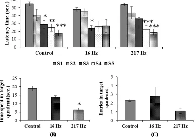
There was significant reduction observed in the latency time of all groups on subsequent trials or sessions compared to S1 (Fig. 1A). Probe test results showed that only 217 Hz exposed group has shown significant reduction in time spent in target quadrant as compared to control group. Whereas, 16 Hz exposed mice showed no significant difference compared to control (Fig. 1B). Number of entries in the target quadrant was also found insignificant among groups (Fig. 1 C.).
(A)
(B) (C)
Fig. 1:- Effect of pulsed MW on spatial memory of mice. Significant reduction in latency time among groups showed intact spatial memory (A) (sessions compared to S1 within each group). In Probe test, only 217 Hz showed reduced time spent in target quadrant (B).Whereas, entries in target quadrant of 16 and 217 Hz were insignificant difference from control (C). (S represents sessions, S1, S2, S3, S4 and S5). Data values were expressed as Mean±S.E.M. *p<0.05,**p<0.01, ***p<0.001 considered significant.
**
*
***
*
***
***
Effect of pulsed MW on the expression of Cdc 42, Rac1 and pCREB proteins in hippocampus
We observe no significant difference in the expression of Cdc 42, Rac1 and pCREB between exposed groups compared
to control (Fig.2 A-C).
Fig.2: Effect of pulsed MW irradiation on Cdc-42 (A), Rac1 (B) and pCREB(s-133) (C) expression in hippocampus. The expression of these molecules was found unchanged in hippocampus following pulsed MW exposure. Values expressed as Mean±S.E.M. (pCREB primary antibody (Catalog. No. 87G3) also detects pCREB related protein, ATF-1)
V. DISCUSSION
Present study demonstrated the effects of pulsed MW (16 and 217 Hz) exposure on anxiety& depression like behaviour and spatial memory with any relation to the expression of Rac1, Cdc42 and pCREB molecules. Our results indicated that 16 and 217 Hz exposure did not affect adversely studied behaviour and expression level of investigated proteins
β
actin
Rac1
Cdc42
Control 16 Hz 217 Hz
Control 16 Hz 217 Hz
β
actin
pCREB
ATF-1
A
B
C 25 kDa
21 kDa
42 kDa
OFT results denoted that 217 pulsed MW did not produce any significant change in time in centre and entries. Whereas, only 16 Hz group showed increased centre entries but did not affect time spent in centre suggesting no anxiogenic effect of 16 Hz. Grooming is an index to stress level and was observed insignificant among exposed groups versus control. Similarly, FST data showed no effect of either types of MW on immobility time, indicating no adverse effect on depression like behaviour. MWM results indicated that both the types of MW radiation showed decline in latency time over consecutive sessions and probe data confirmed no adverse effect on spatial memory. However, decrease of time spent in target quadrant was noted only in 217 Hz. Though, there are scarce findings and limited knowledge pertaining to pulsed MW induced effect on behavioural functions in rodents as well as health effects in human [29-31]. In addition to this, some studies noted the effect of pulsed MW or mobile phone radiation resulting in alteration of neuronal activity and cognitive function in humans [32-34]. Contrary to this, some studies also negate the findings of pulsed MW or GSM signals effects on spatial memory on non-spatial tasks suggesting that these exposure types did not affect such behaviour [16].
At molecular level, we have detected no significant change in the expression level of Rac1, Cdc 42 and pCREB. Western blot analysis showed that cytoskeleton remodelling proteins Rac1 and Cdc 42 was found unchanged following pulsed MW exposure. It can be suggested that neuronal actin organization remain unchanged complying with no disruption in the studied behavioural functions (anxiety & depression like behaviour and spatial memory). Rac1 and Cdc 42, GTPase molecule participates in the regulation of actin skeleton and also modulates synaptic plasticity [18]. Further, expression of both proteins was reported abundant in cortex, hippocampus and thalamic region of brain, suggesting their critical role in neuronal functions and behaviour. The knockout study for Rac 1 protein has lend deeper understanding in the regulation of cortical and hippocampal neurons development and hence deletion laid the basis for impaired memory functions by compromising plasticity in hippocampus (i.e. reduced spine size and density) [35]. Our results suggested that pulse MW exposure did not induce any change in expression level of active CREB (i.e. phosphorylated at serine 133 residues). Though, mobile phone exposure related effects on the pCREB signalling pathway is not well understood. Earlier studies have stemmed evidences regarding the mobile phone exposure effects on genomic response or gene expression [36, 37]. Some recent reports have stated that mobile phone exposure delimit the neurodegeneration via CREB activation (phosphorylation). An investigation of Inoue et al [38] also showed beneficial effect of mobile phone exposure enhanced neurite outgrowth and neurosurvival of cultured cells, mentioning the exposure induced activation of p38 MAPK (mitogen activated protein kinases) pathway via CREB activation that facilitatesneurite outgrowth.
VI. CONCLUSION
Above results summarize that behavioural functionsalong with associated proteins of neuronal plasticity in hippocampus were unaltered followingpulsed MW irradiation. Overall, our findings suggest that pulsed MW radiation of 2.45 GHz at SAR 0.017 W/Kg did not induceany anxiety & depression like behaviour,spatial memory and the unaltered level of cytoskeleton/nuclear proteins in hippocampus have strengthened behavioural findings. Therefore, it might be possible that in our designed experimental specifications of pulsed MW exposure, there is no suchadverse effect of pulsed MW on hippocampus dependent behavioural functions.
ACKNOWLEDGEMENT
REFERENCES
[1]. Hossmann KA, and Hermann DM,“Effects of electromagnetic radiation of mobile phones on the central nervous system” Bioelectromagnetics, Vol. 24 no. 1, 49-62, 2003.
[2]. LeunerB,andGould E,“Structural plasticity and hippocampal function”Annu Rev Psychol, Vol. 61,111–140, 2010.
[3]. BannermanDM, Sprengel, R, Sanderson, DJ, McHugh, SB, Rawlins, JNP, Monyer, H and Seeburg, PH, “Hippocampal synaptic plasticity, spatial memory and anxiety” Nature Reviews, Neuroscience, Vol. 15, no.3, 181, 2014.
[4]. Bannerman DM, Rawlins, JNP, McHugh, SB, Deacon, RMJ, Yee, BK, Bast, T, Zhang, W-N, Pothuizen, HHJ and Feldon, J, “Regional dissociations within the hippocampus—memory and anxiety” Neuroscience and Biobehavioral Reviews, Vol. 28, 273–283, 2004.
[5]. Bas O, Odaci, E, Kaplan, S, Acer, N, Ucok, K and Colakoglu, S, “900 MHz electromagnetic field exposure affects qualitative and quantitative features of hippocampal pyramidal cells in the adult female rat” Brain Res,Vol. 1265, 178-185, 2009.
[6].Grigor'evIuG, Luk'ianovaSN, MakarovVP, and RynskovVV, “Total bioelectric activity of various structures of the brain in low-intensity microwave irradiation” RadiatsBiolRadioecol, Volume 35 no. 1, 57-65, 1995.
[7]. McEwen BS, “The plasticity of the hippocampus is the reason for its vulnerability,” SeminNeurosci, Vol. 6, 239–246, 1994.
[8].Ikinci, A, Odaci, E, Yilidirum, M, Kaya, H, Akça, M, Hancı, H, Aslan, A, Fikret, O and Baş, O,The Effects of prenatal exposure to a 900 megahertz electromagnetic field on hippocampus morphology and learning behavior in rat pups, NeuroQuantology, 11(4), 582-590, 2013. [9].Eulitz C, Ullsperger P, Freude G, andElbert T, “Mobile phones modulate response patterns of human brain activity” Neuroreport, Volume 9 no.
14, 3229-3232 (1998).
[10].Borbely, AA, Huber, R, Graf, T, Fuchs, B, Gallmann, E, Achermann, P, “Pulsed high-frequency electromagnetic field affects human sleep and sleep electroencephalogram”NeurosciLett, Vol. 275, no.3, 207- 210, 1999.
[11]. D'Costa H, Trueman G, Tang L, Abdel-rahman U, Abdel-rahman W, Ong K, and Cosic I, “Human brain wave activity during exposure to radiofrequency field emissions from mobile phones” AustralasPhysEngSci Med. Vol. 26, no. 4, 162-167, 2003.
[12]. Mohammed HS, Fahmy HM, Radwah NM, and Elsayed AA, “Non-thermal continuous and modulated electromagnetic radiation fields effects on sleep EEG of rats” J Adv Res. Vol. 4, no. 2,181-187, 2013.
[13].AboulEzz, HS, Khadrawy, YA, Ahmed NA, Radwan NM, El BakryMM,“The effect of pulsed electromagnetic radiation from mobile phone on the levels of monoamine neurotransmitters in four different areas of rat brain”Eur Rev Med Pharmacol Sci. Vol. 17, no.13, 1782-1788, 2013. [14].Fragopoulou AF, Miltiadous P, Stamatakis A, Stylianopoulou F, Koussoulakos SL, and Margaritis LH, “Whole body exposure with GSM
900MHz affects spatial memory in mice” Pathophysiology Vol. 17 no. 3,179-187, 2010.
[15].Cassel JC, Cosquer B, Galani R and Kuster,N, “Whole-body exposure to 2.45 GHz electromagnetic fields does not alter radial-maze performance in rats” Behav Brain Res.Vol. 155, no.1, 37-43,2004.
[16]. Dubreuil D, Jay T, and Edeline JM, “Head-only exposure to GSM 900-MHz electromagnetic fields does not alter rat's memory in spatial and non-spatial tasks” Behav Brain Res. Vol. 145, no. 1-2,51-61, 2003.
[17].Cosquer B, Galani R, Kuster N, and Cassel JC, “Whole-body exposure to 2.45 GHz electromagnetic fields does not alter anxiety responses in rats: a plus-maze study including test validation” Behav Brain Res.Vol. 156, no.1,65-74, 2005.
[18].Haditsch U, Leone DP, Farinelli M, Chrostek-Grashoff A, Brakebusch, C, Mansuy, IM, McConnell, SK and Palmer TD, “A central role for the small GTPase Rac1 in hippocampal plasticity and spatial learning and memory” Mol Cell Neurosci. Vol. 41, 409–419,2009.
[19].Olenik C, Barth H, Just I, Aktories K, and Meyer DK, “Gene expression of the small GTP-binding proteins RhoA, RhoB, Rac1, and Cdc42 in adult rat brain” Molecular brain research Vol. 52, no.2, 263-269, 1997.
[20]. Hua ZL, Emiliani FE, and Nathans J, “Rac1 plays an essential role in axon growth and guidance and in neuronal survival in the central and peripheral nervous systems” Neural development Vol. 10, no.1, 21,2015.
[21]. Sakamoto K, Kate K, and Karl O, “CREB: a multifaceted regulator of neuronal plasticity and protection” Journal of neurochemistry Vol. 116, no.1, 1-9, 2011
[22]. Blendy JA, “The role of CREB in depression and antidepressant treatment”Biological psychiatry Vol. 59, no.12, 1144-1150, 2006. [23]. Walton MR, and Dragunow M, “Is CREB a key to neuronal survival?” Trends in neurosciences” Vol. 23, no.2, 48-53, 2000.
[24].Gandhi OP, Hunt EL, and D’Andrea JA, “Deposition of electromagnetic energy in animals and in models of man with and without grounding and reflector effects” Radio Sci.Vol. 12, 39-47, 1977.
[25].Kumar M, Singh SP, and Chaturvedi CM, “Chronic nonmodulated microwave radiations in mice produce anxiety-like and depression-like behaviours and calcium- and NO-related biochemical changes in the brain” ExpNeurobiol. Vol.25, no.6, 318-327, 2016.
[26].Prut L, and Belzung C, “The open field as a paradigm to measure the effects of drugs on anxiety-like behaviors: a review” Eur J Pharmacol. Vol. 463, 3-33, 2003.
[27].Porsolt RD, Le Pichon M, and Jalfre M, “Depression: a new animal model sensitive to antidepressant treatments” Nature Vol. 266, 730-732, 1977.
[28].Morris R, “Developments of a water-maze procedure for studying spatial learning in the rat” J Neurosci Methods Vol. 11, 47–60, 1984. [29].Cobb BL, Jauchem JR, and Adair ER,“Radial arm maze performance of rats following repeated low level microwave radiation exposure”
Bioelectromagnetics Vol. 25, no.1, 49-57, 2004.
[30]. Sienkiewicz ZJ, Blackwell RP, Haylock RG, Saunders RD, and Cobb BL, “Low-level exposure to pulsed 900 MHz microwave radiation does not cause deficits in the performance of a spatial learning task in mice”BioelectromagneticsVol. 21, no.3, 151-158, 2000.
[30]. Lu Y, Xu S, He M, Chen C, Zhang L, Liu C, Chu F, Yu Z, Zhou Z, and Zhong M, “Glucose administration attenuates spatial memory deficits induced by chronic low-power-density microwave exposure” PhysiolBehav Vol. 106, no.5, 631-637, 2012.
[32].Carrubba S, Frilot C, Chesson AL Jr, and Marino AA,“Mobile-phone pulse triggers evoked potentials”NeurosciLett. Vol. 469, no.1, 164-168, 2010.
[33].Fritzer G, Göder R, Friege L, Wachter J, Hansen V, Hinze-Selch D and AldenhoffJB,“Effects of shortand long-term pulsed radiofrequency electromagnetic fields on night sleep and cognitive functions in healthy subjects” Bioelectromagnetics Vol. 28, no. 4, 316-325, 2007. [34]. Kramarenko AV, and Tan U, “Effects of high-frequency electromagnetic fields on human EEG: A brain mapping study”Int J Neurosci, Vol.
113, no.7, 1007-1019, 2003.
[35].Bongmba OY, Martinez LA, Elhardt ME, Butler K, and Tejada-Simon MV,“Modulation of dendritic spines and synaptic function by Rac1: A possible link to Fragile X syndrome pathology” Brain research, Vol. 1399, 79-95, 2011.
[36].Belyaev IY, Koch CB, Terenius O, Roxstrom-Lindquist K, Malmgren LG, Sommer LG, Salford LG, and Persson BR, “Exposure of rat brain to 915MHz GSM microwaves induces changes in gene expression but not double stranded DNA breaks or effects on chromatin conformation” BioelectromagneticsVol. 27, 295–306, 2006.
[37]. Fritze K, Wiessner C, Kuster N, Sommer, C, Gass, P, Hermann, DM, Kiessling, M, and Hossmann, KA, “Effect of global system for mobile communication microwave exposure on the genomic response of the rat brain” Neuroscience Vol. 81,627–639,1997.