1
Effect of Resveratrol on Reactive Oxygen Species-Induced Cognitive Impairment in Rats with Angiotensin II-Induced Early Alzheimer’s Disease
Yu-Te Lin, MD, PhD1,2; Yi-Chung Wu, MD, MS3; Gwo-Ching Sun, MD, PhD4; Chiu-Yi Ho, MS5; Tzyy-Yue Wong, PhD6; Ching-Huang Lin, MD1; Hsin-Hung Chen, PhD6; Tung-Chen Yeh, MD, PhD7; Chia-Jung Li, PhD8; Ching-Jiunn Tseng, MD, PhD5,6,9; Pei-Wen Cheng, PhD6,10,11
Affiliation:
1
Section of Neurology, Kaohsiung Veterans General Hospital, Taiwan; 2Center for Geriatrics and Gerontology, Kaohsiung Veterans General Hospital, Kaohsiung, Taiwan; 3Section of
Neurology, Zouying Branch of Kaohsiung Armed Forces General Hospital Kaohsiung,
Taiwan;4Department of Anesthesiology, Kaohsiung Medical University Hospital, Kaohsiung Medical University, Kaohsiung, Taiwan; 5Department of Biomedical Science, National Sun Yat-Sen University, Kaohsiung, Taiwan; 6Department of Medical Education and Research, Kaohsiung Veterans General Hospital, Kaohsiung, Taiwan; 7Department of Internal Medicine, Division of Cardiology, Kaohsiung Veterans General Hospital, Kaohsiung, Taiwan;
8
Research Assistant Center, Show Chwan Memorial Hospital, Changhua, Taiwan;
9
Department of Pharmacology, National Defense Medical University Hospital, China Medical
University, Taichung, Taiwan; 10Yuh-Ing Junior College of Health Care & Management, Kaohsiung, Taiwan; 11Shu-Zen Junior College of Medicine and Management, Kaohsiung, Taiwan
Conference paper:
The manuscript is conference paper of 18th World Congress of Basic and Clinical
Pharmacology (WCP2018); Abstract submission ID: 20345; poster number: PO1-2-78.
Correspondence:
Ching-Jiunn Tseng, MD, PhD
Department of Medical Education and Research, Kaohsiung Veterans General Hospital, 386
Ta-Chung 1st Rd., Kaohsiung, Taiwan 813
E-mail: [email protected]
Tel: 886-7-3422121 ext. 1505; Fax: 886-7-3468056
or
Pei-Wen Cheng, PhD
Department of Medical Education and Research, Kaohsiung Veterans General Hospital,
Kaohsiung, Taiwan
2
E-mail:
Telephone: 886-7-3422121 ext. 1593; Fax: 886-7-3468056
Yu-Te Lin and Yi-Chung Wu contributed equally to this work.
Abbreviations:
AD, Alzheimer’s disease; Ang II, Angiotensin II; A42, Fbrillar -amyloid; BP, Blood pressure. BDNF , Brain-derived neurotrophy factor; CNS, Central nervous system.
ICV, intracerebroventricular. NTS, Nucleus tractus solitarii. NADPH oxidase,
nicotinamide adenine dinucleotide phosphate-oxidase; MCI, mild cognitive
3 Abstract
Recent studies have indicated that several anti-hypertensive drugs may delay the
development and progression of Alzheimer’s disease (AD). However, the
relationships among AD, hypertension, and oxidative stress remain to be elucidated.
In the present study, we aimed to determine whether treatment with resveratrol
reduces reactive oxygen species (ROS) generation in the brain, thereby reducing
cognitive impairment in rats with angiotensin II (Ang-II)-induced early AD. Male
WKY rats with Ang-II-induced AD were treated with losartan or resveratrol for 2
weeks. Our results revealed that treatment with resveratrol (10 mg/kg/day) decreased
blood pressure, increased levels of brain-derived neurotrophic factor (BDNF) in the
hippocampus, and decreased ROS production in the nucleus tractus solitarius (NTS)
in the Ang-II groups. In addition, inhibition of TauT231 phosphorylation in the
hippocampus using resveratrol significantly abolished Ang-II-induced expression of
Aprecursors, active caspase 3, and glycogen synthase kinase 3 (GSK-3)Y216 while
increasing AktS473 phosphorylation. Notably, resveratrol reversed impairments in
hippocampal-dependent contextual memory induced by deleting NADPH oxidase and
NOX2. Overall, our results suggest that resveratrol exerts neuroprotective effects
against memory impairment and hippocampal damage in a rat model of early stage
4
represent a pharmacological option for patients with hypertension at a risk of AD
5 Introduction
Alzheimer's disease (AD) is an age-dependent neurodegenerative disorder associated
with abnormal energy metabolism, representing the most common neurodegenerative
disorder worldwide [1, 2]. AD is characterised neuropathologically by the formation
of senile plaques and neurofibrillary tangles, and clinically by the progressive
deterioration of memory and other cognitive functions [3], which cannot be prevented
using currently available treatments [4]. The pathogenesis of AD is considered a
multifactorial neurodegenerative process, in which several pathways are damaged due
to oxidative stress injury, abnormal energy processing, mitochondrial dysfunction,
and inflammation [5, 6]. Moreover, recent studies have reported that hypertension is a
major risk factor for the development of AD in old age [7]. The nucleus tractus
solitarius (NTS) is located in the dorsal medulla of the brainstem, which is the
primary integrating centre for cardiovascular regulation and other autonomic
functions of the CNS. Specifically, increases in circulating and brain levels of
angiotensin II (Ang-II), a primary effector peptide of the
renin–angiotensin–aldosterone system, not only play important roles in the genesis of
arterial hypertension, but also in the pathophysiology of AD [8]. Therefore, a
relationship appears to exist among hypertension, cerebrovascular disease, and
6
it remains to be determined whether hippocampal changes result from pre-existing
hypertension, or whether hypertension and brain pathology reflect a central defect
associated with AD [10].
Excessive binding of Ang-II to the AT1 receptor in the brain negatively affects
cognition by blocking hippocampal long-term potentiation (LTP) and enhancing
-amyloid production [11, 12]. In particular, such binding may lead to widespread
neuronal injury, including increases in reactive oxygen species (ROS), activation of
NADPH oxidases, blood-brain barrier (BBB) breakdown, decreased access to
nutrients, and -amyloid toxicity in the cerebral vasculature [11, 13, 14].
Resveratrol (3,5,4′-trihydroxy-trans-stilbene) is a stilbenoid, a type of natural
phenol that may exert beneficial effects in a variety of human diseases such as type 2
diabetes, obesity, and cancer [15]. Resveratrol has attracted increasing attention due to
its neuroprotective effects, which include facilitating anti-amyloidogenic cleavage of
β-amyloid precursor protein, promoting clearance of neurotoxic Aβ peptides, and
reducing oxidative stress [16, 17]. In addition, accumulating evidence indicates that
resveratrol attenuates oxidative imbalances by simultaneously enhancing ROS
production and downregulating key antioxidant enzymes such as copper-zinc
superoxide dismutase (SOD1) and manganese superoxide dismutase (SOD2) [18].
7
and enhances SOD2 expression, thereby negatively regulating Racl-induced NADPH
oxidase levels in the NTS during oxidative stress-associated hypertension[19].
Previous studies have further suggested that oxidative imbalances and the resultant
neuronal damage play a critical role in the initiation and progression of AD [20].
NAD(P)H oxidase plays a role in multiple central autonomic networks associated
with Ang-II-induced ROS in neurons [21]. In our previous study, spontaneously
hypertensive rats (SHRs) were treated with losartan or tempol for 2 weeks, following
which we observed significant decreases in systolic blood pressure (SBP) and ROS
generation in the NTS [22]. Therefore, in the present study, we aimed to determine
whether treatment with resveratrol improves ROS generation in the brain, thereby
reducing cognitive impairment in rats with Ang-II-induced early AD.
Materials and Methods
Experimental chemicals
All experimental chemicals were purchased from Sigma-Aldrich (St. Louis, MO,
USA), unless otherwise indicated.
Animals
8
Animal Facility (NSCAF; Taipei, Taiwan) and housed in the animal room of
Kaohsiung Veterans General Hospital (VGHKS; Kaohsiung, Taiwan). Both the
NSCAF and VGHKS are internationally certified by the Association for Assessment
and Accreditation of Laboratory Animal Care (AAALACi). Rats housed in the animal
room of VGHKS were fed in a specific pathogen-free (SPF) room. SPF facilities are
designed to maintain rodents in an environment that is free of certain infectious
organisms that are pathogenic and/or capable of interfering with research objectives.
The rats were kept in individual cages in a light-controlled room (12-hour
light/12-hour dark cycle), the temperature of which was maintained between 23°C
and 24°C. The rats were given normal rat chow (Purina; St. Louis, MO) and tap water
ad libitum. All animal research protocols were approved by the Animal Research
Committee, and the institutional review board at VGHKS approved all study
procedures (VGHKS-2018-A002). The study was performed in accordance with
approved guidelines and conducted in compliance with the Declaration of Helsinki.
The rats were housed in an animal room at VGHKS and randomly divided into
the following four groups, with six rats in each group: (1) the sham group, in which
each rat received an intracerebroventricular (ICV) injection of artificial cerebrospinal
fluid (aCSF) and an oral dose (1 ml/kg body weight (BW)) of distilled water once
9
received an ICV injection of angiotensin II (14.4 g/l) and an oral dose of distilled
water; (3) the WKY + Ang-II + losartan group, in which rats treated with losartan (10
mg/kg BW, Chunghwa Yuming Healthcare, Taiwan) received an ICV injection of
angiotensin II; (4) the WKY + Ang-II + resveratrol group, in which rats treated with
resveratrol (10 mg/kg BW) received an ICV injection of angiotensin II. All surgeries
were performed in an effort to minimise animal suffering.
After 14 days, rats were euthanised using CO2 in accordance with the 2013
AVMA Guidelines for the Euthanasia of Animals. All rats were killed using 100%
CO2, with death occurring within 2 to 5 minutes. Brains were immediately dissected
on ice, frozen, and maintained at −20°C until assayed.
ICV injection procedure
ICV infusion experiments were performed following a stabilisation period of at least
30 minutes after the insertion of the microinjector into the ventricular-guided cannula.
Blood pressure was monitored for 3 days after drug infusion. As a vehicle control, we
analysed the effect of an ICV injection of aCSF (142 mmol/L NaCl, 5 mmol/L KCl,
10 mmol/L glucose, and 10 mmol/L HEPES, pH 7.4). Ang-II (14.4 g/l/per day) was
dissolved in aCSF. Resveratrol was initially dissolved in DMSO and then diluted in
10
prior to injection. The daily ICV drug infusions were performed over a 2-min period
and delivered as a single bolus of a final volume of 5 μL from day 0 to day 14.
BP measurement
Using a previously described tail-cuff method (CODA, Kent Scientific Corporation,
USA), we measured SBP and heart rate prior to losartan or resveratrol treatment (day
0). Measurements were also obtained 3, 7, 11, and 14 days after surgery, as well as
prior to sacrifice. All measurements were performed between 08:00 am and 12:00 am.
In this method, the reappearance of pulsation on the digital display of the BP cuff was
detected using a pressure transducer and was amplified and recorded as the SBP.
During measurement, we obtained a rapid series of 10 individual readings, the highest
and lowest of which were discarded, while the remaining eight were averaged.
Morris water maze (MWM)
Cognitive and behavioural functions were assessed using the MWM task. Briefly, a
circular pool (diameter: 180 cm) was filled with tap water maintained at 24 ± 1℃.
The escape platform (diameter: 12 cm) was submerged 2 cm below the surface of the
water. Each rat underwent four trials per day over four consecutive days (day 3 to day
11
selected starting points (north, south, east, or west pole). Upon locating the platform,
the rat was allowed to remain there for 20 s before being returned to its cage. If the rat
did not find the platform within 90 s, the time was recorded as 90 s, following which
the rat was guided to the platform and allowed to remain there for 20 s. The escape
latency and swim speed were measured using a video tracking system (EthoVision XT,
Noldus, USA). After the last training trial, rats were subjected to a probe trial in which
the platform was removed. Animals were placed in the pool at the same pole and
allowed to swim for 2 min. The time that each animal spent in the quadrant that had
previously contained the hidden platform was recorded.
Measurement of A40 and A42 in the hippocampus and post-hypothalamus
We measured levels of soluble A40 and A42 in the hippocampus and
post-hypothalamus using Ultrasensitive Rat A40 and A42 ELISA kits (Arigo
Biolaboratories Corp, Taiman), in accordance with manufacturer instructions.
Expression levels were detected using a Biochrom Anthos Zenyth 200rt Microplate
Reader (Cambridge, UK).
Immunoblotting analysis
12
tissues in lysis buffer containing a protease inhibitor cocktail and a phosphatase
inhibitor cocktail. The sample was subsequently incubated for 1 h at 4°C. The protein
extracts (20 g/sample based on the BCA protein assay, Pierce Chemical Co.,
Rockford, IL, USA) were resolved on a 6% polyacrylamide gel and transferred to a
PVDF membrane (GE Healthcare, Buckinghamshire, UK). The membranes were
incubated in the appropriate anti-P-TauT231 (ab151559), anti-P-AktS473 (4060, Cell
Signaling Technology, Beverly, MA), anti-P-GSK-3bS9 (05-643, EMD Millipore,
Billerica, MA), anti-P-GSK-3Y216 (ab75745), anti-amyloid precursor protein
(ab12266), anti-gp91-phox (sc5827, Santa Cruz Biotechnology, Dallas, TX), anti-Tau
(ab80579), anti-Akt (9272), anti-p67-phox (sc7663), anti-GSK-3 (07-389),
anti-p47-phox (sc14015), anti-p22-phox (sc11712), or anti-caspase 3 (9662)
antibodies. The membranes were then incubated in an HRP-labelled goat anti-rabbit
secondary antibody at 1:10,000. The membranes were developed using an ECL-Plus
detection kit (GE Healthcare).
Immunohistochemistry analysis
For immunohistochemistry experiments, initial specimen processing and staining
were performed as described earlier [22]. Sections were first stained with
13
with horse anti-mouse AP. Briefly, sections were incubated with both primary
antibodies (Anti–P-TauT231, anti-P-TauT181, or anti-BDNF) overnight at 4°C. Slides
were then washed three times with PBS, following which they were incubated for 1 h
at room temperature (RT) with a combination of biotinylated HRP-conjugated goat
anti-rabbit (1:200) secondary antibodies. After three washes with PBS, the sections
were incubated with ABC complex (1:50) for 30 min at RT. After three washes with
PBS, sections were visualised using the DAB substrate kit (Vector Laboratories,
Burlingame, CA, USA) and counterstained with haematoxylin. After rinsing in PBS,
the sections were dehydrated using a graded series of ethanol and xylene. The sections
were then photographed using an Olympus microscope equipped with a Nikon Cool
Scan 995 digital camera (Nikon, Tokyo, Japan).
Statistical analyses
All data are expressed as the mean ± standard error of the mean (SEM). Paired t-tests
were used to compare baseline and post-treatment BP measurements, while a one-way
analysis of variance (ANOVA) with Scheffe’s post hoc testing was employed to
evaluated differences among the groups. The level of statistical significance was set at
14 Results
BDNF levels and superoxide imbalances contributed to hypertension and early
AD
Previous studies have suggested that BDNF-deficient mice are more susceptible to
stress-induced oxidative damage, as indicated by the direct association between
indicators of oxidative stress and BDNF levels in the brain [23]. Oxidative stress is an
important pathogenic factor in the development of hypertension. Angiotensin
receptors (AT1R) play a pivotal role in the development and maintenance of
hypertension during oxidative stress [24]. Accumulating evidence indicates that
BDNF and its interaction with ROS may be crucial for several symptoms of
neurodegenerative and neuropsychiatric conditions. Thus, we aimed to determine
whether ROS levels were significantly increased in the NTS of mice with
Ang-II-induced AD, and whether such increases led to down-regulation of BDNF
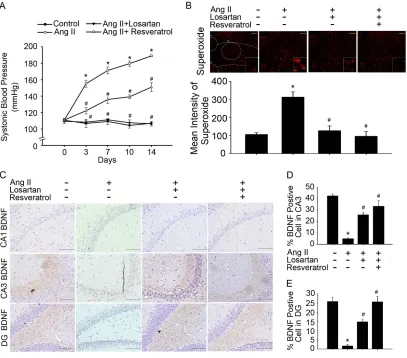
release in the hippocampus. In addition to increased SBP, rats with Ang-II-induced
AD exhibited significantly higher levels of superoxide in the NTS and hippocampal
areas CA1, CA3, and DG than those in the control group. Furthermore, treatment with
losartan or resveratrol reversed these effects (Fig. 1A, B and Fig 2 A, B). Interestingly,
treatment with losartan or resveratrol markedly increased BDNF levels in the
15 Fig. 1).
To investigate whether resveratrol limits ROS production via inhibition of
NADPH oxidases during Ang-II-induced early AD, we examined the expression of
NADPH oxidase subunits and SOD when both Ang-II and resveratrol were
administered. Our results indicated that resveratrol abolished increases in the
expression of NOX2, the NADPH oxidase subunit p67-phox, and reduced levels of
SOD2 in the hippocampus (Fig. 2C and D). These results indicate that the elimination
of ROS may be required to increase BDNF levels and activate the depressor response.
Taken together, these findings suggest that resveratrol mitigates oxidative stress,
normalises BDNF levels, and reduces BP in Ang-II-induced early AD.
Resveratrol impaired the activity of A precursors, active caspase 3, and
GSK-3-Tau by normalising renal AT1R signalling in the hippocampus of rats
with Ang-II-induced early AD
Accumulating evidence has indicated that patients with AD exhibit significantly
higher levels of anti-AT1R and tau than healthy controls [25]. However, the
angiotensin system increases the permeability of the BBB, induces oxidative stress in
the microcirculation of the brain, and leads to an increase in A and tau pathologies
16
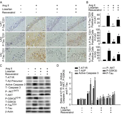
hippocampus of rats with Ang-II-induced early AD. ICH experiments demonstrated
that treatment with losartan or resveratrol influenced the expression of phosphorylated
TauT231 in hippocampal areas CA1, CA3, and DG in rats with Ang-II-induced AD
(Fig. 3A and B). Immunoblot analyses of proteins extracted from the hippocampus
demonstrated that treatment with losartan or resveratrol decreased the expression of
AT1R, A precursors, and active caspase 3 in Ang-II-induced groups. Similarly,
GSK-3Y216 expression and TauT231 phosphorylation in the hippocampus were
significantly attenuated by treatment with losartan or resveratrol. The addition of
losartan or resveratrol increased Akt activation, as well as hyperphosphorylation of
critical Akt substrates, in Ang-II-induced groups (Fig. 3C and D). Resveratrol
treatment resulted in favourable shifts in Ang-II-induced expression of AT1R, A
precursors, active caspase 3, GSK-3, and tau activation in the hippocampus. These
results suggest that resveratrol attenuated Ang-II-induced tau pathologies, thereby
improving Akt activation and attenuating down-regulation of the AT1R–A–caspase
3–GSK-3 signalling pathway.
Resveratrol treatment improved spatial learning and memory in rats with
Ang-II-induced early AD
17
AD. Some patients with early-onset familial AD exhibit elevated levels of A42/A40
[27]. To investigate whether resveratrol limits AT1R signalling to contribute to
improvements in spatial learning and memory during Ang-II-induced early AD, we
examined the expression of A42/A40 and MWM results in rats treated with
losartan or resveratrol. Rats with Ang-II-induced AD exhibited significantly higher
levels of A42/A40 in the hippocampus than controls (Fig. 4A, histograms 1 and 2).
Interestingly, treatment with losartan or resveratrol markedly inhibited A42/A40
expression in the hippocampus of rats with Ang-II-induced AD (Fig. 4A, histograms 3
and 4). While all rats exhibited progressive decreases in escape latency during the
MWM task (Figure 4B), Ang-II-treated rats took significantly longer to reach the
platform than controls (P<0.01), suggestive of notable cognitive damage. Furthermore,
this increase in escape latency was significantly ameliorated by losartan and
resveratrol treatment (day 5, day 6, and 7, P<0.05 vs. Ang-II groups). While rats with
Ang-II-induced AD spent significantly less time in the central area than control
groups, rats treated with losartan spent significantly more time in this area than other
rats with Ang-II-induced AD. No significant differences were observed between
losartan and resveratrol treatment in the Ang-II groups (Fig. 4C). The rats exhibited
similar motor capabilities, as swim speed did not differ between the groups (Fig. 4D),
18
results suggest that resveratrol and losartan attenuated Ang-II-induced impairments in
hippocampal-dependent and contextual memory.
Discussion
In the present study, we aimed to determine whether treatment with resveratrol
improves ROS generation and cognitive function in rats with Ang-II-induced early
AD. Our results revealed that treatment with resveratrol for 2 weeks decreased BP,
increased levels of BDNF in the hippocampus, and decreased ROS production in the
NTS in the Ang-II groups. Overall, our results suggest that resveratrol exerts
neuroprotective effects against memory impairment and hippocampal damage in a rat
model of early stage AD by reducing oxidative stress.
Alzheimer’s Disease International (ADI) has determined that, in the next 30
years, the number of patients with AD will more than quadruple in India, China, other
countries in Asia, Australasia, and Oceania, from approximately 16 million in 2010 to
approximately 61 million by 2050. The total worldwide costs of dementia in 2010
were estimated to be $604 billion (USD) (70% of which occur in Europe and North
America), representing approximately 1% of the global gross domestic product [28].
A previous population-based study revealed that hippocampal atrophy (HA) is
19
AD [29]. Several studies have indicated that the clinical end points of AD are
strongest in those who have never been treated for hypertension. Additional studies
have demonstrated that treatment with antihypertensive medication reduces the risk
associated with high BP [30, 31]. In the present study, we observed that central BP is
regulated by ROS levels in the NTS, which are thought to contribute to
down-regulation of BDNF expression in both the NTS and hippocampus of rats with
Ang-II-induced AD. In addition, our results suggest that resveratrol not only
attenuated increases in superoxide levels in the NTS and increased BDNF expression,
but also increased the antioxidant capacity of the NTS in rats with hypertension. Our
findings further demonstrated that Ang-II increased the generation of superoxide and
the activity of the Aβ–caspase 3–Akt–GSK-3β-Tau pathway by positively regulating
NADPH oxidase levels. Such changes were also accompanied by decreases in SOD2
and BDNF expression in the hippocampus. However, treatment with resveratrol
improved cognitive function in rats with Ang-II-induced early AD by abolishing ROS
generation and reducing activity of the Aβ–caspase 3–Akt–GSK-3β-Tau pathway
activity by negatively regulating NADPH oxidase and NOX2 levels. Therefore, our
findings suggest that early treatment with resveratrol lowers oxidative stress,
preserves SOD function, and attenuates the development of hypertension (Fig. 5).
20
associated with atrophy of the hippocampus and temporal lobe, as well as an
increased risk of cognitive decline, suggesting that such patients are at increased risk
of developing AD [29]. Indeed, neurofibrillary tangles, senile plaques, and neuronal
lesions have been observed in patients with hypertension [32]. While such findings
suggest a relationship among hypertension, cerebrovascular disease, and decreased
cognitive function [9], it remains to be determined whether hippocampal changes are
the consequences of pre-existing hypertension, or whether hypertension and brain
pathology reflect a central defect in patients with AD [10].
Some studies have suggested that SHRs and rats with DOCA-salt-induced
hypertension exhibit low BDNF expression and deficient neurogenesis in the
hippocampus. However, treatment with oestrogens may normalise brain parameters
(i.e., BDNF levels) by decreasing peripheral BP in both rat groups [33, 34].
In the present study, treatment with losartan or resveratrol not only normalised
BP and superoxide levels in the NTS, but also markedly increased BDNF levels in the
hippocampus and NTS of rats with Ang-II-induced AD (Fig. 1D and E and
Supplementary Fig. 1). These results indicate that the elimination of ROS may be
required to increase BDNF levels and activate the depressor response. To our
knowledge, our study provides the most conclusive evidence that resveratrol improves
21
A and tau pathologies in rats with Ang-II-induced early AD. ROS in the brain are
thought to contribute to the neuropathogenesis of hypertension by enhancing
sympathetic nervous system activity. The key mechanism for reduced nitric oxide
(NO) bioavailability is oxidative stress [35, 36]. Oxidative stress can be defined as
increased bioactivity of ROS relative to antioxidant defences [37]. In accordance with
our present findings, our previous studies demonstrated that resveratrol decreases BP
better than rosuvastatin, abolishes ROS generation, and enhances activity of the
ERK1/2-RSK-nNOS pathway by activating AMPK to negatively regulate
Racl-induced NADPH oxidase levels in the NTS during oxidative stress-associated
hypertension[38].
While current pharmacological approaches simply provide symptomatic
improvement rather than prevent or delay cognitive decline, many commonplace
medications are being re-evaluated for their potential benefits among patients with
AD. Treatment of vascular risk factors has been associated with a reduced incidence
of AD and slower cognitive decline in patients with AD. Angiotensin converting
enzyme inhibitors (ACEIs) and angiotensin receptor blockers (ARBs) are widely
prescribed as antihypertensive drugs, acting on the renin–angiotensin system (RAS).
Some research indicates that they may be superior to other antihypertensive drugs
22
vascular and amyloid pathways [26]. Consequently, several high-quality longitudinal
studies have explored the precise influence of hypertensive drugs targeting the RAS
among patients with AD. However, our results suggested that resveratrol exerts
effects similar to those of the Ang-II receptor antagonist losartan by attenuating
Ang-II-induced tau pathologies, improving Akt activation, and attenuating
down-regulation of the AT1R–A–caspase 3–GSK-3 signalling pathway (Fig. 3).
Resveratrol targets the CNS, can cross the BBB, and induces neuroprotective effects
[39]. Notably, Vingtdeux et al. observed that resveratrol is present in the brain
following oral administration, indicating that it may exert direct effects in patients
with neurological disorders [40].
Our results demonstrated that treatment with resveratrol for 2 weeks decreased
BP, attenuated ROS production in the NTS, and increased BDNF levels in the
hippocampus of rats with Ang-II-induced early AD. In addition, inhibition of TauT231
phosphorylation in the hippocampus using resveratrol significantly abolished
Ang-II-induced expression of Aprecursors, active caspase 3, and glycogen synthase
kinase 3 (GSK-3)Y216 while increasing AktS473 phosphorylation. Interestingly,
resveratrol reversed impairments in hippocampal-dependent and contextual memory
induced by deletion of NADPH oxidase and NOX2.
23
memory impairment and hippocampal damage in a rat model of early stage AD by
reducing oxidative stress. These novel findings indicate that resveratrol may represent
a pharmacological option for patients with hypertension at risk for the development of
AD during old age. Furthermore, our findings may aid in the identification of
molecular targets for recovering memory pathways, potentially leading to the
development of new therapeutic strategies.
Author contributions
The study was conceived and designed by Pei-Wen Cheng, Yu-Te Lin, Yi-Chung Wu,
Ching-Jiunn Tseng. Pei-Wen Cheng conducted most of the experiments with
assistance from Hsin-Hung Chen, Chia-Jung Li, and Chi-Cheng Lai. The paper was
written by Pei-Wen Cheng, with contributions from Gwo-Ching Sun, Tung-Chen Yeh,
and Ching-Huang Lin.
Funding and Disclosures
This work was supported by funding from the National Science Council
MOST104-2320-B-075B-003-MY3, MOST106-2320-B075B-001) and Kaohsiung
Veterans General Hospital (VGHKS 106-157, VGHKS107-175) (to P.-W. C. and
24 Acknowledgements
We gratefully acknowledge Ya-Chu Chuang for technical assistance.
References
[1] Yin, F.; Sancheti, H.; Patil, I.; Cadenas, E. Energy metabolism and inflammation in brain aging and Alzheimer's disease. Free radical biology & medicine 100:108-122; 2016.
[2] Salminen, A.; Haapasalo, A.; Kauppinen, A.; Kaarniranta, K.; Soininen, H.; Hiltunen, M. Impaired mitochondrial energy metabolism in Alzheimer's disease: Impact on pathogenesis via disturbed epigenetic regulation of chromatin landscape. Prog Neurobiol 131:1-20; 2015.
[3] Arendt, T.; Stieler, J. T.; Holzer, M. Tau and tauopathies. Brain Res Bull
126:238-292; 2016.
[4] Salloway, S.; Correia, S. Alzheimer disease: time to improve its diagnosis and treatment. Cleve Clin J Med 76:49-58; 2009.
[5] Kim, G. H.; Kim, J. E.; Rhie, S. J.; Yoon, S. The Role of Oxidative Stress in Neurodegenerative Diseases. Exp Neurobiol 24:325-340; 2015.
[6] Cabezas-Opazo, F. A.; Vergara-Pulgar, K.; Perez, M. J.; Jara, C.; Osorio-Fuentealba, C.; Quintanilla, R. A. Mitochondrial Dysfunction Contributes to the Pathogenesis of Alzheimer's Disease. Oxid Med Cell Longev 2015:509654; 2015.
[7] Ashby, E. L.; Kehoe, P. G. Current status of renin-aldosterone angiotensin system-targeting anti-hypertensive drugs as therapeutic options for Alzheimer's disease. Expert Opin Investig Drugs 22:1229-1242; 2013.
[8] Munoz-Durango, N.; Fuentes, C. A.; Castillo, A. E.; Gonzalez-Gomez, L. M.; Vecchiola, A.; Fardella, C. E.; Kalergis, A. M. Role of the Renin-Angiotensin-Aldosterone System beyond Blood Pressure Regulation: Molecular and Cellular Mechanisms Involved in End-Organ Damage during Arterial Hypertension. Int J Mol Sci 17; 2016.
25
[10] Saavedra, J. M. Opportunities and limitations of genetic analysis of hypertensive rat strains. J Hypertens 27:1129-1133; 2009.
[11] Faraco, G.; Park, L.; Zhou, P.; Luo, W.; Paul, S. M.; Anrather, J.; Iadecola, C. Hypertension enhances Abeta-induced neurovascular dysfunction, promotes beta-secretase activity, and leads to amyloidogenic processing of APP. J Cereb Blood Flow Metab 36:241-252; 2016.
[12] Dai, H. L.; Hu, W. Y.; Jiang, L. H.; Li, L.; Gaung, X. F.; Xiao, Z. C. p38 MAPK Inhibition Improves Synaptic Plasticity and Memory in Angiotensin II-dependent Hypertensive Mice. Scientific reports 6:27600; 2016.
[13] Bloch, S.; Obari, D.; Girouard, H. Angiotensin and neurovascular coupling: beyond hypertension. Microcirculation 22:159-167; 2015.
[14] Ali, M. R.; Abo-Youssef, A. M.; Messiha, B. A.; Khattab, M. M. Tempol and perindopril protect against lipopolysaccharide-induced cognition impairment and amyloidogenesis by modulating brain-derived neurotropic factor, neuroinflammation and oxido-nitrosative stress. Naunyn Schmiedebergs Arch Pharmacol 389:637-656; 2016.
[15] Cottart, C. H.; Nivet-Antoine, V.; Laguillier-Morizot, C.; Beaudeux, J. L. Resveratrol bioavailability and toxicity in humans. Molecular nutrition & food research 54:7-16; 2010.
[16] Richard, T.; Pawlus, A. D.; Iglesias, M. L.; Pedrot, E.; Waffo-Teguo, P.; Merillon, J. M.; Monti, J. P. Neuroprotective properties of resveratrol and derivatives. Ann N Y Acad Sci 1215:103-108; 2011.
[17] Braidy, N.; Jugder, B. E.; Poljak, A.; Jayasena, T.; Mansour, H.; Nabavi, S. M.; Sachdev, P.; Grant, R. Resveratrol as a Potential Therapeutic Candidate for the Treatment and Management of Alzheimer's Disease. Curr Top Med Chem
16:1951-1960; 2016.
[18] Francini, F.; Castro, M. C.; Schinella, G.; Garcia, M. E.; Maiztegui, B.; Raschia, M. A.; Gagliardino, J. J.; Massa, M. L. Changes induced by a fructose-rich diet on hepatic metabolism and the antioxidant system. Life Sci 86:965-971; 2010.
[19] Cheng, P. W.; Lee, H. C.; Lu, P. J.; Chen, H. H.; Lai, C. C.; Sun, G. C.; Yeh, T. C.; Hsiao, M.; Lin, Y. T.; Liu, C. P.; Tseng, C. J. Resveratrol Inhibition of Rac1-Derived Reactive Oxygen Species by AMPK Decreases Blood Pressure in a Fructose-Induced Rat Model of Hypertension. Sci Rep 6:25342; 2016.
[20] Wang, X.; Wang, W.; Li, L.; Perry, G.; Lee, H. G.; Zhu, X. Oxidative stress and mitochondrial dysfunction in Alzheimer's disease. Biochim Biophys Acta
1842:1240-1247; 2014.
26
96:659-666; 2005.
[22] Cheng, W. H.; Lu, P. J.; Ho, W. Y.; Tung, C. S.; Cheng, P. W.; Hsiao, M.; Tseng, C. J. Angiotensin II inhibits neuronal nitric oxide synthase activation through the ERK1/2-RSK signaling pathway to modulate central control of blood pressure. Circulation research 106:788-795; 2010.
[23] Kocisova, E.; Petr, M.; Sipova, H.; Kylian, O.; Prochazka, M. Drop coating deposition of a liposome suspension on surfaces with different wettabilities: "coffee ring" formation and suspension preconcentration. Phys Chem Chem Phys 19:388-393; 2016.
[24] Lara, L. S.; McCormack, M.; Semprum-Prieto, L. C.; Shenouda, S.; Majid, D. S.; Kobori, H.; Navar, L. G.; Prieto, M. C. AT1 receptor-mediated augmentation of angiotensinogen, oxidative stress, and inflammation in ANG II-salt hypertension. Am J Physiol Renal Physiol 302:F85-94; 2012.
[25] Giil, L. M.; Kristoffersen, E. K.; Vedeler, C. A.; Aarsland, D.; Nordrehaug, J. E.; Winblad, B.; Cedazo-Minguez, A.; Lund, A.; Reksten, T. R. Autoantibodies Toward the Angiotensin 2 Type 1 Receptor: A Novel Autoantibody in Alzheimer's Disease. J Alzheimers Dis 47:523-529; 2015.
[26] Tian, M.; Zhu, D.; Xie, W.; Shi, J. Central angiotensin II-induced Alzheimer-like tau phosphorylation in normal rat brains. FEBS Lett 586:3737-3745; 2012.
[27] Chang, Y. J.; Chen, Y. R. The coexistence of an equal amount of Alzheimer's amyloid-beta 40 and 42 forms structurally stable and toxic oligomers through a distinct pathway. FEBS J 281:2674-2687; 2014.
[28] Trojanowski, J. Q.; Arnold, S. E.; Karlawish, J. H.; Naylor, M.; Brunden, K. R.; Lee, V. M. A model for improving the treatment and care of Alzheimer's disease patients through interdisciplinary research. Alzheimers Dement 8:564-573; 2012.
[29] Korf, E. S.; White, L. R.; Scheltens, P.; Launer, L. J. Midlife blood pressure and the risk of hippocampal atrophy: the Honolulu Asia Aging Study. Hypertension 44:29-34; 2004.
[30] Kril, J. J.; Patel, S.; Harding, A. J.; Halliday, G. M. Patients with vascular dementia due to microvascular pathology have significant hippocampal neuronal loss. J Neurol Neurosurg Psychiatry 72:747-751; 2002.
[31] Ye, R.; Hu, Y.; Yao, A.; Yang, Y.; Shi, Y.; Jiang, Y.; Zhang, J. Impact of renin-angiotensin system-targeting antihypertensive drugs on treatment of Alzheimer's disease: a meta-analysis. Int J Clin Pract 69:674-681; 2015.
27
[33] Pietranera, L.; Lima, A.; Roig, P.; De Nicola, A. F. Involvement of brain-derived neurotrophic factor and neurogenesis in oestradiol neuroprotection of the hippocampus of hypertensive rats. J Neuroendocrinol 22:1082-1092; 2010.
[34] Jazbutyte, V.; Arias-Loza, P. A.; Hu, K.; Widder, J.; Govindaraj, V.; von Poser-Klein, C.; Bauersachs, J.; Fritzemeier, K. H.; Hegele-Hartung, C.; Neyses, L.; Ertl, G.; Pelzer, T. Ligand-dependent activation of ER{beta} lowers blood pressure and attenuates cardiac hypertrophy in ovariectomized spontaneously hypertensive rats. Cardiovasc Res 77:774-781; 2008.
[35] Pattillo, C. B.; Bir, S.; Rajaram, V.; Kevil, C. G. Inorganic nitrite and chronic tissue ischaemia: a novel therapeutic modality for peripheral vascular diseases. Cardiovascular research 89:533-541; 2011.
[36] Torregrossa, A. C.; Aranke, M.; Bryan, N. S. Nitric oxide and geriatrics: Implications in diagnostics and treatment of the elderly. Journal of geriatric cardiology : JGC 8:230-242; 2011.
[37] Kregel, K. C.; Zhang, H. J. An integrated view of oxidative stress in aging: basic mechanisms, functional effects, and pathological considerations. American journal of physiology. Regulatory, integrative and comparative physiology 292:R18-36; 2007. [38] Yeh, T. C.; Shin, C. S.; Chen, H. H.; Lai, C. C.; Sun, G. C.; Tseng, C. J.; Cheng, P. W. Resveratrol regulates blood pressure by enhancing AMPK signaling to downregulate a Rac1-derived NADPH oxidase in the central nervous system. J Appl Physiol (1985)
125:40-48; 2018.
[39] Quincozes-Santos, A.; Gottfried, C. Resveratrol modulates astroglial functions: neuroprotective hypothesis. Annals of the New York Academy of Sciences 1215:72-78; 2011.
29
Fig. 1. Downregulation of BDNF levels was associated with increased superoxide
expression in rats with Ang-II-induced early Alzheimer’s disease (AD). (A) Time
course of systolic blood pressure (SBP) after intracerebroventricular administration of
angiotensin II (Ang-II) for 2 weeks. The filled circles (●) represent the WKY group,
the open circles (○) represent the Ang-II group, the inverted filled triangles (▼)
represent the Ang-II + losartan group, and the open triangles (△) represent the Ang-II
+ resveratrol group. SBP was measured on days 0, 4, 7, 11, and 14. The data are
30
Ang-II group. (B) Confocal microscopy analysis of DHE-treated brain sections in the
nucleus tractus solitarius (NTS) after treatment with losartan or resveratrol.Bar graph
showing the superoxide production ratio after treatment with Ang-II and/or losartan or
resveratrol. Note the significant decrease in Ang-II-induced superoxide production
after the administration of losartan or resveratrol. (C) In situ qualitative analysis of
BDNF-immunopositive cells in the hippocampus of AD model rats. Scale bar, 200
mm. (D-E) Bar graph showing BDNF-expressing cells after treatment with Ang-II
and/or losartan or resveratrol. Note the significant increase in Ang-II-induced BDNF
production after the administration of losartan or resveratrol. The percentage of
BDNF-positive cells was determined by counting the BDNF-expressing cells in each
hemisphere of the hippocampus (CA1 and CA3) at ×200 magnification. These values
were divided by the total number of cells in the same paraffin section. BDNF:
31
Fig. 2. Resveratrol abolished Ang-II-induced increases in superoxide and
NADPH oxidase activity and reduced SOD2 activity in the hippocampus of AD
model rats. (A) Confocal microscopy analysis of DHE-treated brain sections in the
hippocampus after treatment with losartan or resveratrol.(B) Bar graph showing the
superoxide production ratio after treatment with Ang-II and/or losartan or resveratrol.
Note the significant decrease in Ang-II-induced superoxide production after the
32
demonstrating decreased expression of the NADPH oxidase subunit p67-phox and
decreases in the NOX2 ratio in the hippocampus of Ang-II-treated rats following
treatment with losartan or resveratrol. The level of SOD2 protein in the hippocampus
was significantly increased following treatment with losartan or resveratrol. (The
values are presented as the mean ± SEM; n = 6. *P < 0.05 vs. the WKY group. #P <
33
Fig. 3. Resveratrol attenuated Ang-II-induced A precursor and caspase
3-Akt-GSK-3-Tau pathwaysin the hippocampus of AD model rats. (A) In situ
qualitative analysis of P-TauT231-immunopositive cells in the hippocampus of AD
model rats. Scale bar, 200 mm. (B) Bar graph showing P-TauT231-expressing cells
after treatment with Ang-II and/or losartan or resveratrol. Note the significant
decrease in Ang-II-induced P-TauT231 production after the administration of losartan
34
the P-TauT231-expressing cells in each hemisphere of the hippocampus (CA1, CA3,
and DG) at ×200 magnification. These values were divided by the total number of
cells in the same paraffin section. (C) Immunoblot demonstrating decreased levels of
the proteins T-AT1R, T-A precursor, T-active-caspase 3, P-GSK-3Y216, and
P-TauT231 in the hippocampus after treatment with Ang-II and/or losartan or
resveratrol. (D) Quantitative immunoblot analysis demonstrating reductions in
T-AT1R, T-A precursor, T-active-caspase 3, P-GSK-3Y216, and P-TauT231 expression
in the hippocampus of rats with Ang-II-induced AD following treatment with losartan
or resveratrol. The values are presented as the mean ± SEM; n = 6. *P < 0.05 vs. the
WKY group. #P < 0.05 vs. the Ang-II group. AD: Alzheimer’s disease; Ang-II:
35
Fig. 4. Resveratrol reversed impairments in hippocampal-dependent and
contextual memory in rats with Ang-II-induced early Alzheimer’s disease (AD).
(A) Bar graph showing the A42 production ratio after treatment with Ang-II and/or
losartan or resveratrol. Note the significant decrease in Ang-II-induced A42
production after the administration of losartan or resveratrol. (B) Bar graph showing
the latency to find the hidden platform. (C) Bar graph showing the time spent in the
central area. (D) Bar graph showing the swim speed. Note that learning and memory
deficits were also reversed in Ang-II-treated rats after losartan or resveratrol
administration. The values are presented as the mean ± SEM; n=6. *P<0.05 vs. the
36
Fig. 5. Resveratrol attenuated ROS-induced cognitive impairments in rats with
Ang-II-induced early Alzheimer’s disease (AD).
Ang-II not only increased the generation of superoxide and the activity of the
A–caspase 3–Akt–GSK-3β–Tau pathway by positively regulating NADPH oxidase
levels, but also attenuated SOD2 and BDNF expression in the hippocampus (black
line). However, treatment with resveratrol improved cognitive impairments in rats
with Ang-II-induced early AD by abolishing ROS generation and reducing activity of
37
oxidase and NOX2 levels (red line). ROS: reactive oxygen species; BDNF: