Ras/Raf signalling in
Primary cells
Marie Harrlsingh
A thesis submitted fo r the degree
o f D octor o f Philosophy
Medical Research Council Laboratory of
Molecular and Cellular Biology,
ProQuest Number: U643164
All rights reserved
INFORMATION TO ALL USERS
The quality of this reproduction is dependent upon the quality of the copy submitted.
In the unlikely event that the author did not send a complete manuscript and there are missing pages, these will be noted. Also, if material had to be removed,
a note will indicate the deletion.
uest.
ProQuest U643164
Published by ProQuest LLC(2016). Copyright of the Dissertation is held by the Author.
All rights reserved.
This work is protected against unauthorized copying under Title 17, United States Code. Microform Edition © ProQuest LLC.
ProQuest LLC
789 East Eisenhower Parkway P.O. Box 1346
Abstract
O ncogenic activation of the Ras gene has been implicated in many human tumours. H owever, despite the ability of Ras to transform immortal cell lines, activated Ras is growth inhibitory in primary cells. W e have previously shown that activation of Ras/Raf signalling in primary Schwann cells results in a proliferative arrest due to the induction of the cyclin dependent kinase inhibitor (CDKI) In this thesis I exam ine the m echanisms involved in p21^'^^ induction and the roles of other CDKIs in the Ras/Raf induced cell-cycle arrest. I show that R af activation is also associated with the induction of the CDKI p i h o w e v e r , in contrast to other primary cell types, p i l e v e l s decrease and the induction of p l9 ^ ”^^is not associated with p53 stabilisation.
Acknowledgements
I would like to thank Alison Lloyd for her guidance and constant enthusiasm for all matters scientific. In the lab I would like to thank Nicole and Phil for their friendship, support and technical help over the years.
I would like to acknowledge the work of my collaborators at the Breakthrough Breast cancer centre - Alan Ashworth and Afshan McCarthy. At the LMCB I collaborated with Anne Mudge on the experiments involving Dorsal root ganglion-Schwann cell co-cultures and Denise M alcolm was involved in carrying out some of the W estern blotting analyses and PCR screening for transgenic mice. Thanks also to David Parkinson for proof reading and helpful discussions; Mark Shipman for technical assistance with the microscopes, Mike Corder for helping with my computer traumas and everyone at the LMCB for creating a great working environment.
Thanks to all my friends for their understanding and support, especially Joanna Holbrook, and M artha Betson who helped with proof reading and formatting amongst other things, and Louise Rollick for making me laugh.
Table of Contents
Abstract_________________________________________________________________________ 2 Acknowledgements_______________________________________________________________ 3 Table of Contents________________________________________________________________ 4 List of Figures and Tables_________________________________________________________ 7
Chapter 1: Introduction__________________________________________________________ 10 1.1. Regulation of G l-S phase transition____________________________________________ 13 1.2. Ras signalling_______________________________________________________________ 16
Activation o f R a s _______________________________________________________________________________ 16 Ras signalling in tu m origen esis__________________________________________________________________19 Ras signalling pathw ays_________________________________________________________________________20 PI3K sig n a llin g _____________________________________________________________________________ 20 Raf/M APK s ig n a llin g ______________________________________________________________ :_________ 21 Regulation o f G l-S phase cell cycle progression by Ras signalling_______________________________ 24
1.3. Ras signalling in primary cells________________________________________________ 24
CDKI induction and fun ction ____________________________________________________________________ 25 p53 activation and functions_____________________________________________________________________ 27 Regulation o f p53 levels and sta b ility _________________________________________________________ 27 Downstream signalling pathways from p 5 3 ____________________________________________________ 30 Regulation o f p53 activity by plO'^'^^__________________________________________________________ 31 The mechanism s o f the oncogenic Ras induced cell cycle a rr est____________________________________ 33 M ouse em bryonic fibroblasts (M E F s)_________________________________________________________ 33 Human diploid fibroblasts (HDFs)_____________________________________________________________ 37 K ératinocytes________________________________________________________________________________ 38 Schwann c e l l s _______________________________________________________________________________39
1.4. Neurofibromatosis type 1 (N F l)_______________________________________________ 39
Incidence and sym ptom s _______________________________________________________________________ 39 Neurofibroma structure and tumour progression __________________________________________________ 40 Expression o f N FI and functions o f Neurofibromin________________________________________________42 Neurofibromin as a tumour suppressor___________________________________________________________ 44 In vitro cellular studies__________________________________________________________________________45 N eu ron s_____________________________________________________________________________________45 Fibroblasts or perineural c e l l s ________________________________________________________________ 46 Schwann c e l l s ______________________________________________________________________________ 46
l.S.Schwann cell differentiation___________________________________________________ 48
Krox-20 and Krox-24 ________________________________________________________________________66
Schwann cell-axon interactions____________________________________________________68
Schwann cell-axon interactions during peripheral nerve degeneration and regeneration _______________ 68 Regulation o f Schwann behaviour; dedifferentiation, demyelination and re-differentiation__________ 69 The importance o f Schwann cells in peripheral nerve regeneration________________________________ 72
Chapter Two; Materials and M ethods_____________________________________________ 74
2.1. C ell c u ltu r e______________________________________________________________________________ 74 Primary Schwann cells ______________________________________________________________________ 74 NSARaf-1 :ER c e l l s _______________________________________________:___________________________ 74 GFP-NSARaf-1 :ER c e lls _____________________________________________________________________ 74
NIH 3T3 cells, phoenix cells and perineural fibroblasts_________ 75
Dorsal root g a n g lia ______________________________________________________________________ ,___ 76 2.2 Tissue culture assays _______________________________________________________________________ 76 DRG and Schwann cell or fibroblast co-cultures________________________________________________ 76 Time-lapse m icro sco p y ______________________________________________________________________ 77 Differentiation assays _______________________________________________________________________ 77 M EK inhibitor assays ________________________________________________________________________77 D N A synthesis a ssa y _________________________________________________________________________78 U V treatment o f ce lls_________________________________________________________________________78 Transfection o f D N A into Schwann cells_______________________________________________________ 78 Determination of the stability of p 2 l‘^''’' protein using cyclohexam ide_____________________________ 79 2.3 R N A /D N A m anipulation.____________________________________________________________________ 79 Generation o f the pRafTR 1RES- EGFP transgenic con stru ct____________________________________ 79 Ligation o f D N A fragments into v ec to rs_______________________________________________________ 82 Restriction enzym e digests ___________________________________________________________________ 83 Bacterial Transformation_____________________________________________________________________ 83 Plasm id D N A extraction _____________________________________________________________________ 84 Genom ic D N A ex tra ctio n ____________________________________________________________________ 84 Southern Blotting____________________________________________________________________________ 85 R NA extraction______________________________________________________________________________86 Northern Blotting____________________________________________________________________________ 86 Probe labelling_______________________________________________________________________________ 88 cD N A synthesis______________________________________________________________________________89 PC R ________________________________________________________________________________________ 89 Sem i -quantitative RT-PCR ___________________________________________________________________90 2.4 Other techniques____________________________________________________________________________ 91 Phase contrast m icroscopy____________________________________________________________________ 91 Im m unofluorescence_________________________________________________________________________ 91 Staining cells for (3-galactosidase a ctiv ity ______________________________________________________ 92 Protein extraction____________________________________________________________________________ 92 Western B lo ttin g ____________________________________________________________________________ 94 Statistical a n alysis___________________________________________________________________________ 95
Chapter 3: Characterising the Raf induced cell cycle arrest and the effects of Raf activation on Schwann cell differentiation____________________________________________________96
R af activation can block or reverse PO expression______________________________________________109 R af activation causes downregulation o f PO m RNA l e v e l s _____________________________________ 111 Raf activation results in Schwann cell dedifferentiation ________________________________________115 R af activation can alter the expression o f transcription factors associated with differentiation 120 R af activation in differentiating or differentiated cells is associated with induction of cyclin
dependent kinase inhibitors, cyclin D1 and p l 9 ^ ' ' ____________________________________________ 121 Inhibition o f the M APK pathway prevents Schwann cell dedifferentiation in response to Raf
activation. _________________________________________________________________________________ 127 Decreasing the activity o f the M APK pathway is insufficient to cause differentiation_____________ 128
Chapter 4: Examining the effects of Raf activation on Schwann cell-axon interactions. _ 133
Schwann cells recognise and associate with DRG axon s________________________________________135 Perineural fibroblasts do not recognise and associate with DRG axons over tim e _________________ 138 Confirming that the Schwann cells in DRG-Schwann cell co-cultures are NSRafER c e l l s _________138 Schwann cells with activated Raf are still able to rapidly associate with a x o n s___________________ 140 The majority o f Schwann cells with Raf activated prior to association remain stably associated with DRG a x o n s ________________________________________________________________________________ 142 Raf activation in Schwann cells stably associated with axons does not cause the majority o f these cells to lose axon contact____________________________________________________________________ 145 Individual Schwann cells with activated Raf can dissociate from DRG axons_____________________145 Individual Schwann cells with activated Raf exhibit increased motility _________________________ 149 Myelination assays _________________________________________________________________________ 153
Chapter 5: Generation of RafTR transgenic m ice___________________________________ 154
Cloning strategy and construct te s tin g ___________________________________________________________155 PCR cloning RafTR into LX SN 3_____________________________________________________________ 155 Subcloning RafTR into p IRES2-EGFP_______________________________________________________ 155 Construction o f the pPG6 RafTR 1RES-EGFP expression v e c t o r _______________________________ 158 Examination o f the RafTR transgenic m i c e ______________________________________________________ 162 Generation o f potential transgenic mice and screening for the presence o f the transgene in gD N A _ 162 Investigation o f transgene expression in the two lines o f transgenic m ic e ________________________ 162
Chapter 6: Discussion___________________________________________________________ 169
Characterising the Raf induced arrest in primary Schwann cells ___________________________________ 169 Examining the effects o f Raf activation on Schwann cell differentiation____________________________ 174
Examining the implications o f Raf activation on Schwann cell behaviour during Wallerian
degeneration _______________________________________________________________________________ 174 Examining the implications o f Raf activation on Schwann cell behaviour during development and axon regeneration follow ing in ju ry___________________________________________________________177 Potential relevance o f Raf activation in Schwann cells for N F l__________________________________182
List of Figures and Tables
Fig. 1.1. Fig. 1.2. Fig. 1.3. Box. 1.1. Fig. 1.4. Table 1.1.
Fig. 1.5. Fig. 1.6. Fig. 1.7. Fig. 1.8. Fig. 1.9. Fig.1.10. Fig.1.11. Fig.1.12. Table 2.1. Table 2.2. Table 2.3. Table.2.4. Table 2.5. Table 2.6.
Genetic changes associated with Colorectal Tumorigenesis 10 Regulation of the G l-S phase transition 15 Ras activation and downstream signalling 18
Ras-mediated activation of Raf 23
p53 activation and downstream targets 29
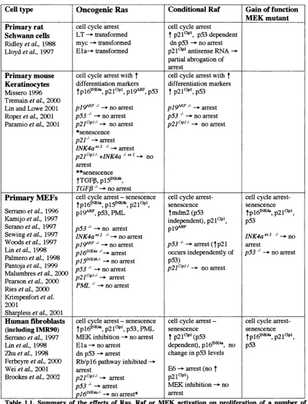
Summary of the effects of Ras, R af or MEK activation on 34 proliferation of a number of primary cell types
Induction of a cell cycle arrest by oncogenic Ras in 35 primary fibroblasts
The structure of normal peripheral nerves and neurofibromas 41 of proteins
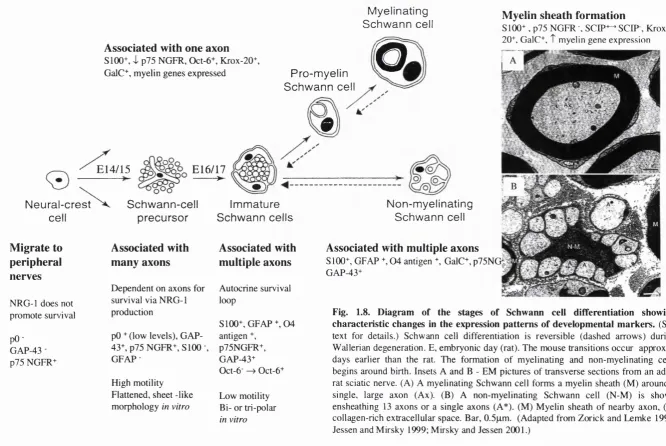
Sequence similarity of Neurofibromin to the Ras-GAP family 41 Diagram of the stages of Schwann cell differentiation showing 49 characteristic changes in the expression patterns of
developmental markers
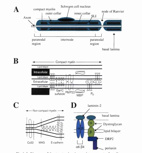
Diagrams of the structural regions and components of myelin Diagram to summarise the relationship between mutations in pO, PMP-22 and connexin 32 and inherited demyelinating neuropathies
56 57
62
70
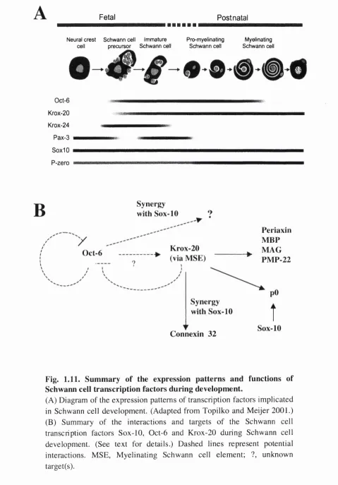
80 Summary of the expression patterns and functions of
Schwann cell transcription factors during development Diagram of the main events that occur during Wallerian degeneration and axonal regeneration
Primer sequences and reaction conditions used for PCR and RT-PCR/ SQ-RT-PCR
Solutions used in genomic DNA extraction and Southern blotting 85 Northern blotting solutions and wash conditions 87 Origin of the DNA fragments used for probe labelling 8 8
Antibodies used for W estern blotting and immunofluorescence 93 Solutions for protein extraction and Western blotting 94
Fig.3.1. Activation of Raf results in a cell cycle arrest associated with the 98 induction of cyclin dependent kinase inhibitors
Fig.3.2. Activation of Raf results in increased p21"^'^' mRNA expression 101 Fig.3.3. Induction of p i 9^^^ by Raf is not associated with increased 103
levels of p53
Fig.3.4. Raf activation is associated with decreased and pl9^^’^'^'’ 106 protein levels, however p i m R N A levels remain constant
Fig.3.5. Raf activation does not causepO expression 110 Fig.3.6. Raf activation can block ^0 expression. 112
Fig.3.7. Raf activation can reversepO expression 113
Fig.3.8. Upregulation o f f 0 levels in primary Schwann cells is unaffected 114
by the addition of tamoxifen or (3-estradiol
Fig.3.9. Raf activation results in decreasedfO mRNA levels 116 Fig.3.10. Raf activation can block or reverse Schwann cell differentiation 118
-downregulation of myelination markers
Fig.3.11. Expression patterns of some of the transcription factors involved 122 in Schwann cell differentiation to the myelinating form
Fig.3.12. The effect of Raf activation on transcription factors involved in 123 Schwann cell differentiation
Fig.3.13. The effect of Raf activation on the levels of cyclin D l, p i 9^*^^ 126 and the cyclin dependent kinase inhibitors plS^^^'^^ and p27^^
Fig.3.14. Inhibition of the MAPK pathway is associated with Schwann 129 cell differentiation and prevents Raf from blocking differentiation Fig.3.15. Raf activation is unable to reverse differentiation in the presence 130
of a MEK inhibitor
Fig.3.16. Decreasing the activity of the MAPK pathway is insufficient 131 to cause differentiation
Fig.4.4. Schwann cells with activated Raf are still able to rapidly associate 143 with axons
Fig.4.5. The majority of Schwann cells with prior activation of Raf remain 144 stably associated with axons
Fig.4.6. Schwann cells remain associated with axons following Raf 146 activation
Fig.4.7. Schwann cells with activated Raf can dissociate from DRG axons 147 Fig.4.8. Raf activation is associated with increased Schwann cell motility 151
Fig.5.1. The RafTR is specifically activated by Tmx addition 156 Fig.5.2. Diagram of the pRafTR 1RES-EGFP cloning strategy and 157
construct testing
Fig.5.3. Diagram of the pPG6 RafTR IRES-EGFP cloning strategy 159
Fig.5.4. The pPG6 RafTR IRES-EGFP is active m under 160
differentiating conditions
Fig.5.5. pPG6 RafTR IRES-EGFP is detectable in 957 and 963 163
transgenic mice
Fig.5.6 . Germline transmission is observed in both parental lines 165
Fig.5.7. The 963 line expresses RafTR mRNA 166
Fig.6 .1. Model of the mechanism of the oncogenic Ras/Raf induced 177
cell cycle arrest in primary Schwann cells
Fig.6 .2. Models for the role of Raf/MAPK signalling during the 179
Chapter 1: Introduction
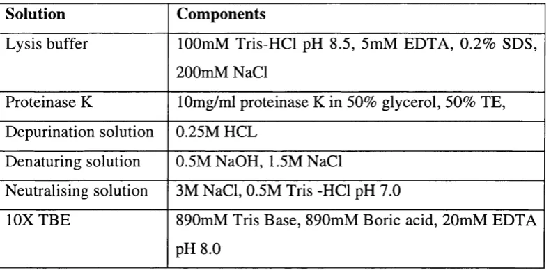
T um o rig e n esis is thought to be a multistep process, requiring several successive genetic changes for cellular transform ation. This requirem ent has been most well docum ented for colorectal cancer, which usually develops over several decades and appear to require at least seven different genetic lesions, including those resulting in the activation of K-ras and loss of p53 function (reviewed by Kinzler and Vogelstein, 1996, see Fig. 1.1.). M oreover, these m utations appear to occur in a specific sequence and the num ber of lesions can be correlated with the stage of tum our development.
A PC K -R A S D C C /Ü P C 4 /
J V18? p53
O th er C hanges?
N orm al E p ith eliu m
D y sp ia stic A C E
Early A d e n o m a
Interm ediate A d en o m a
L ate
A d e n o m a
H
C arcin o m a M etastasis
I xRii
w
NTAIK D eficiencyF ig.l .1. Genetic changes associated with Colorectal Tum origenesis
A PC m u la fio n s initiale the n eop lastic p r o c e s s with su b s eq u e n t mut ation o f the in d ic ated g e n e s res ulting
in tu m ou r p ro g res sio n . M is -m a tc h repair ( M M R ) d e f i c i e n c y a c c e le r a tes this p r o ces s. K -R a s is activated by a s i n g l e mutation. p 5 3 inac tivation m a y also on ly require o n e mut ation s in c e mutant proteins m ay act
in a d o m in a n t n e g a t iv e m ann er to inhib it the r em ain in g w ild - t y p e alle le. T h e other g e n e s ind icat ed are
tumour su pp re ss or g e n e s that require tw o g e n e tic e v en ts (on e per a lle le) for ina ctivation. T h e D C C , C P C 4
and J V J 8 g e n e s are c a n d id a te tum ou r s u p p r e s s o r g e n e s for c o lo r e c t a l n e o p la s ia that are lo ca te d on c h r o m o s o m e 1 8q 21. O ther g e n e t ic c h a n g e s have a lso be en d e s c r ib ed in a sm all nu m b e r o f colo r ec ta l
can cer s. (A d a p te d K in zle r and V o g e l s t e i n , 199 6.)
lu vivo s u p p o rt for the m u ltistep nature of tu m o rig e n e s is has been p ro v id e d using
molecules are acting synergistically. However, since the tumours are clonal and form in a stochastic manner this would suggest that deregulated expression of myc and oncogenic H- ras is still insufficient for tumour formation and additional changes are required prior to malignant tumour formation.
The requirem ent for m ultiple genetic changes in tumour formation is also supported by work in vitro using primary cells and immortalized cell lines. In primary cells, expression of a single oncogene is insufficient to cause cellular transformation (Land et a i , 1983; Ruley, 1983). However, co-expression of at least two oncogenes, or a single oncogene in association with loss of a tumour suppressor gene function, facilitates transformation (Land
et a i, 1983; Ruley, 1983; Ridley et a i, 1988; Tanaka et a i , 1994; Serrano et a i, 1997). For example oncogenic H-ras is unable to transform primary Schwann cells or fibroblasts, whereas oncogenic H-ras and myc, or oncogenic H-ras and loss of p53 function, cooperate to transform the primary cells. In contrast established cell lines can be transformed by a single oncogene due to the acquisition of a num ber of genetic changes during the immortalisation procedure. For example NIH 3T3 cells (an immortal mouse fibroblast cell line) are transformed by oncogenic Ras, due to the deletion o f the INK4a and INK4b loci that encodes the cell cycle regulators p t ^'NK4a pj^iNK4b the tumour suppressor
P I9ARF (Sej^ano et a l , 1993; Quelle et a l, 1995a; Kamijo et a l, 1997; Sewing et a l, 1997;
Malumbres et a l , 2000).
Rodent cells and human cells appear to differ in their susceptibility to transformation in vitro. Human cells are not transform ed by two co-operating oncogenes and appear to require additional changes compared to rodent cells for successful transformation (Hahn and W einberg, 2002). Recently Hahn and colleagues have achieved transform ation of human cells in vitro by expressing SV40 large T antigen (LT), which inhibits the Rb and p53 pathways; the catalytic subunit of telomerase, hTERT; oncogenic Ras and SV40 small t antigen (ST), which inhibits protein phosphatases 2A (Hahn et a l , 1999; Hahn et a l ,
2002). Thus transform ation of human cells appears to require changes in at least five separate pathways.
The resistance of primary cells to transformation by a single oncogene has been proposed to act as a protective mechanism to prevent unregulated proliferation in response to a single genetic alteration. The mechanism of this resistance appears to vary between oncogenes, but it is becom ing clear that the tum our promoting function of an oncogene is frequently balanced by an inhibitory effect, which must be overcome by additional mutations to allow tumorigenesis (Evan and Vousden, 2001). These inhibitory effects include induction of a cell cycle arrest or apoptosis. For example, Myc expression is tightly regulated in normal cells, where it prom otes proliferation in the appropriate m itogenic environm ent, but deregulated Myc signalling is often observed in tumour cells. However, deregulated Myc expression is also able to promote apoptosis (Evan et a l , 1992). Understanding of the mechanisms underlying the resistance of primary cells to transformation may enable us to reactivate these inhibitory pathways as a form of cancer therapy.
Thesis Aims
Many prim ary cell types, including Schwann cells, undergo a G1 cell cycle arrest in response to oncogenic R as/R af (Ridley et a l, 1988; Lloyd et al., 1997). However, the mechanism underlying the resistance of primary cells to transformation by oncogenic Ras is still under examination and appears to differ between cells types and species (see below). In this thesis I shall present work aimed at investigating the mechanism of the p53 dependent Ras/Raf induced cell cycle arrest in primary Schwann cells. In addition, since oneogenic Ras/Raf activity is associated with the induction of a more differentiated phenotype in certain cell types (Bar-Sagi and Feramisco, 1985; Halfar et a l , 2001; Roper et a l , 2001), I have exam ined the effects of R af activation on Schwann cell differentiation. We have chosen to use Schwann cells as a primary cell type for this research because Schwann cells are easily obtained from rat sciatic nerves and ean be cultured in vitro for prolonged periods without undergoing transformation or senescence (Brockes et a i , 1979; M athon et a i,
In this introduction I shall begin by presenting a brief overview of the regulation of the G l- S-phase cell cycle transition. I will then summarise the mechanisms of Ras signalling, including the effects of Ras on cell cycle progression. As I am interested in the oncogenic R as/Raf induced p53 dependent cell cycle arrest in prim ary Schwann cells, which is associated with CDKI induction, I shall review the regulation of p53 activity and CDKI regulation. I will then discuss the mechanisms involved in mediating the cell cycle arrest in response to oncogenic Ras/Raf signalling in a number of primary cells. I will then discuss the genetic disorder Neurofibrom atosis type 1 (N F l) and conclude with an account of Schwann cell developm ent, focusing on differentiation to the m yelinating form and the roles of Schwann cells in peripheral nerve regeneration.
1.1. Regulation of G l-S phase transition
The somatic cell cycle is divided into four phases. DNA replication occurs during S-phase, with division of the cell and its components into two usually equal daughter cells during M- phase (mitosis). These phases are separated by two gap phases, G1 and G2, during which the cell accumulates the components required for DNA synthesis or mitosis and increases in size. Progression through the cell cycle is regulated at numerous stages by a series of checkpoints that prevent premature transition to the next phase or arrest the cell in response to abnormalities such as m isalignment of chromosomes or DNA damage (reviewed by Malumbres and Pellicer, 1998).
Following mitogenic stimuli cells progress through G1 and, on passing the restriction point in late G l, becom e irreversibly committed to S-phase entry and a new round of division. Transition through the cell cycle is controlled by a number of cyclin dependent kinases (CDK), which function at different stages of the cell cycle. CDKs are serine/threonine kinases that require cyclins for their activity. The cyclin-CDK complexes are regulated by many mechanisms including cyclical changes in cyclin levels, and inhibitory and activating phosphorylation of the CDKs (Malumbres and Barbacid, 2001). Cyclin D-CDK4/6 and cyclin E-CDK2 activity are required to initiate S-phase entry, whilst cyclin A-CDK2 is active in late G l and S-phases (Dulic et ah, 1992; M atsushime et a l, 1992; Pagano et a l ,
1992; Meyerson and Harlow, 1994). In line with their critical role in regulating G l-Sphase
transition the G l cyclins and CDKs are frequently found to be deregulated in human cancers (Malumbres and Pellicer, 1998).
The activity o f cyclin-CD K complexes is also regulated by cyclin dependent kinase inhibitors CDKIs, which are divided into two families of related molecules- the Cip/Kip proteins and the INK4 proteins (reviewed by Sherr and Roberts, 1995; Sherr, 1999). The CDKIs m ediate the cell cycle arrest in prim ary cells in response to oncogenic Ras signalling (Lloyd et al., 1997; Serrano et al., 1997; Malumbres et al., 2000). The Cip/Kip family comprises p21^'^% p27^^^ and p57’^^^, which bind both the cyclin and CDK subunits to preferentially inhibit the activity of G l cyclin-CDK complexes (Harper et a l, 1993 X iong et a l , 1993; el-Deiry et a l , 1994; Polyak et a l , 1994a; Polyak et a l , 1994b Toyoshima and Hunter, 1994; Zhang et a l , 1994b; Lee et a l , 1995; Matsuoka et a l, 1995 LaBaer et a l , 1997). However, p21'^‘'’' and p27’^‘’’’ have also been isolated in active CDK complexes and have been implicated in regulating the assembly and localisation of active type D cyclin-C D K 4 complexes (Zhang et a l , 1994a; LaBaer et a l , 1997; Cheng et a l ,
1999). The INK4 family of CDKIs consists of pl5'""""\ p i8''"'""' and p l 9 ^ T
which specifically bind to and inhibit CDK4/6, preventing their association with D type cyclins (Serrano et a l , 1993; Hannon and Beach, 1994; Chan et a l , 1995; Guan et a l ,
1994; Guan et a l , 1996, Hirai et a l, 1995). The INK4 CDKIs also induce the displacement of C ip/K ip proteins from active cyclin D-CDK com plexes onto CDK2 containing complexes (McConnell et a l , 1999).
The G 1-S-phase transition is summarised in Fig. 1.2. In the absence of mitogenic stimuli cells are quiescent (non-dividing) and cyclin D-CDK activity is low, allowing Rb to inhibit the activity of the E2F transcription factors to prevent the expression of genes required for cell cycle progression (Sherr, 1993), (Dyson, 1998) Mitogens induce cyclin D expression and the formation of cyclin D-CDK4/6 complexes (Sherr, 1993; Aktas et a l, 1997; Cheng
(Active)
Mitogenic signais
(Inactive) CDKI Phospho
And degradatic
rylation
Cip/Kip
CDKI sequestration
E-K2 Activation
Cip/Kip
Positive feedback Phosphoryiatio
S-phase entrv
Fig.1.2 Regulation of the Gl-S phase transition.
been proposed to displace histone deacetylase (HDAC) from the Rb-E2F complex and induce the partial activation of E2F transcription factors, resulting in the expression of specific genes such as cyclin E (Harbour et a i, 1999; Zhang et a i , 2000). The active cyclin E-CDK2 complexes then cooperate with cyclin D-CDK4/6 to fully phosphorylate and inactivate Rb, leading to the transcription of targets required for DNA replication and irreversible commitment to S-phase entry (Dyson, 1998; Lundberg and W einberg, 1998). The cyclin D1-CDK4/6 complexes also facilitate cyclin E-CDK2 activity by sequestering Cip/Kip proteins, which are able to inhibit cyclin E-CDK2 activity (Harper et al., 1993; Polyak et a i , 1994b; Cheng et a i, 1998). In addition, phosphorylation of p27’^^’ by Cyclin E-CDK2 complexes results in the targeting of p27 for degradation (Sheaff et a i , 1997).
1.2. Ras signalling
The ras genes were first discovered as the transforming agents of Harvey and Kirsten murine sarcom a viruses (reviewed by M alumbres and Pellicer, 1998). The Ras family consists of K-Ras A and K-Ras B, which are encoded by different exons for the last 25 amino acids, H-Ras, and N-Ras. The Ras proteins are highly homologous to each other across most of the protein sequence, however, the Ras proteins may have different cell specific activities since their expression patterns vary and different Ras molecules are specifically activated in certain types of tumour (Bos, 1989, see below; M alumbres and Pellicer, 1998). Moreover, N-Ras and H-Ras knockout mice are developmentally normal, whilst K -R a s nulls die from liver defects and anaemia in utero (Um anoff et al., 1995; Johnson et al., 1997).
Activation of Ras
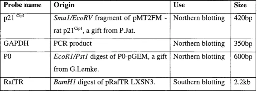
Ras proteins act as m olecular switches, which are activated in response to extracellular stimuli and m ediate signal transduction into the cytoplasm (Fig. 1.3). In response to mitogenic stimuli inactive GDP bound Ras is activated by the exchange of GDP for GTP, facilitating interaction with effector molecules and downstream signalling. Ras proteins have low intrinsic GTPase activity and the hydrolysis of GTP to GDP is promoted by interaction with GTPase activating proteins (GAPs), resulting in Ras inactivation (Trahey and M cCormick, 1987; Adari et al., 1988; Scheffzek et a l, 1997; Scheffzek et at., 1998). A number of RasGAPs have been identified in mammalian cells, including pl20R asG A P and Neurofibromin (Trahey and M cCormick, 1987; Adari et a l , 1988; Ballester et a l , 1990; M artin et al., 1990; Xu et al., 1990). The other group of Ras regulatory proteins are the guanine exchange factors (GEFs or guanine dissociation stimulators, GDS), which are thought to increase the dissociation of GDP by inducing a conformational change in Ras, facilitating the binding of GTP and thus promoting Ras activation (Quilliam et al., 1995; Boriack-Sjodin et al., 1998; W ittinghofer, 1998). The major mammalian GEFs are thought to be mSos 1 and 2, which are homologues of the Drosophila Son of Sevenless protein (Simon et al., 1991; Bonfini et al., 1992; Bowtell et al., 1992; Chardin et al., 1993).
Ras signalling is activated in response to a variety of extracellular stimuli including growth factors, hormones, cytokines and neurotransmitters that interact with cell surface receptors such as receptor tyrosine kinases (RTKs, for example the EGF receptor), non-RTK associated receptors or G-protein coupled seven transmembrane receptors (reviewed by Campbell et al., 1998). The pathway mediating Ras activation in response to growth factor signalling has been well characterised (Fig 1.2.). For example, binding of EGF to the EGF homodimer stimulates receptor autophosphorylation of tyrosine residues in the cytoplasmic domain, which form binding sites for the Src homology 2 domain (SH2) of the adaptor protein Grb2 (Lowenstein et a l , 1992). Grb2 also contains two SH3 domains that bind a proline-rich m otif in the carboxy-term inal of Sos and translocate the GEF to the PM to transiently activate Ras (Lowenstein et a l , 1992; Chardin et al., 1993). Ras can also be activated in response to integrin signalling which is thought to be important for mediating
Inactive Ras
Ras GAP RTK
Active Ras
Ras GDP She
Grb2
SOS (Ras GEF)
Raf PI3K RaiGDS
BAD MEK
ERK Apoptosis
Elk-1
Sos and Vav Rai
AKT
/ \
PAK _
Forkhead Transcription factors
:
ApoptosisRBPl
CDC42
Gene expression Gene expression Actin cytoskeleton
Fig.l.3. Ras activation and downstream signalling.
anchorage signals (M ainiero et a l, 1997; W ary et al., 1998; review ed Giancotti and Ruoslahti, 1999; Cullen and Lockyer, 2002).
Ras signalling in tumorigenesis
Ras activity is tightly regulated in the cell under normal conditions; however, mutations resulting in constitutive activation of Ras are frequently detected in human tumours. The incidence o f R a s mutation varies betw een tum our types, for exam ple over 90% of pancreatic tumours and about 50% of colon tumours have mutated Ras, compared to less than 10% o f breast tumours (Bos, 1989). M utations in specific R a s genes are also associated with different types of tumour. N-Ras is mutated in melanomas, whilst K-Ras
mutations are found in adenocarcinomas of the lung, pancreas and colon. M oreover, a mouse model with somatic activation of oncogenic K-ras is predisposed to the development of early onset lung cancer, supporting a causal role for mutations of specific Ras genes in the formation of certain types of cancer (Johnson et a l , 1997). In addition, in certain human tumours mutations have been detected in other components of the Ras signalling pathway, resulting in elevated levels of Ras signalling in the absence of Ras mutation. For example in the inherited genetic disorder Neurofibromatosis type 1 (N Fl), mutation of the
N F l gene, which encodes a putative RasGAP, results in elevated Ras signalling in Schwann cells and is associated with the developm ent of benign neurofibrom as and malignant peripheral nerve sheath tumours (Lakkis and Tennekoon, 2000). Overexpression of the neuregulin growth factor receptor erbB2, which can activate Ras signalling, is associated with tumours including breast, lung, pancreas and colon cancers (Yarden and Sliwkowski, 2001). Ras signalling is not only important for tumour formation, but is also required for tumour maintenance (Chin et at., 1999). In vitro, oncogenic Ras is able to transform immortal rodent cell lines, resulting in the loss of contact inhibition of growth, anchorage independent proliferation and mitogen independent proliferation (DeFeo et a l ,
1981; Inoue et a l , 1996; Newbold and Overell, 1983). The oncogenic Ras induced loss of growth control is likely to be important for its role in tumour development.
Ras signalling pathways
Ras signalling has been implicated in numerous cellular processes including differentiation (Bar-Sagi and Feram isco, 1985; Freeman, 1998; Sternberg and Han, 1998; Jiang et a l ,
1999); cell growth (Prober and Edgar, 2000); cell cycle progression in response to mitogen and integrin signalling (Dobrowolski et al., 1994; Aktas et al., 1997; (Giancotti and Ruoslahti, 1999)); cell survival and apoptosis (Downward, 1998; Halfar et al., 2001). The ability of activated Ras to specify such a variety of different cellular outcomes may be mediated by the activation of different downstream effector pathways, of which the best characterised are the Raf/MAPK (mitogen activated protein kinase), PI3K (phosphoinositol 3-kinase) and RalGDS pathways (reviewed by Campbell et al., 1998, Fig. 1.3.). RalGDS and the related proteins Rif and Rgl are GEFs for the GTPases Ral A and Ral B (Wolthuis
et a l, 1997; reviewed by Bos, 1998). Activated Ral has been shown to interact in vitro with Ral-binding protein (RalBP), which is a GAP for the Rho-like GTPases Ccdc42 and Rac, and thus may have effects on the actin cytoskeleton. I shall now discuss Ras activation of Raf/MAPK and PI3K signalling in more detail.
PI3K signalling
PI3K consists of a p i 10 catalytic subunit and a p85 regulatory subunit (Campbell et al.,
1998). PI3K interacts directly with Ras through the p i 10 catalytic subunit, which activates PI3K phosphorylation of inositol lipids to produce PI(3 ,4)P2 and PI(3,4 ,5)P3 in response to
stimuli such as growth factors (Rodriguez-Viciana et al., 1994; (Campbell et al., 1998). PI3K signalling has been implicated in a num ber of Ras induced cellular processes including cell cycle progression, transcription and cell survival (Downward, 1998) Datta et al., 1999; M edem a et al., 2000; Jones and Kazlauskas, 2001). PI3K can also be activated independently of Ras signalling (Toker and Cantley, 1997, Kahn, 1998; Giancotti and Ruoslahti, 1999).
The PI3K effector AKT/PKB (protein kinase B) is suggested to regulate many of the cellular effects of PI3K (reviewed by Alessi and Cohen, 1998; Datta et a l , 1999). AKT is activated by growth factors, such as PDGF, in a PI3K dependent manner (Franke et a l ,
1995). PI(3 ,4 )P2 and PI(3,4 ,5 )P3, appear to promote translocation of AKT to the PM where
i ) and possibly a modified form of PD K l in complex with another kinase PRK2 (Alessi et al., 1996; Alessi et al., 1997; Stokoe et al., 1997; Datta et al., 1999). AKT targets include GSK3 (glycogen synthase kinase 3), which is inactivated by AKT phosphorylation leading to the activation of glycogen synthesis and the Forkhead transcription factors, which are inhibited by AKT, preventing the expression of pro-apoptotic or cell cycle arrest promoting proteins such as Fas and p27^‘P’ respectively (Datta et al., 1999; Kops et al., 1999; M edem a et al., 2000). AKT can also promote cell survival by phosphorylating Bad on S e ri36, which inhibits the pro-apoptotic activity of Bad by inducing dissociation from BcIxl and association with 14-3-3 (Datta et al., 1997; del Peso et al., 1997). PI3K activation of A KT is antagonized by the tum our suppressor PTEN (Phosphatases and Tensin homologue deleted on chromosome Ten) (Stambolic et al., 1998; W hang et a i , 1998; reviewed by Yamada and Araki, 2001).
Raf/MAPK signalling
The R af family consists of the serine/threonine kinases Raf-1, A -Raf and B-Raf, which interact directly with the effector domain of Ras-GTP (Vojtek et al., 1993; Warne et al.,
1993; Zhang et al., 1993). Raf signalling has been shown genetically to specifically mediate a num ber of Ras activities including differentiation in the Drosophila eye and vulval development in C.elegans, demonstrating the importance of Raf as a Ras effector molecule (Dickson et a i , 1992; Han et al., 1993; Freeman, 1998; Sternberg and Han, 1998). Ras induced R af signalling has also been implicated in regulating transcription and mammalian cell differentiation, proliferation, apoptosis and survival (Kolch et al., 1991; Bruder et al.,
1992; W ood et al., 1993; Marshall, 1995; Coolican et al., 1997; Downward, 1998; Mikula
et al., 2001; H user et al., 2001). In addition, interaction between the R af and PI3K pathw ays has been shown to co-operatively m ediate Ras signalling under certain circumstances, whilst in others the two pathways can have opposing effects (Kolch et al.,
1991; D atta et al., 1997; del Peso et al., 1997; Fang et al., 1999; Rommel et al., 1999; Zimmermann and M oelling, 1999; Jones and Kazlauskas, 2001).
Raf activation is a multistep process that requires the co-ordinated interaction of a number of proteins and is summarised in Box 1.1. Recently mammalian scaffolding proteins such as IN K interacting protein (JIP-1) and MEK protein l(M P l) have been suggested to
localise m olecules involved in M APK signalling, facilitating their interaction and potentially regulating the specificity of signalling in response to particular stimuli (Schaeffer et al., 1998; Stewart et al., 1999; W hitm arsh et al., 1998). These signalling complexes may also contain regulatory molecules such as R af kinase inhibitor protein (RKIP) (Yeung et al., 1999).
Ras induced activation of Raf initiates a MAPK cascade whereby R af (a M APKK kinase) phosphorylates and activates the MAPK kinases, M EK 1/2, which then phosphorylate ERK 1/2 (M APK) (Howe et al., 1992; Moodie et al., 1993). However, it has also been reported that R af can signal independently of MEK (M ikula et al., 2001; Huser et al.,
2001). Activated ERK 1/2 can translocate to the nucleus and regulate transcription by phosphorylating a number of targets including the Ets transcription factors (Wasylyk et al.,
1998), the M APK interacting kinases M N K l/2 (Fukunaga and Hunter, 1997; Waskiewicz
et a l, 1997) and the M APK activated protein kinase (MAPKAPs), which phosphorylate a number of substrates such as CREB and CBP (Tan et al., 1996; Xing et al., 1996; Dalby et al., 1998). In addition, phosphorylation of the anti-proliferative protein Tob by ERK 1/2 has recently been implicated in Ras mediated proliferation and transformation (Suzuki et al.,
2002).
Interestingly, Ras/Raf/M APK signalling can have different effects in the same cell type depending on the intensity and duration of signalling. For example in PC 12 cells (a phaeochromocytoma cell line) EGF addition is associated with transient M APK activation, which is m ediated by R as/R af signalling and results in proliferation (Bar-Sagi and Feramisco, 1985; W ood et al., 1993; Cowley et al., 1994) Marshall, 1995; York et al.,
Raf-1
14-3-3
CNK 14-3-3
Kinase
No signal Signal
Box 1.1. Ras-mediated activation o f Raf.
Conserved region 1 (C R l) of Raf (shown in colour) contains two Ras binding domains, the RED, and the cysteine rich domain (CRD), which is obscured by 14- 3-3 binding, stabilizing the inactive conformation. The kinase domain is located in CR3, which constitutively associates with the heat-shock proteins Hsp90 and p50 (Schulte 1995). Ras activation facilitates interaction with the Raf RED and translocation of Raf to the PM, which may result in a conformational change that displaces 14-3-3 and exposes the CRD to Ras binding. 14-3-3 proteins then interact with the Raf carboxy terminal kinase domain and may be required to stabilise Raf in an active conformation (Tzivion 1998). Phospholipids (PS), Ksr (kinase suppressor of Ras) and CNK (connector enhancer of KSR) may also be involved in Raf activation and/ or downstream signalling (Thierren et al, 1995, 1996; Stokoe and McCormick 1997; Denouel-Galy et a l, 1998; Thierren et al,
1998; Yu et al, 1998; Muller et a l, 2001). Activation of Raf may also involve phosphorylation by Src family kinases, Janus activated kinases or PAK3 (JAKs, Marais et al, 1995; Xia et a l, 1996; King et al, 1998). (Adapted from Morrison and Cutler 1997; Campbell et a l, 1998.)
Regulation of G l-S phase cell cycle progression by Ras signalling
Stimulation of quiescent cells by mitogens, such as PDGF, and integrins results in rapid Ras activation and cell cycle entry (Satoh et a l, 1990; Giancotti and Ruoslahti, 1999). Ras activity is required at several points during the G 1-S-phase interval to inactivate Rb (Mulcahy et al., 1985; Dobrowolski et a l , 1994; Mittnacht et at., 1997; Peeper et al., 1997; Takuwa and Takuwa, 1997). During early G l, mitogenic stimulation of Ras activates the Raf/M EK/ERK pathway leading to transcription of the cyclin D l gene and assembly of cyclin D1-CDK4/6 complexes (Aktas et al., 1997; Kerkhoff and Rapp, 1997; Peeper et al.,
1997; Cheng et a l, 1998). During the later stages of G l, however, elevated PI3K levels are required for cell cycle progression (Gille and Downward, 1999; Jones and Kazlauskas, 2001). Ras dependent PI3K activation of AKT has been demonstrated to stabilize cyclin D l by inhibiting the phosphorylation of cyclin D l on Thr 286 by GSK3|3, preventing nuclear export and degradation of cyclin D l (Diehl et al., 1998). Thus mitogenic stimulation of Ras results in elevated levels o f active cyclin D l complexes, which facilitates cell cycle progression (Peeper et al., 1997; Cheng et al., 1998). Moreover, Ras activation reduces the expression of p27^^' via PI3K/AKT induced inhibition of AFX-like Forkhead transcription factor activity and promotes p27’^^'degradation, which may be mediated by cyclin E-CDK2 phosphorylation of p27^‘P' (Aktas et al., 1997; Takuwa and Takuwa, 1997; Medema et al.,
2000).
1.3. Ras signalling in primary cells
Oncogenic Ras is able to transform immortal cell lines, how ever, in prim ary cells oncogenic Ras causes a cell cycle arrest and a second co-operating oncogene or loss of a tumour suppressor gene function is required for transformation (Ridley et al., 1988; Lloyd
et a l , 1997; Serrano et al., 1997; Zindy et al., 1998). In many primary cell types the oncogenic Ras induced cell cycle arrest is caused by the induction of the CDKIs p lb '^ ’^'^^ and p21*^‘P’, with p21^'^^ induction occurring in a p53 dependent manner. This response has been proposed to act as a protective mechanism to prevent unregulated proliferation in response to a single genetic alteration (Lloyd et al., 1997; Serrano et al., 1997; (Malumbres
overcome the oncogenic Ras induced cell cycle arrest during tumorigenesis in vivo (Kinzler and Vogelstein, 1996; Lloyd et al., 1997; Serrano et a i , 1997; Kamijo et a i , 1997). I will now discuss the regulation of the CDKIs in response to oncogenic Ras/Raf signalling and their involvem ent in human tumour formation. I will then present an overview of the regulation of p53 activity and discuss the current ideas regarding the mechanisms involved in the induction of the cell cycle arrest in a number of primary cell types.
CDKI induction and function
p21^'P' has been implicated in mediating the induction of a G1 cell cycle arrest, and/ or a G2/M arrest, in response to DNA damage in a p53 dependent manner (Xiong et al., 1993; el-Deiry et al., 1994; Chan et al., 2000). In addition, in many primary cell types, oncogenic Ras/Raf expression results in the induction of p21^‘P’, leading to a cell cycle arrest (Lloyd et al., 1997; Serrano et al., 1997). The oncogenic Ras induced cell cycle arrest is p53 dependent in some primary cell types, which may reflect a requirement for p53-mediated transcription of p21^''"' (Lloyd et al., 1997; Serrano et al., 1997; Roper et al., 2001). However, oncogenic R as/Raf can also induce p21^'P’ independently of p53 under certain circumstances and this process may be negatively regulated by Rho signalling (Sewing et al., 1997; Woods et al., 1997, Olson et al., 1998; Kivinen et al., 1999; Gartel et al., 2000). In addition, p21^'^' is expressed independently of p53 during developm ent and may be involved in m ediating the cell cycle arrest preceding terminal differentiation (Macleod et al., 1995; Zhang et al., 1995; Di Cunto F, 1998; Harvat et al., 1998).
Mutations resulting in the complete loss of p21^‘^* function are rarely observed in human cancers; however, mutations in p53 are very common and are associated with reduced levels of p21^'P’ (Gao et al., 1995; Hollstein et al., 1991; Tanaka et al., 1996; Sherr, 1999). In agreem ent with these findings in hum ans, p21^‘^l-/~ mice are not predisposed to spontaneous tum our development, unlike p53-/- mice (Donehower et al., 1992; Deng et al.,
1995). However, p21^‘^l-/- kératinocytes have an increased proliferative potential and are susceptible to transformation by oncogenic Ras, suggesting that p21^'^^ is also able to act as a tum our suppressor under certain circumstances, although it is unclear whether this function also exists in human cells (Missero et al., 1996). In addition, Zhou and colleagues
have recently shown that some cancers associated with erbB2 mutations may overcome the
anti-proliferative effects of high levels of p2 1^'^' by promoting translocation of p2 1^^ho the
cytoplasm (Zhou et a l, 2001).
The signals regulating the INK4 proteins remain unclear, p i i s expressed in young mice and has been implicated in mediating the TGF(3 induced cell cycle arrest, although p l5iNK4b is not essential for this process (Hannon and Beach, 1994; Zindy et al., 1997b;
Zindy et al., 1997a; Latres et al., 2000). pig^b;K4c ^^giNK4d expressed during
development and may be involved in terminal differentiation (Zindy et al., 1997b; Zindy et al., 1997a; Phelps, 1998). p i6^biK4a not expressed during development in the mouse, but is
widely expressed at increasing levels in older animals (Zindy et al., 1997a; Zindy et a l ,
1997b). In vitro p l6^ ’^'^® is induced with progressive cell population doublings and has been
suggested to form part of a mechanism that prevents unlim ited cellular replication by inducing a cell cycle arrest (Alcorta et al., 1996).
pl6iNK4a has also been implicated in the induction of a cell cycle arrest in response to oncogenic Ras activation in some cell primary types (Serrano et al., 1997; Zhu, 1998; Lin and Lowe, 2001). However, the importance of p i6^b(K4a ^ mediator of the Ras induced
arrest appears to vary between cell types. For example, p i6^ ^ ^ null MEFs are still capable
of undergoing a cell cycle arrest in response to oncogenic Ras, whilst in plb^^’^'*^ null HDFs this arrest is abrogated (Serrano et a l , 1996; Kamijo et al., 1997; Krimpenfort et al., 2001 ; Sharpless et al., 2001; Brookes et al., 2002). Induction o f pi^iNK4a response to
Ras/Raf/M APK activation in human diploid fibroblasts (HDFs) is p53 independent (Wei et al., 2001) and may be mediated by the balance between the transcriptional activators E tsl/2 and their repressor Id l (Ohtani et a l, 2001; reviewed by Zebedee and Hara, 2001). Another possible regulator of transcription at the INK4a locus is the transcriptional repressor bmi-1, which may prevent p l6^^"^® expression during developm ent (Jacobs et a l , 1999). In
The INK4a locus, which encodes is unusual in that it also encodes another protein, plQARF, alternative reading frame (Quelle et al., 1995b, see below), plb^^*^"^® and P I9 ARF encoded by exon l a and Ip respectively, but share a common exon 2. The
IN K 4 a locus is frequently m utated in human cancers or inactivated by méthylation, although it can be difficult to determine whether these changes affect p i o r pl9**^ activity (review ed by Ruas and Peters, 1998; Sharpless and DePinho, 1999). Germline m utations in p i 6 have been identified in fam ilial m elanom a and pancreatic adenocarcinoma patients. INK4a mutant mice, which produce an unstable p i p r o t e i n , do not form tumours within the first year of life, however INK4a mutant mice that are also heterozygous for p i 9^^^ are susceptible to developing melanomas, suggesting that p i 9^^^ is haplo-insufficient for tumour suppression in this genetic environment (Krimpenfort et a l,
2001). In contrast, lNK4a ex la -/- (plô^^*^"^^ null) mice have an increased incidence of spontaneous tumours and are susceptible to tumour formation following treatment with carcinogens (Sharpless et al., 2001). In addition, lNK4a e xla -/- MEFs are immortalized in vitro at an accelerated rate. In contrast, p i a n d rarely mutated in human cancers (Ruas and Peters, 1998). In mice loss of pl9^^’^'‘'‘ function does not appear to be associated with tumour formation (Zindy et al., 2000), however p l 8 ‘^^"^''-/- mice develop pituitary hyperplasia, amongst other tumours, and may exhibit gigantism and organomegaly (Franklin et al., 1998; Latres et al., 2000). p i m u t a t i o n s also occur in human cancers, but are usually associated with a large homozygous deletion that also affect the lN K 4a
locus (Ruas and Peters, 1998). p i 5'^^'^'’-/- mice have a have a slight increase in spontaneous tumour formation supporting the role of as a tum our suppressor (Latres et al.,
2000).
p53 activation and functions
Regulation of p53 levels and stability
p53 is one of the most frequently mutated genes in human tumours (Hollstein et al., 1991). Both Li-Fraum eni patients, who inherit m utations in p 5 3 , and p 5 3 null mice are predisposed to tum our formation, supporting the role of p53 as a tum our suppressor (Malkin et al., 1990; Donehower et a i, 1992). Moreover, mice expressing a stable mutant of p53 in the active wild-type conformation have an enhanced resistance to tumorigenesis
(Tyner et al., 2002). p53 function may also be inactivated in the absence of p53 mutation in some tumours by overexpression of mdm-2, deletion of pl9**^, p53 mislocalisation and viral oncoproteins including adenovirus E lb , SY40 LT and the papillomavirus E6 protein
(Vogelstein et al., 2000). The p53 family also includes p63 and p73 (reviewed by Yang et a l , 2 0 0 2).
p53 is maintained at low levels in the cell under normal circumstances by the interaction of mdm-2 with the p53 transcriptional activator domain, which inhibits p53 activity and targets p53 for proteosomal degradation (Momand et al., 1992; Oliner et al., 1993; Honda
et al., 1997, Haupt et al., 1997; Kubbutat et al., 1997; Roth et al., 1998; Tao and Levine, 1999a). Yap and colleagues have recently demonstrated that mdm-2 inhibits the apoptotic and trans-repressor functions of p53 mainly by targeting p53 for degradation, whilst this is not necessary for the inhibition of p53 transcriptional activity by mdm- 2 (Yap et al., 2 0 0 0).
Moreover, since mdm-2 is a transcriptional target of p53, this interaction creates a negative feedback loop whereby p53 activation results in increased levels of mdmd-2, promoting p53 degradation and thus returning p53 levels to basal (Barak et al., 1993; Wu et al., 1993; Haupt et al., 1997; Kubbutat et al., 1997). M dm-2 expression can also be regulated independently of p53 by the Ras/Raf/MAPK pathway (Ries et al., 2000).
p53 is activated by many stimuli, in addition to oncogenic stress, including DNA damage, hypoxia and rN TP depletion (summarised in Fig. 1.4, reviewed by Giaccia and Kastan, 1998; Abraham, 2001). These stimuli induce a range of post-translational modifications that stabilise and activate p53, in part by regulating the interaction between mdm-2 and p53 (Avantaggiati et al., 1997; Gu et al., 1997; Shieh etal., 1997; Banin et al., 1998; Canman et al., 1998; Sakaguchi et al., 1998; W aterman et al., 1998; Gostissa et al., 1999; Liu et al.,
1999; Rodriguez et al., 1999; Chehab et al., 2000; Hirao et al., 2000; Shieh et a i, 2000; Ito
rNTP depletion DNA Damage Oncogenes DNA-PK P300/CBP pCAF Redox ATM ATR Chk2 Chkl Hypoxia
p53 phosphorylation H IF la binding
p53 jM dm -2
p 53^ y + accessory Proteins
I e .g .p 3 3 IN G , A S P P l/2 Mdm-2 Targets Noxa PIDD Fas Bax p53AIP Mdm-2 p2lCipi 14-3-30
Reprimo TSPl Maspin GD-ALF BAll p53R2
DNA repair G1 arrest G2 arrest apoptosis Prevention of new blood vessel formation
Fig.1.4. p53 activation and downstream targets.
p53 is maintained in a transcriptionally inactive state in the cell by interaction with mdm-2, which also targets p53 for proteosomal degradation. Following stimuli such as DN A damage or oncogenic stress p53 is covalently modified by acétylation, sumoylation and/or phosphorylated by a number o f protein kinases depending on the nature o f the stimuli.This results in p53 activation and/or stabilisation. Examples o f m olecules that are proposed to modify p53 are shown. Mdm-2 may also be inactivated by interaction with or phosphorylation. Active p53 transactivates expression o f a variety o f target genes leading to a diverse array o f potential outcomes, depending on the original stimulus and cell type. Selected exam ples o f target genes are shown. Dashed ai rows indicate potential interactions. See text for details. (Adapted from Giaccia and Kastan 1999; V ogelstein et a l, 2000.)
Oncogenic stress has been shown to stabilise and activate p53 by an alternative mechanism involving the induction of p i F o r example, in primary mouse embryonic fibroblasts (M EFs) oncogenic Ras expression induces a p53 dependent cell cycle arrest, which also requires pl9**^ activity (Kamijo et al., 1997; Palmero et al., 1998, see below). PML, the product o f the P M L gene of prom yelocytic leukaem ia, has also been im plicated in regulating p53 activity in response to oncogenic Ras and DNA damage (Guo et al., 2000; Ferbeyre, 2000; Pearson et al., 2000). In primary fibroblasts oncogenic Ras upregulates PM L expression and PM L overexpression is able to induce a p53 dependent cell cycle arrest, sim ilar to oncogenic Ras (Ferbeyre, 2000; Pearson et al., 2000). In MEFs, oncogenic Ras has been found to induce the re-localisation of p53 and CBP to PML nuclear bodies, where they form a trimeric complex with PML that may be involved in mediating p53 activation by promoting acétylation on lysine 382 (Pearson et al., 2000). Moreover, the oncogenic Ras induced cell cycle arrest and acétylation of p53 are impaired in PML -/- M EFs, confirm ing the im portance of this protein in mediating the Ras induced p53 dependent arrest. Interestingly oncogenic Ras expression in PM L -/- MEFs is still able to induce similar levels of p53 although this is associated with reduced expression of the p53 transcriptional target p21^'P', suggesting that PML is required for p53 activation but not stabilisation. In contrast to M EFs, in human diploid fibroblasts (HDFs), it has been suggested that Ras induced upregulation of PML results in p53 activation by promoting phosphorylation of serine 15 (Ferbeyre, 2000). The histone deacetylase S IR T l, which is recruited to PML nuclear bodies following oncogenic Ras expression, has recently been found to antagonise PM L induced acétylation o f p53 and repress p53-m ediated transactivation (Vaziri et al., 2001; Langley e t a l , 2002).
Downstream signalling pathways from p53
M anfredi, 2002). Interestingly, c-Jun has recently been shown to negatively regulate p53 association with the p21‘^‘P’ promoter following UV treatment, and thus may downregulate p21‘^‘P' expression to allow cell cycle re-entry after successful DNA repair (Shaulian et a l,
2000). The effect of p53 activation appears to vary between cell type and stimuli to some extent, but it remains unclear how the outcome of p53 activation is determined (Lane, 2001). However, the identification of A S P P l/2 (apoptosis stim ulating protein of p53) suggests that accessory proteins may target p53 activity to specific promoters to induce apoptosis (Samuels-Lev et al., 2001).
Regulation of p53 activity by
M utations of the INK4a locus that selectively affect p i 9^^^ (or p i 4^^^ in humans) are much less common than mutations affecting both proteins or just pl6IN K 4a, although a recent study suggests that the p i 9^*^^ promoter can also be inactivated by méthylation (Robertson and Jones, 1998; Ruas and Peters, 1998; Zhang and Xiong, 1999). However, the importance of p i 9^^'' as a tumour suppressor has been clearly demonstrated by
pl9^^^-/-mice, which are predisposed to spontaneous tumour formation (Kamijo et a i, 1997. p i 9^*^^ expression is induced by hyperproliferative signals including overexpression of myc, adenovirus E l a, v-Abl, resulting in apoptosis (Radfar et a l , 1998; de Stanchina et a l ,
1998; Zindy et a l , 1998). In contrast, p 19^^^ overexpression or induction of pl9**^ by oncogenic Ras or overexpression of p-catenin is associated with a cell cycle arrest (Quelle
et a l , 1995b; Palmero e t a l , 1998).
The induction of p i 9^'^'' in response to oncogenic stress and certain types of DNA damage is thought to promote p53 activation (Quelle et a l , 1995b; Kamijo et a l , 1997; Palmero et a l , 1998; Stott, 1998; Damalas et a l, 2001; Khan et a l , 2000). Overexpression of pl9^*^^ in m ouse fibroblasts is associated with p53 stabilisation and transactivation of p21‘^‘'^', resulting in a G1 and/or G2/M cell cycle arrest (Quelle et a l, 1995b; Kamijo et a l, 1997; Stott, 1998). p i 9^^^ interacts with mdm-2 and by antagonising mdm-2 function is able to stabilise p53 levels and promote p53 activity (Pomerantz et a l, 1998; Zhang, 1998; Honda and Yasuda, 1999). pl9*^^ is thought to sequester mdm-2 in the nucleolus, preventing ubiquitination and export of p53 and facilitating p53 activation (Honda and Yasuda, 1999;