78:12 (2016) 89–94 | www.jurnalteknologi.utm.my | eISSN 2180–3722 |
Jurnal
Teknologi
Full Paper
FABRICATION
AND
CHARACTERIZATION
OF
PCL/HA/PPY
COMPOSITE
SCAFFOLD
USING
FREEZE-DRYING
TECHNIQUE
Sharon Kalu Joseph Ufere
a, Naznin Sultana
a,b*a
Faculty of Bioscience and Medical Engineering, Universiti Teknologi
Malaysia, 81310 UTM Johor Bahru, Johor, Malaysia
b
Advanced Membrane Technology Research Center (AMTEC),
Universiti Teknologi Malaysia, 81310 UTM Johor Bahru, Johor,
Malaysia
Article history Received 21 August 2016 Received in revised form
1 October 2016 Accepted 3 November 2016
*Corresponding author
[email protected]
Graphical abstract
Abstract
Bone tissue regeneration and healing could be notably quickened via applying electrical stimuli in the defected area. Hence, a conductive tissue engineering scaffold that is capable of delivering the electrical stimuli is greatly desirable. In this study, electrically conductive scaffold was fabricated by using a biocompatible conductive polymer, polypyrrole (PPY) in the optimized nanocomposite scaffold of Polycaprolactone (PCL) and Hydroxyapatite (HA) using freeze–drying technique. The scaffolds were evaluated by using a number of techniques. The morphology of the scaffolds was observed and analyzed using a scanning electron microscope (SEM). Composite scaffolds with suitable pore size distribution were obtained by freezing the polymer solution mixture at -18ºC, by controlling the polymer and solvent phase crystallization. The results showed that the average pore sizes were decreased from 123.7μm for PCL scaffolds to 91.6μm with the incorporation of HA nanoparticles. Electrical conductivity of the scaffolds was evaluated using a digital multimeter. The wettability and porosity of the scaffolds were increased with the incorporation of Polypyrrole than Polycaprolactone scaffold. The newly fabricated PCL/HA/PPY scaffold showed good prospect to be employed for bone tissue engineering applications.
Keywords: Bone Tissue Engineering, Polypyrrole, Freeze-Drying, Conductive scaffold
Abstrak
Penyembuhan dan pertumbuhan semula tisu tulang dapat dipercepatkan terutamanya melalui penggunaan ransangan elektrik di kawasan bermasalah. Oleh itu, perancah konduktif kejureteraan tisu yang berupaya menghantar rangsangan elektrik amat dicari. Dalam kajian ini, perancah konduktif elektrik dihasilkan dengan menggunakan polimer bioserasi konduktif, polypyrrole (PPY) di dalam nanokomposit Polycaprolactone (PCL) dan Hydroxyapatite (HA) yang telah dioptimumkan menggunakan teknik beku-pengeringan. Perancah ini telah dinilai menggunakan beberapa teknik. Morfologi perancah telah deperhatikan dan dinilai menggunakan mikroskop electron imbasan. Perancah komposit dengan distribusi saiz liang yang sesuai telah diperoleh melalui pembekuan campuran larutan polimer pada -18ºC, yang telah dikawal melalui penghabluran fasa pelarut dan polimer. Keputusan menunjukkan saiz liang berkurang dengan penggabungan nanopartikel HA. Konduktiviti elektrik daripada perancah telah dinilai mengunakan multimeter digital. Kadar kebolehbasahan dan kadar liang sampel telah diperhatikan meningkat naik dengan penggabungan perancah Polypyrrole berbanding hanya dalam Polycaprolactone. Oleh itu, ciri-ciri perancah PCL/HA/PPY yang telah dikaji dalam kajian inin menunjukkan prospek yang bagus bagi kejuruteraan tisu tulang dan mungkin boleh digunakan untuk aplikasi kejuruteraan tisu tulang.
Kata kunci: Kejuruteraan Tisu Tulang, Polypyrrole, Beku-pengeringan, Perancah konduktif
1.0 INTRODUCTION
In orthopedics, one of the challenging and serious problems includes bone healing and regeneration. These days, a lot of people are suffering from different kinds of bone defects. Usually to tackle these defects, autogenic/allogeneic bone grafts and implants are being utilized. Nevertheless, these procedures have several disadvantages. It is well known that in autogenic process, a secondary surgery is required to obtain donor bone from the patient’s own bone and this could lead to delay in recovery. Similarly, use of allograft bone could lead to rejection, pathogen transmission and immune response. Hence, these need prompted the production of artificial bone tissue material, surgical reconstruction, transplantation and artificial prostheses use [1]. Tissue engineering tries to offer a new remedy to tissue loss. A feasible alternative to bone grafting procedure known as bone tissue engineering has emerged which is reliable and economical, is intended to overcome several restrictions of present bone grafts. In tissue engineering scaffold the principal aim is to use engineering principles to provoke and stimulate the natural healing process of bone [2]. The strategy behind these scaffolds is that they act as short-term substrates providing basic support for cells to attach and thrive, and also helps to retain functions of cells. Performance of the scaffolds is basically determines the success of the bone tissue engineering [3, 4]. A scaffold requires high porosity, interconnectivity and appropriate pore sizes to aid cell attachment, in-growth and mass transport of nutrients [5]. Most often, biodegradable polymers are chosen to produce tissue engineering scaffolds which help to eradicate the worries of long-term biocompatibility and similarly to avoid additional surgery. Therefore, by choosing appropriate materials and fabrication techniques, mechanical and degradation properties of the scaffold could be obtained.
Polycaprolactone (PCL), a biodegradable polymer,
possesses notable toughness and good
biocompatibility [6]. It has been approved by FDA for specific uses in human body [5, 7]. However, as with most biodegradable polymeric scaffolds, PCL surface has an intrinsic hydrophobic characteristics which renders it unfavorable for cell growth which does not permit cell attachment on the surface of the polymer [8]. On the other hand, Hydroxyapatite (HA) is an inorganic constituent of bone with general formula Ca10(PO4)6(OH)2. This biologically active calcium
phosphate ceramic is used in surgery to substitute and mimic bone. It is compatible with human body owing to its biological features. Apart from being the main constituent of bone, HA has a definite affinity towards several adhesive proteins, and also it’s directly involved in bone cell differentiation and hardening.
Because of these suitable properties, HA has been greatly studied over the past thee decades. However, HA has poor biomechanical characteristics; it is brittle, has low fatigue strength and also has low flexibility
which renders it insufficient for major load bearing applications [1, 9]. For providing new, desirable biocomposite for a particular application, blending of synthetic polymer and ceramics is one of the most effective methods [10]. Ceramic and polymer composites show the best features of both constituent that is the toughness of polymer and stiffness of ceramic [5] over scaffolds consisting of either only polymer or only ceramics [7]. Studies have shown that combining polymer (PCL) and ceramics (HA) will enhance mechanical properties, biodegradability as well as improve osteoconductivity [1, 7].
Conductive polymers (CPs), because of its satisfactory biocompatibility have been studied and utilized in several biomedical applications since 1980s [11]. CPs mediate electrical stimulation and is likely to be the stimulating factor to enhance bone regeneration. In earlier studies, it was shown that the addition of CP could enhance both the mechanical strength, the biodegradability of scaffolds including their in vitro biocompatibility [12, 13]. Polypyrrole (PPY) is used today in numerous applications, including biosensors, computer displays, corrosion protection, microsurgical tools, and drug delivery systems, and as a biomaterial in neural tissue engineering and blood conduits [14]. PPY possesses many exceptional qualities, such as good in vitro and in vivo biocompatibility, chemical stability, low oxidative potential and high conductivity [[14, 15].
Most studies on scaffolds intended for bone Tissue Engineering are typically concentrated on enhancing the properties of the scaffolds especially in respect to their chemical and mechanical properties [16, 17]. However, in order to combine the tissue engineering methods with the impression of improving the bone regeneration and healing using electrical stimuli, the electrical property of the scaffolds are being adjusted. Therefore, to enhance the electrical property of the scaffolds, compositions of biocompatible conductive polymer (CP) was employed. This study reports the development of a new composite scaffold PCL/HA/PPY utilizing freeze-drying technique. Firstly, PCL and PCL/HA composite scaffolds were fabricated with the incorporation of different amount of HA. Based on the characterization results, PCL/HA scaffold with 10% (w/w) HA was found to be suitable. Then PPY was added to render it conductive. The electrically
conductive composite scaffolds were then
characterized utilizing Scanning electron microscopy (SEM), Energy dispersive X-ray spectroscopy (EDX) and contact angle.
2.0 METHODOLOGY
2.1 Materials
was also purchased from Sigma-Aldrich (UK). The solvent 1,4 Dioxane and other chemicals used were all of analytical grade.
2.2 Methods
2.2.1 Preparation of Composite Solutions
Composite scaffolds of PCL/HA and PCL/HA/PPY were fabricated by the following procedures. PCL solution was prepared by dissolving 1g of PCL in 10ml of 1,4 Dioxane into a centrifuge tube. In order to obtain homogeneous solution, the mixture was stirred using magnetic stirrer for 3 hours at 50C until the polymer was completely dissolved. The solution was then transferred into a glass vial. Then 0.1g HA nanoparticles were incorporated into 1g of PCL and was dissolved into 1,4 Dioxane. Utilizing a hand-held homogenizer, the solution was dispersed onto the polymer solution. The same procedure was repeated and different amount of PPY (5%, 10% and 15% w/w) were incorporated into the mixture of 10% PCL/HA and was mixed properly with a homogenizer to produce a homogeneous solution.
2.2.2 Freeze–Drying
The prepared PCL, PCL/HA and different percentage of PCL/HA/PPY were placed into separate glass vials. The glass vials containing the solutions were then moved into a freezer at a preset temperature of -18C for overnight to solidify the solution. The frozen solution was then transferred into a freeze-drying vessel (Labconco –Freezone, USA) and freeze- dried for 48 hours to remove the solvents.
2.2.3 PCL, PCL/HA, and PCL/HA/PPY Characterization
Microstructures of all the fabricated scaffolds were studied using a scanning electron microscope (SEM, Table Top TM3000). The morphology and structures were also examined using Field Emission Scanning Electron Microscope (FESEM, SU8020, Hitachi). The pore size was measured using Image J software. At least 20 pore sizes were measured and their averages were calculated.
2.2.4 Energy Dispersive X- ray Spectroscopy
By using an energy dispersive X-ray spectroscopy (FESEM, SU8020, Hitachi), the scaffolds were examined to ascertain the existence of elements in the scaffolds. The distribution of HA nanoparticles in composite scaffolds were also determined.
2.2.5 Density and Porosity Measurement
Liquid displacement technique was used to measure the density (g/cm3) and porosity (%) of the scaffolds according to the methods described in previous study [18]. Ethanol was used as displacement liquid. The weight of scaffolds (w) was measured and was dipped
into a known volume (V1) of ethanol in a measuring
cylinder for 5 min. The total volume of ethanol and ethanol-impregnated scaffold was recorded as V2.
Subsequently, the scaffold was removed from the measuring cylinder and the remaining volume of ethanol in the measuring cylinder was recorded as V3.
By using the equations below, the porosity and density of the scaffolds were estimated.
Total volume of the scaffold:
V = V2 (1)
Density of the scaffold:
d = w/(V2 – V3) (2)
Porosity () of the scaffold was:
= (V1 – V3)/(V2 – V3) (3)
3.0 RESULTS AND DISCUSSION
3.1 Morphology and EDX Analysis of the Scaffolds
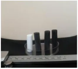
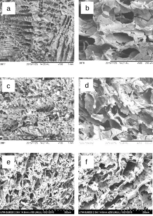
Figure 1 shows the physical appearance of scaffolds. Figure 2 represents the SEM micrographs of PCL, PCL/HA and PCL/HA/PPY composite scaffolds at different concentrations produced through freeze drying technique revealing the porous structure of the scaffold. Using this technique, comparatively large, three dimensional (3D) and homogeneous scaffolds were produced. The scaffolds were easy to handle.
Figure 1 Physical appearance of scaffolds
Many parameters influence the composite
immiscible phases, in order to achieve scaffolds with preferred porous structure and properties. In both PCL/HA and PCL/HA/PPY composite scaffolds, HA nanoparticle agglomeration were not detected. Homogenizer was used at high speed to reduce the agglomeration and to disperse the HA nanoparticles in the polymer pore walls. The distinctive pore morphology of the scaffolds was as a result of the solid-liquid phase separation and the heat transfer during freezing as well as freeze-drying process.
Figure 2 SEM micrographs of scaffolds, (a, b) PCL, (c, d)PCL/HA and (e, f) PCL/HA/PPY at different magnifications
Through suitable parameters, the scaffolds
produced had interconnected pores and large pore sizes. The pore sizes were observed to decrease when HA was incorporated with PCL compared to pure PCL scaffold. It was observed that by incorporating 10% PPY into 10%HA/PCL, the pore sizes of the PCL/HA/PPY scaffold continued to decrease when compared to PCL and PCL/HA scaffold.
Figure 3 shows the EDX analysis of PCL, PCL/HA and PCL/HA/PPY confirming the presence of elements in each scaffold. Calcium and Phosphorus that were presented in HA nanoparticles was observed on the pore walls of PCL/HA, and PCL/HA/PPY composite scaffolds.
Element Weight % Weight % σ Atomic %
Carbon 64.810 0.831 71.042 Oxygen 35.190 0.831 28.958
(a)
Element Weight % Weight % σ Atomic %
Carbon 64.318 0.808 71.368 Oxygen 33.359 0.816 27.789 Phosphorus 0.721 0.092 0.310
Calcium 1.602 0.124 0.533
(b)
Element Weight % Weight % σ Atomic %
Carbon 62.936 0.863 70.455 Nitrogen 0.000 0.000 0.000
Oxygen 33.713 0.874 28.333 Phosphorus 0.885 0.106 0.384
Calcium 2.466 0.157 0.827
(c)
Figure 3 EDX spectra and elemental compositions of the scaffolds (a) PCL, (b) PCL/HA (c) PCL/HA/PPY
10% (v/v) and 15% (v/v) PPY added in PCL/HA were examined using Energy Dispersive X-ray spectroscopy (EDX) to ascertain the existence of Nitrogen (N) element on the scaffolds.The existence of N element established that the mixing was effective, owing to the fact that N can only exist in PPY, not in PCL and HA. The percentage of N was very low; this may be due to
a
b
c
d
the low concentrations used in the process. EDX was also used to confirm the presence of other elements such as Carbon (C), Oxygen (O), Calcium (Ca), and Phosphorous (P) in the PCL/HA/PPY composite. The presence of C element was 65%, O element 30%, Ca. element 2.9, P element 0.6% and N element 0.0%.
3.2 Pore size Analysis of the Scaffolds
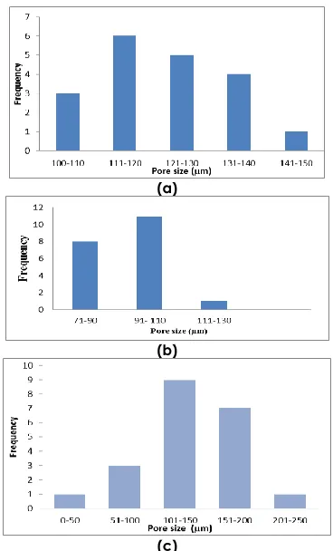
Figure 4 represents the distributions of pore sizes of PCL, PCL/HA and PCL/HA/PPY composite scaffolds. 20 pore sizes were taken and the average pore size was calculated. Average pore sizes of composite scaffolds are shown in Figure 4 (a, b and c). The pore sizes were in the range between 109.6µm and 147.6μm and with the average of 123.7μm for PCL scaffold whereas for PCL/HA were between 77.3μm and 113.6μm and average pore size was 91.6μm. PCL/HA/PPY had pore sizes ranging from 50μm to 250μm with average of 130.4µm. It was observed that 10% PPY incorporated into 10% HA/PCL and pure PCL had larger pore sizes compared to PCL/HA. However, in this study both pure PCL and PCL/HA/PPY attained the minimum pore size required for cell vascularization, transport and migration [20].
3.3 Elemental Mapping of the Scaffolds
Figure 5 represents the elemental mapping of PCL/HA/PPY composite scaffold revealing the elements presented in PCL/HA/PPY scaffold. The elements are represented with different colors to show their abundance and concentration. The elements presented included carbon, oxygen, calcium, phosphorous and nitrogen. The most abundant element was carbon while the least was nitrogen.
3.4 Swelling Studies
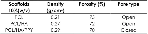
Table 1 shows the porosity and density of the fabricated scaffold. The porosity and density of 10% PCL, 10% HA/PCL and 10% PPY/10%HA/PCL scaffolds were evaluated utilizing liquid displacement method with ethanol. It was observed that ethanol infiltrated inside the pores of the scaffolds but caused neither shrinkage nor swelling of the composites scaffolds.
The fabricated scaffold had porosity more than 65%. The density of the scaffolds increased and the porosity decreased about 5% after incorporation of PPY conductive polymer. According to earlier report, the preferred porosity for cell penetration is between 60–90% [21]. In this study, the results disclosed that the scaffolds had porosity within the preferred range.
(a)
(b)
(c)
Figure 4 The pore size distribution of the scaffolds (a) PCL, (b) PCL/HA and (c) PCL/HA/PPY
Table 1 Density and porosity measurement of scaffolds
Scaffolds 10%(w/v)
Density (g/cm3)
Porosity (%) Pore type
PCL 0.21 75 Open PCL/HA 0.27 72 Open PCL/HA/PPY 0.29 70 Closed
4.0 CONCLUSIONS
By combining conductive polymer PPY, synthetic polymer PCL and bioceramic HA, it was possible to fabricate an electrically conductive composite scaffold for the application of bone tissue engineering using the freeze-drying technique. The range of pores of PCL/HA/PPY scaffold was 50-250μm. EDX analysis confirmed the presence of Nitrogen in the PCL/HA/PPY scaffold. It was observed that the density increased and the porosity decreased with the incorporation of HA and PPy. The properties of the scaffolds in this study were promising to be used in bone tissue engineering applications.
Acknowledgement
This research was supported by HiCOE grant (4J191) and GUP Tier 1 (12H24). The authors fully acknowledged Ministry of Higher Education (MOHE) and Universiti Teknologi Malaysia, RMC, FBME and AMTEC for the supports.
References
[1] Chen, L., Hu, J., Shen, X. and Tong, H. 2013. Synthesis And
Characterization Of Chitosan–Multiwalled Carbon
Nanotubes/Hydroxyapatite Nanocomposites For Bone Tissue Engineering. Journal of Materials Science: Materials in Medicine. 24(8): 1843-1851.
[2] Sultana, N., Mokhtar, M., Hassan, M. I., Mad Jin, R., Roozbahani, F., Khan, T. H. 2015. Chitosan-based
Nanocomposite Scaffolds For Tissue Engineering
Applications. Materials and Manufacturing Processes. 30(3): 373-278
[3] Vacanti, J. P. and Langer, R. 1999. Tissue Engineering: The Design And Fabrication Of Living Replacement Devices For Surgical Reconstruction And Transplantation. The Lancet. 354: S32-S34.
[4] Lanza, R., Langer, R. and Vacanti, J. P. eds. 2011. Principles of Tissue Engineering. Academic Press.
[5] Sultana, N. and Wang, M. 2007. Fabrication And
Characterisation Of Polymer And Composite Scaffolds Based On Polyhydroxybutyrate And Polyhydroxybutyrate-Co-Hydroxyvalerate. Key Engineering Materials. 334: 1229-1232.
[6] Jin, R. M., Sultana, N., Baba, S., Hamdan, S. and Ismail, A. F. 2015. Porous PCL/Chitosan and nHA/PCL/Chitosan Scaffolds
For Tissue Engineering Applications: Fabrication And Evaluation. Journal of Nanomaterials. 1-8.
[7] Kim, J. Y., Lee, T. J., Cho, D. W. and Kim, B. S. 2010. Solid Free-Form Fabrication-Based PCL/HA Scaffolds Fabricated With A Multi-Head Deposition System For Bone Tissue Engineering. Journal of Biomaterials Science, Polymer Edition. 21(6-7): 951-962.
[8] Choong, C., Triffitt, J. T. and Cui, Z. F. 2004. Polycaprolactone Scaffolds For Bone Tissue Engineering: Effects Of A Calcium Phosphate Coating Layer On Osteogenic Cells. Food and Bioproducts Processing. 82(2): 117-125.
[9] Sultana, N. and Kadir, M. R. A. 2011. Study Of In Vitro Degradation Of Biodegradable Polymer Based Thin Films And Tissue Engineering Scaffolds. African Journal of Biotechnology. 10(81): 18709-18715.
[10] Cheng, M., Deng, J., Yang, F., Gong, Y., Zhao, N. and Zhang, X. 2003. Study On Physical Properties And Nerve Cell Affinity Of Composite Films From Chitosan And Gelatin Solutions. Biomaterials. 24(17): 2871-2880.
[11] Guimard, N. K., Gomez, N. and Schmidt, C. E. 2007. Conducting Polymers In Biomedical Engineering. Progress in Polymer Science. 32(8): 876-921.
[12] Mozafari, M., Vashaee, D., Tayebi, L. and Mehraien, M. 2012. Electroconductive Nanocomposite Scaffolds: A New Strategy Into Tissue Engineering And Regenerative Medicine. INTECH Open Access Publisher. 369-392.
[13] Li, M., Guo, Y., Wei, Y., MacDiarmid, A. G. and Lelkes, P. I. 2006. Electrospinning Polyaniline-Contained Gelatin Nanofibers For Tissue Engineering Applications. Biomaterials. 27(13): 2705-2715.
[14] Balint, R., Cassidy, N. J. and Cartmell, S. H. 2014. Conductive Polymers: Towards A Smart Biomaterial For Tissue Engineering. Acta biomaterialia. 10(6): 2341-2353.
[15] Chronakis, I. S., Grapenson, S. and Jakob, A. 2006. Conductive Polypyrrole Nanofibers Via Electrospinning: Electrical And Morphological Properties. Polymer. 47(5): 1597-1603.
[16] Chen, Q. Z., Thompson, I. D. and Boccaccini, A. R. 2006. 45S5 Bioglass®-derived Glass–Ceramic Scaffolds For Bone Tissue Engineering. Biomaterials. 27(11): 2414-2425.
[17] Yoshikawa, H. and Myoui, A. 2005. Bone Tissue Engineering With Porous Hydroxyapatite Ceramics. Journal of Artificial Organs. 8(3): 131-136.
[18] Hsu, Y. Y., Gresser, J. D., Trantolo, D. J., Lyons, C. M., Gangadharam, P. R. and Wise, D. L. 1997. Effect Of Polymer Foam Morphology And Density On Kinetics Of In Vitro Controlled Release Of Isoniazid From Compressed Foam Matrices. Journal of Biomedical Materials Research. 35(1): 107-116.
[19] Sultana, N. and Wang, M. 2008. Fabrication of HA/PHBV Composite Scaffolds Through The Emulsion Freezing/Freeze-Drying Process And Characterisation Of The Scaffolds.
Journal of Materials Science: Materials in Medicine. 19(7): 2555-2561.
[20] Cannillo, V., Chiellini, F., Fabbri, P. and Sola, A. 2010. Production of Bioglass® 45S5–Polycaprolactone Composite Scaffolds Via Salt-Leaching. Composite Structures. 92(8): 1823-1832.
[21] Chong, E. J., Phan, T. T., Lim, I. J., Zhang, Y. Z., Bay, B. H., Ramakrishna, S. and Lim, C. T. 2007. Evaluation Of Electrospun PCL/Gelatin Nanofibrous Scaffold For Wound
Healing And Layered Dermal Reconstitution. Acta